

Improved Detection of Target Metabolites in Brain Tumors with Intermediate TE, High SNR, and High Bandwidth Spin-Echo Sequence at 5T
- PMID: 38453417
- PMCID: PMC11288575
- DOI: 10.3174/ajnr.A8150
Improved Detection of Target Metabolites in Brain Tumors with Intermediate TE, High SNR, and High Bandwidth Spin-Echo Sequence at 5T
Abstract
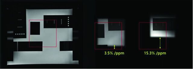
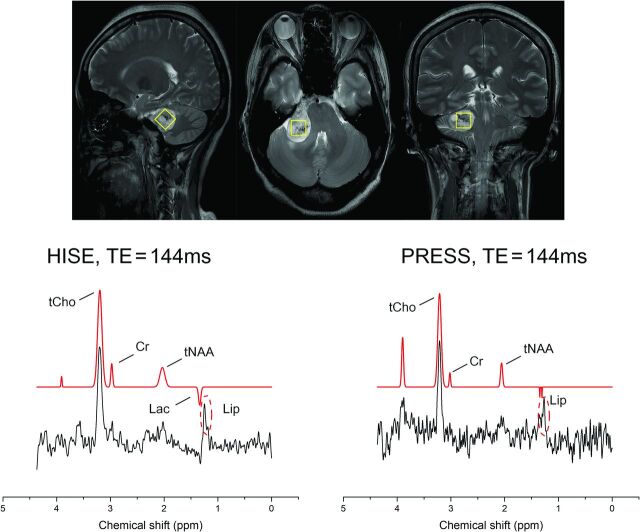
Background and purpose: Due to high chemical shift displacement, challenges emerge at ultra-high fields when measuring metabolites using 1H-MRS. Our goal was to investigate how well the high SNR and high bandwidth spin-echo (HISE) technique perform at 5T for detecting target metabolites in brain tumors.
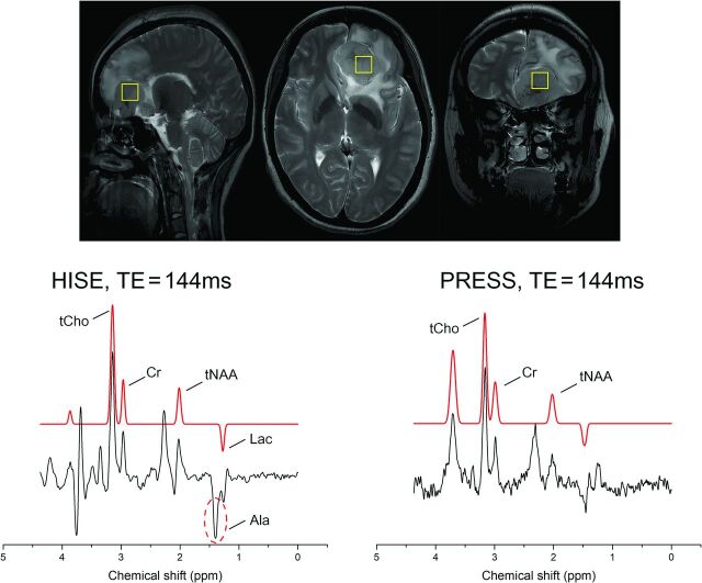
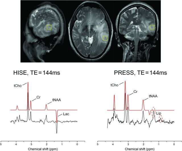
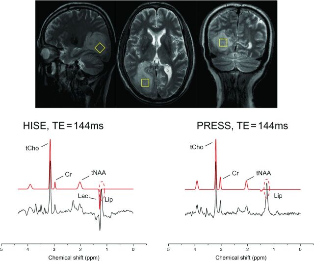
Materials and methods: Twenty-six subjects suspected of having brain tumors were enrolled. HISE and point-resolved spectroscopy (PRESS) single-voxel spectroscopy scans were collected with a 5T clinical scanner with an intermediate TE (TE = 144 ms). The main metabolites, including total NAA, Cr, and total Cho, were accessed and compared between HISE and PRESS using a paired Student t test, with full width at half maximum and SNR as covariates. The detection rate of specific metabolites, including lactate, alanine, and lipid, and subjective spectral quality were accessed and compared between HISE and PRESS.
Results: Twenty-three pathologically confirmed brain tumors were included. Only the full width at half maximum for total NAA was significantly lower with HISE than with PRESS (P < .05). HISE showed a significantly higher SNR for total NAA, Cr, and total Cho compared with PRESS (P < .05). Lactate was detected in 21 of the 23 cases using HISE, but in only 4 cases using PRESS. HISE detected alanine in 8 of 9 meningiomas, whereas PRESS detected alanine in just 3 meningiomas. PRESS found lipid in more cases than HISE, while HISE outperformed PRESS in terms of subjective spectral quality.
Conclusions: HISE outperformed the clinical standard PRESS technique in detecting target metabolites of brain tumors at 5T, particularly lactate and alanine.
© 2024 by American Journal of Neuroradiology.
Figures
Similar articles
-
Comparison of 1.5T and 3T 1H MR spectroscopy for human brain tumors.Korean J Radiol. 2006 Jul-Sep;7(3):156-61. doi: 10.3348/kjr.2006.7.3.156. Korean J Radiol. 2006. PMID: 16969044 Free PMC article.
-
Single voxel proton MR spectroscopy findings of typical and atypical intracranial meningiomas.Eur J Radiol. 2006 Oct;60(1):48-55. doi: 10.1016/j.ejrad.2006.06.002. Epub 2006 Jul 17. Eur J Radiol. 2006. PMID: 16844335
-
Utilization of glutamate/creatine ratios for proton spectroscopic diagnosis of meningiomas.Neuroradiology. 2007 Feb;49(2):121-7. doi: 10.1007/s00234-006-0167-z. Epub 2006 Nov 4. Neuroradiology. 2007. PMID: 17086406 Clinical Trial.
-
Evaluation of the diagnostic utility on 1.5T and 3.0T 1H magnetic resonance spectroscopy for temporal lobe epilepsy.BMC Med Imaging. 2023 Nov 14;23(1):185. doi: 10.1186/s12880-023-01136-w. BMC Med Imaging. 2023. PMID: 37964218 Free PMC article.
-
Proton MR spectroscopic characteristics of pediatric pilocytic astrocytomas.AJNR Am J Neuroradiol. 1998 Mar;19(3):535-40. AJNR Am J Neuroradiol. 1998. PMID: 9541314 Free PMC article. Review.
References
MeSH terms
Substances
LinkOut - more resources
Full Text Sources
Medical