Review
. 2024 Mar 8;9(1):55.
doi: 10.1038/s41392-024-01769-5.

Ferroptosis in cancer: From molecular mechanisms to therapeutic strategies

Affiliations
Review

Ferroptosis in cancer: From molecular mechanisms to therapeutic strategies

Qian Zhou et al. Signal Transduct Target Ther. .

Abstract

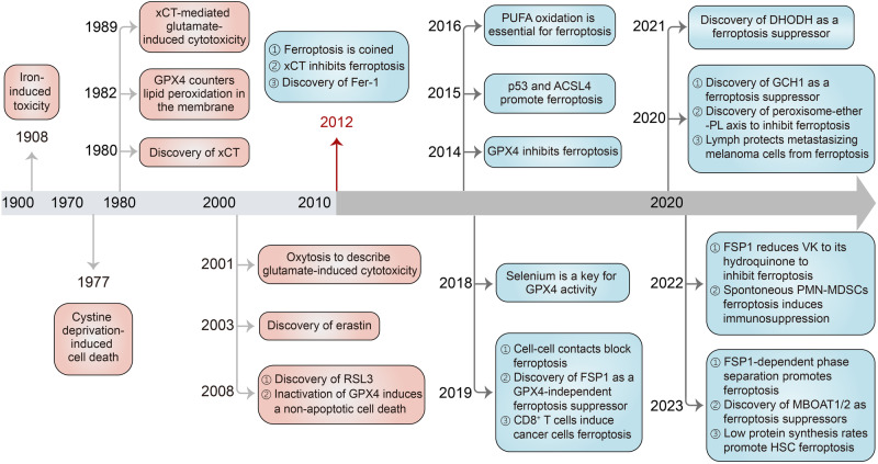
Ferroptosis is a non-apoptotic form of regulated cell death characterized by the lethal accumulation of iron-dependent membrane-localized lipid peroxides. It acts as an innate tumor suppressor mechanism and participates in the biological processes of tumors. Intriguingly, mesenchymal and dedifferentiated cancer cells, which are usually resistant to apoptosis and traditional therapies, are exquisitely vulnerable to ferroptosis, further underscoring its potential as a treatment approach for cancers, especially for refractory cancers. However, the impact of ferroptosis on cancer extends beyond its direct cytotoxic effect on tumor cells. Ferroptosis induction not only inhibits cancer but also promotes cancer development due to its potential negative impact on anticancer immunity. Thus, a comprehensive understanding of the role of ferroptosis in cancer is crucial for the successful translation of ferroptosis therapy from the laboratory to clinical applications. In this review, we provide an overview of the recent advancements in understanding ferroptosis in cancer, covering molecular mechanisms, biological functions, regulatory pathways, and interactions with the tumor microenvironment. We also summarize the potential applications of ferroptosis induction in immunotherapy, radiotherapy, and systemic therapy, as well as ferroptosis inhibition for cancer treatment in various conditions. We finally discuss ferroptosis markers, the current challenges and future directions of ferroptosis in the treatment of cancer.


Conflict of interest statement

The authors declare no competing interests.

Figures

Fig. 1
Fig. 1
History of research on the discovery and development of ferroptosis. The term ferroptosis was coined in 2012, but the understanding of ferroptosis can be traced back as early as 1908. Since 2012, there has been a flourishing development in the research of ferroptosis and its regulatory mechanisms. ACSL4 acyl-CoA synthetase long-chain family member 4, DHODH dihydroorotate dehydrogenase, Fer-1 ferrostatin-1, FSP1 ferroptosis suppressor protein 1, GCH1 GTP cyclohydrolase 1, GPX4 glutathione peroxidase 4, HSC hematopoietic stem cells, MBOAT1/2 membrane-bound O-acyltransferase domain-containing 1 and 2, PL phospholipid, PMN-MDSC polymorphonuclear myeloid-derived suppressor cell, PUFA polyunsaturated fatty acid, VK vitamin K
Fig. 2
Fig. 2
Molecular mechanisms of ferroptosis. Ferroptosis is driven by PUFA-PLs synthesis, lipid peroxidation and iron toxicity. Major defense systems of ferroptosis include the GPX4 antioxidant system, FSP1/ubiquinol (CoQH2), DHODH/CoQH2, GCH1/tetrahydrobiopterin (BH4) systems, monounsaturated fatty acid (MUFA)-PLs synthesis, and the ESCRT-III-mediated membrane repair systems. When ferroptosis-promoting activities significantly surpass the detoxification capabilities provided by the defense systems, a fatal accumulation of lipid peroxides on the cellular membranes ultimately results in membrane rupture and ferroptotic cell death. ABCB7 ATP binding cassette subfamily B member 7, ACC acetyl-CoA carboxylase, ALOX lipoxygenase, CISD1 CDGSH iron sulfur domain 1, CoQ coenzyme Q, Cys cysteine, Cys2 cystine, FTMT ferritin mitochondrial, GCL glutamate-cysteine ligase, GSH glutathione, GSSG oxidized glutathione, iPLA2b phospholipase A2 group VI, LIP labile iron pool, LPCAT3 lysophosphatidylcholine acyltransferase 3, NAD(P)H nicotinamide adenine dinucleotide phosphate, POR cytochrome P450 oxidoreductase, SCD1 stearoyl-CoA desaturase 1, SFA saturated fatty acid, SLC25A37, solute carrier family 25 member 37, SLC25A28 solute carrier family 25 member 28, SLC40A1 solute carrier family 40 member 1, STARD7 StAR-related lipid transfer domain containing 7, TF transferrin, TFR1 transferrin receptor, VDAC voltage-dependent anion channel. This figure was created with BioRender.com
Fig. 3
Fig. 3
Cancer-related pathways in ferroptosis. a RAS signaling governs upregulation of SCL7A11, FASN, and FSP1 to evade ferroptosis, establishing a targetable vulnerability. b NRF2 protects cancer cells from ferroptosis primarily through transcriptional regulation of downstream target genes involved in iron metabolism, GSH metabolism and ROS detoxification enzymes. c mTOR signaling primarily inhibits the sensitivity to ferroptosis through autophagy, promoting GPX4 protein synthesis, and upregulating the SREBP1/SCD and KEAP1/NRF2 axis. d Hypoxia plays a dual role in regulating ferroptosis by inducing the expression of its primary regulators HIF1α and HIF2α. e EMT reshapes the metabolic status granting mesenchymal tumor cells vulnerability to ferroptosis. f p53 transcriptionally suppresses SLC7A11 expression or modulates metabolism-related genes to promote ferroptosis. g The YAP/TAZ pathway plays a crucial role in regulating cell density-mediated and D-lactate-induced ferroptosis. h Ferroptosis serves as a type of autophagy-dependent cell death involving ferritinophagy, lipophagy, mitophagy, clockophagy, and chaperone-mediated autophagy. i Mitochondrial TCA cycle, ETC and glutamate are required for cystine deprivation-induced ferroptosis. PPP generate NADPH to implicate in ferroptosis process. Energy stresses facilitate tumor defense against ferroptosis by activating AMPK to enhance ACC-mediated MUFA formation. 4EBP 4E (eIF4E)-binding proteins, α-KG α-Ketoglutaric acid, ACSL5 acyl-CoA synthetase long chain family member 5, AKT AKT serine/threonine kinase, ASS1 argininosuccinate synthase 1, AKR1C1 aldo-keto reductase family 1 member C1, ANGPTL4 angiopoietin-like 4, ARNTL aryl hydrocarbon receptor nuclear translocator like, AMPK protein kinase AMP-activated catalytic subunit alpha 1, ATM ataxia-telangiectasia mutated, BAMBI BMP and activin membrane bound inhibitor, BRAF B-Raf proto-oncogene, serine/threonine kinase, CDKN1A cyclin dependent kinase inhibitor 1A, CDK7 cyclin dependent kinase 7, CHAC1 ChaC glutathione specific gamma-glutamylcyclotransferase 1, DPP4 dipeptidyl peptidase 4, DPP9 dipeptidyl peptidase 9, EGLN2 egl-9 family hypoxia inducible factor 2, EMP1 epithelial membrane protein 1, EMT epithelial-mesenchymal transition, FABP3/7 fatty acid binding protein 3/7, FASN fatty acid synthase, FTH1 ferritin heavy chain 1, GCLC glutamate-cysteine ligase catalytic subunit, GCLM glutamate-cysteine ligase modifier subunit, GFPT1 glutamine--fructose-6-phosphate transaminase 1, GINS4 GINS complex subunit 4, GLS glutaminase, GLUD1 glutamate dehydrogenase 1, HDAC Type-2 histone deacetylase 2, HIF1α hypoxia inducible factor 1 subunit alpha, HIF2α hypoxia inducible factor 2 subunit alpha, HILPDA hypoxia inducible lipid droplet associated, HMOX1 heme oxygenase 1, HSP90 heat shock protein 90, HSC70 heat shock cognate 71 kDa protein, Keap1 Kelch-1ike ECH- associated protein l, KDM5A lysine demethylase 5A, *KRAS mutant KRAS, KRAS, KRAS proto-oncogene, GTPase, LATS1 large tumor suppressor kinase 1, LAMP2A lysosomal-associated membrane protein 2, LC3 MAP1LC3A microtubule associated protein 1 light chain 3 alpha, LDHD lactate dehydrogenase D, LKB1 Lkb1 kinase, MEK MAP kinase-ERK kinase, MDM2 proto-oncogene, MDMX MDM4 regulator of p53, MEX3A mex-3 RNA binding family member A, mTOR rapamycin target protein, MT1G metallothionein 1G, MPC1 mitochondrial pyruvate carrier 1, MST macrophage stimulating, MYC MYC proto-oncogene, bHLH transcription factor, NCOA4 nuclear receptor coactivator 4, NRF2 nuclear factor erythroid 2-related factor 2, NF2 neurofibromin 2, NOX2 NADPH oxidase 2, NOX4 NADPH oxidase 4, OXPHOX oxidative phosphorylation, PI3K phosphoinositide 3-kinase, PPARGC1A PPARG coactivator 1 alpha, PRMT5 protein arginine methyltransferase 5, RAB7A member RAS oncogene family, SCD5 stearoyl-Coenzyme A desaturase 5, SREBP1 sterol regulatory element-binding protein 1, SESN2 sestrin 2, E-cad E-cadherin, SLC40A1 solute carrier family 40 member 1, SLC7A11 solute carrier family 7 member 11, SOD1 superoxide dismutase 1, SQSTM1 sequestosome 1, TAZ Tafazzin, TXNRD1 thioredoxin reductase 1, TCA cycle tricarboxylic acid cycle, WTAP WT1 associated protein, YAP1 Yes1 associated transcriptional regulator, ZEB1 zinc finger E-box binding homeobox 1, ZNF498 zinc finger and SCAN domain containing 25. This figure was created with BioRender.com
Fig. 4
Fig. 4
Ferroptosis-mediated crosstalk in the tumor microenvironment (TME). a Ferroptotic cancer cells in the TME exhibit dual immunoregulatory effects, encompassing both immunostimulatory and immunosuppressive roles. The emission of various immunomodulatory signals by ferroptotic cancer cells activates different immune responses regulating tumor development. b The pro-ferroptotic and anti-ferroptotic impact on cancer cells mediated by immune cells and adipocytes in the TME. c The mechanisms and tumor-modulating effects of ferroptotic immune cells in the TME, including CD8+ T cells, dendritic cells (DCs), natural killer (NK) cells, tumor-associated macrophages (TAMs), regulatory T cells (Tregs), and myeloid-derived suppressor cells (MDSCs). AA arachidonic acid, AGER advanced glycosylation end product-specific receptor, CAF cancer-associated fibroblast, CRT calreticulin, FATP2 fatty acid transport protein 2, FIN ferroptosis inducer, HMGB1 high-mobility group box 1, IFNγ interferon gamma, 8-OHG 8-hydroxy-2-deoxyguanosine, oxLDL oxidized low-density lipoproteins, STING stimulator of interferon genes, TGF-β, transforming growth factor beta, TLR2 Toll-like receptors 2, ULBP UL16 binding protein. This figure was created with BioRender.com
Fig. 5
Fig. 5
Ferroptosis induction for cancer therapy. a Radiotherapy induces ferroptosis to suppress tumor through the following three mechanisms: ① Radiotherapy-induced DNA damage activates the ATM and cGAS/STING/ATF3 axis, leading to SLC7A11 inhibition and subsequent triggering of ferroptosis.② Radiotherapy upregulates the expression of ACSL4, facilitating the PUFA-PLs formation and inducing ferroptosis. ③ RT-MPs induce ferroptosis in neighboring unirradiated cells relying on the bystander effect. After immunotherapy treatment, activated CD8+ T cells release IFNγ, sensitizing tumor cells to ferroptosis by inhibiting SLC7A11, and promoting ACSL4-mediated PUFA-PLs formation, ultimately triggering ferroptosis. Immunotherapy and radiotherapy synergistically inhibit tumors by suppressing SLC7A11. b, c Major systemic drugs and experimental tool compounds for effective treatment of tumors through ferroptosis induction. ATF3 activation transcription factor 3, cGAS cyclic GMP-AMP synthase, cGAMP cyclic 2’,3’-GMP-AMP, DHA dihydroartemisinin, DHODH dihydroorotate dehydrogenase, FSP1 ferroptosis suppressor protein 1, FTH1 ferritin heavy chain 1, GCL glutamate-cysteine ligase, IFNγ interferon gamma, IKE imidazole ketone erastin, RT-MPs irradiated tumor cell-derived microparticles, STING stimulator of interferon genes. This figure was created with BioRender.com

References

    1. Hotchkiss, R. S., Strasser, A., McDunn, J. E. & Swanson, P. E. Cell death. N. Engl. J. Med361, 1570–1583 (2009). 10.1056/NEJMra0901217 - DOI - PMC - PubMed
    1. Galluzzi, L. et al. Essential versus accessory aspects of cell death: recommendations of the NCCD 2015. Cell Death Differ.22, 58–73 (2015). 10.1038/cdd.2014.137 - DOI - PMC - PubMed
    1. Green, D. R. The coming decade of cell death research: five riddles. Cell177, 1094–1107 (2019). 10.1016/j.cell.2019.04.024 - DOI - PMC - PubMed
    1. Dixon, S. J. et al. Ferroptosis: an iron-dependent form of nonapoptotic cell death. Cell149, 1060–1072 (2012). 10.1016/j.cell.2012.03.042 - DOI - PMC - PubMed
    1. Kung, G., Konstantinidis, K. & Kitsis, R. N. Programmed necrosis, not apoptosis, in the heart. Circ. Res. 108, 1017–1036 (2011). 10.1161/CIRCRESAHA.110.225730 - DOI - PubMed

Publication types