. 2024 Mar 7;15(1):2092.
doi: 10.1038/s41467-024-46107-z.

Enhancing prime editor activity by directed protein evolution in yeast

Affiliations

Enhancing prime editor activity by directed protein evolution in yeast

Yanik Weber et al. Nat Commun. .

Abstract

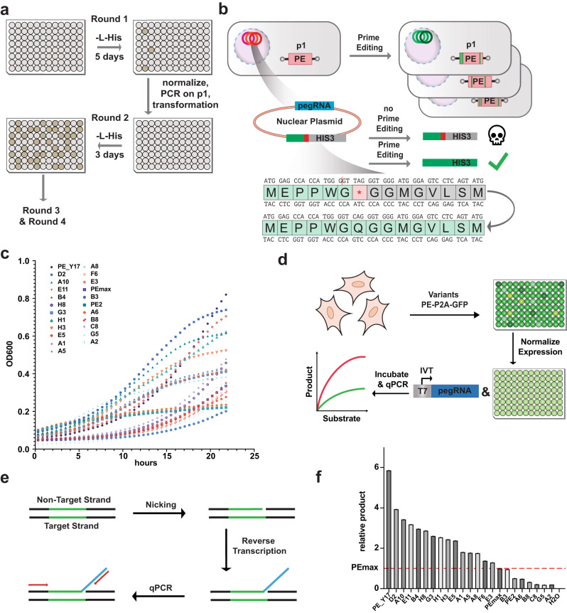
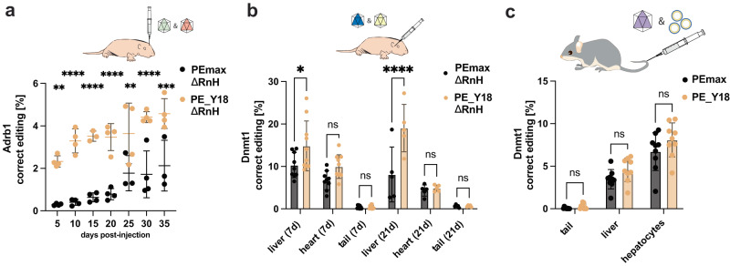
Prime editing is a highly versatile genome editing technology that enables the introduction of base substitutions, insertions, and deletions. However, compared to traditional Cas9 nucleases prime editors (PEs) are less active. In this study we use OrthoRep, a yeast-based platform for directed protein evolution, to enhance the editing efficiency of PEs. After several rounds of evolution with increased selection pressure, we identify multiple mutations that have a positive effect on PE activity in yeast cells and in biochemical assays. Combining the two most effective mutations - the A259D amino acid substitution in nCas9 and the K445T substitution in M-MLV RT - results in the variant PE_Y18. Delivery of PE_Y18, encoded on DNA, mRNA or as a ribonucleoprotein complex into mammalian cell lines increases editing rates up to 3.5-fold compared to PEmax. In addition, PE_Y18 supports higher prime editing rates when delivered in vivo into the liver or brain. Our study demonstrates proof-of-concept for the application of OrthoRep to optimize genome editing tools in eukaryotic cells.


Conflict of interest statement

Y.W. and G.S. have filed a patent application based on evolved PEmax variants as inventors (European Patent Application No. EP24152221.8). G.S. is a scientific advisor to Prime Medicine. The remaining authors declare no competing interests.

Figures

Fig. 1
Fig. 1. Evolution of PE variants with enhanced activity in OrthoRep.
a Parallel evolution of prime editors (PE) by culturing yeast cells in 96 well plates over four subsequent rounds in L-histidine-depleted (-L-His) selection media. In the first round of evolution, outgrowing yeast cells were normalized prior to extraction of the linear plasmid (p1) via PCR and transformation into fresh host cells for the second round. The same procedure was repeated for the third and fourth round. b PE variants containing mutations that increase prime editing rates are enriched in selective conditions: a stop codon in front of the auxotrophic marker Imidazoleglycerol-phosphate dehydratase (HIS3) must be repaired by prime editing on the nuclear plasmid co-expressing the prime editing guide RNA (pegRNA) for successful yeast growth in selective conditions. c Effect of evolved PE variants on yeast growth quantified as optical density at 600 nm (OD600) under selective conditions. The assessed variants contained the following mutations: PE_Y17 (nCas9 S219A, RT K445T), D2 (nCas9 A259D), A10 (RT Y64W, K373R, R389C, L432M, K445T), E11 (RT Y64W), B4 (RT R44H), H8 (nCas9 S219A, RT K445T), G3 (nCas9 A259D, linker G30S, RT R389C, S606A), H1 (nCas9 A259D, RT Y64W), H3 (nCas9 A259D) E5 (nCas9 S219A, RT K445T), A1 (nCas9 A259D, linker G30S, RT R389C, S606A), A5 (nCas9 S219A, RT Y64C, K445T), A8 (nCas9 S219A, linker G30S, RT K445T), F6 (nCas9 A259D, RT R44H), E3 (nCas9 S320R), B3 (nCas9 R71C, A259D, linker G30S, RT K445T), A6 (nCas9 S318N, RT R389C, L432M), B8 (nCas9 S320N,RT K373R), C8 (nCas9 Y132C), G5 (nCas9 A259D,RT K373R), A2 (nCas9 S219A, RT K373R, K445T). Data are displayed as the mean of two individual replicates. d Schematic of the in vitro assay to assess PE kinetics (PEKIN). PE variants are subcloned as self-cleaving peptide fused to a green fluorescent protein (P2A-GFP) and transfected into HEK293T cells. Cells are lysed and PE expression levels are normalized by fluorescence intensity. An in vitro transcribed (IVT) pegRNA forms the RNP with the PE, which is incubated with a synthetic dsDNA substrate. e Schematics illustrating the strategy used to quantify PE activity in PEKIN via quantitative PCR (qPCR). f Quantified prime editing activities of the 21 PE variants isolated after four rounds of evolutions in OrthoRep relative to PEmax. Product formation of the variants is relative to the product formed by PEmax and was performed as a single replicate (n = 1).
Fig. 2
Fig. 2. In vitro characterization of the activity of PE variants using cell lysates and purified proteins.
a Quantification of PE activity with increasing concentrations of dsDNA substrate for PEmax (black), five selected PE variants obtained from OrthoRep (solid lines), and recombined versions of these variants (dashed lines). Individual values are represented from biological replicates (n = 2). b Schematic illustration of constructs that were characterized in PEKIN as purified proteins including the bipartite nuclear localisation signal from the siman virus 40 (bpSV40 NLS) and the nuclear localisation signal (NLS c-Myc) where indicated. PEmax was compared to the evolved variants PE_Y17 and PE_Y18 in a tethered and untethered form. c Quantification of prime editing rates of purified proteins in the PEKIN assay in the tethered (solid lines) and untethered form (dashed lines). Virtual products are calculated from respective Ct values of individual triplicates (n = 3). d Scheme of the assay used to determine nicking activity via double nicking of a synthetic DNA template. Red illustrates the non-target strand, whereas the target strand is illustrated in green. e Schematic of the adapted assay to disentangle reverse transcription activity on a single-stranded DNA (ssDNA) oligo via a complementary RNA template. f Quantification of ∆Ct of double nicked templates relative to the untreated quantified substrate concentrations. Experiments were performed as individual duplicates (n = 2). g Quantification of reverse transcribed product on a ssDNA oligo. Experiments were performed as individual duplicates (n = 2).
Fig. 3
Fig. 3. Comparison of editing rates with evolved PE variants in mammalian cell lines.
a Correct percentage of substitutions assessed by deep sequencing in cells transfected with plasmids encoding for PEmax, PE_Y17, PE_Y18 together with plasmids encoding for the enhanced prime editor guide RNAs (epegRNAs) at different time points on site 1 in HEK293T cells; not significant (ns) P = 0.5476 ****P < 0.0001, ***P = 0.0002, ****P < 0.0001, ***P = 0.0002 and **P < 0.0012 (left to right). b Editing rates of PE_Y17, PE_Y18 and PEmax at other loci in HEK293T cells, analyzed 48 h after plasmid transfection; not significant (ns) P = 0.9462, ns P = 0.2578, **P = 0.0084, ****P < 0.0001, ns P = 0.8254, *P = 0.0478, **P = 0.001, ****P < 0.0001, *P = 0.0341, ***P = 0.0008, ns P = 0.6543, ns P = 0.1331, ns P = 0.1621, ***P = 0.0002, *P = 0.011, **P = 0.0086, ns P = 0.6507, ns P = 0.6442, ns P = 0.7257 and ns P = 0.1566 (left to right). c Editing rates of PEmax, PE_Y17 and PE_Y18 on endogenous loci in K562 cells, analyzed 120 h after transfection; not significant (ns) P = 0.3622, ****P < 0.0001, ns P = 0.8171, ns P = 0.2033, **P = 0.0049, ****P < 0.0001, **P = 0.0022 and ****P < 0.0001 (left to right). d Editing rates of PEmax, PE_Y17 and PE_Y18 on a self-targeting library in K562 cells. The self-targeting library encoding for epegRNAs and their respective target sites was integrated into cells using lentiviral vectors prior to PE plasmid transfection and analysis of editing rates by deep sequencing after 120 h; not significant (ns) P = 0.305 and *P < 0.0486 (left to right). e Editing rates with PE variants encoded on mRNA and nucleofected into HEK293T cell line expressing an epegRNA targeting site 12; not significant (ns) P = 0.1152 and *P = 0.0145 (left to right). f Nucleofection of PEmax, PE_Y17 or PE_Y18 protein into HEK293T cells expressing an epegRNA targeting site 12; not significant (ns) P = 0.6306 and **P = 0.0042 (left to right). g Nucleofection of PE RNPs with a chemically modified pegRNA targeting site 4 in HEK293T cells; **P = 0.0079, ***P = 0.0002 (left to right). Data are displayed as means±s.d. of three independent experiments (n = 3) and were analyzed using a two-way ANOVA using Tukey’s multiple comparisons.
Fig. 4
Fig. 4. In vivo comparison of PE_Y18∆RnH and PEmax delivered via AAV or mRNA-LNP.
a Experimental setup and editing rates at the targeted Adrb1 locus in cortices. Intein-split PEmax∆RnH and PE_Y18∆RnH were packaged into AAV-PHP.eB capsids and injected intracerebroventricular into P1 mice. For PE_Y18∆RnH, four animals were individually treated for each timepoint. For PEmax, time points 5, 10, 15, 20, and 30 days were assessed by four (n = 4) individually treated animals, whereas three (n = 3) animals were treated for 25 and 35 days respectively; **P = 0.0012, ****P < 0.0001, ****P < 0.0001, ****P < 0.0001, **P = 0.0071, ****P < 0.0001 and ***P = 0.0003 (left to right). b Editing rates at the targeted Dnmt1 locus, assessed in different tissues at days 7 and 21 after temporal vein injection of the intein-split PE_Y18∆RnH and PEmax∆RnH packaged in AAV9. For PEmax∆RnH nine animals (n = 9) were individually treated for the time point 7d and five animals (n = 5) for 21d respectively. For PE_Y18∆RnH ten animals (n = 10) were treated for time point 7d and five (n = 5) for 21d respectively; *P = 0.0199, not significant (ns) P = 0.1939, ns P > 0.9999, ****P < 0.0001, ns P > 0.9999 and ns P > 0.9999 (left to right). c Four weeks before single administration of mRNA-LNPs (2 mg/kg PEmax or PE_Y18 mRNA) via the tail vein, male mice were injected with an scAAV9 expressing the epegRNA that targets the Dnmt1 locus. Editing rates after LNP administration were assessed in the tail, liver tissue, and isolated hepatocytes. Organs were analyzed from nine individually treated animals (n = 9) for PEmax and PE_Y18; not significant (ns) P = 0.999, ns P = 0.2932 and ns P = 0.1306 (left to right). Data are displayed as means±s.d. of the indicated and were analyzed using a two-way ANOVA using Tukey’s multiple comparisons.

References

    1. Anzalone AV, et al. Search-and-replace genome editing without double-strand breaks or donor DNA. Nature. 2019;576:149–157. doi: 10.1038/s41586-019-1711-4. - DOI - PMC - PubMed
    1. Anzalone AV, Koblan LW, Liu DR. Genome editing with CRISPR–Cas nucleases, base editors, transposases and prime editors. Nat. Biotechnol. 2020;38:824–844. doi: 10.1038/s41587-020-0561-9. - DOI - PubMed
    1. Nelson JW, et al. Engineered pegRNAs improve prime editing efficiency. Nat. Biotechnol. 2022;40:402–410. doi: 10.1038/s41587-021-01039-7. - DOI - PMC - PubMed
    1. Chen PJ, et al. Enhanced prime editing systems by manipulating cellular determinants of editing outcomes. Cell. 2021;184:5635–5652.e29. doi: 10.1016/j.cell.2021.09.018. - DOI - PMC - PubMed
    1. Doman JL, et al. Phage-assisted evolution and protein engineering yield compact, efficient prime editors. Cell. 2023;186:3983–4002.e26. doi: 10.1016/j.cell.2023.07.039. - DOI - PMC - PubMed