. 2024 Mar 8;9(1):63.
doi: 10.1038/s41392-024-01761-z.

Alpha5 nicotine acetylcholine receptor subunit promotes intrahepatic cholangiocarcinoma metastasis

Affiliations

Alpha5 nicotine acetylcholine receptor subunit promotes intrahepatic cholangiocarcinoma metastasis

Yan Fu et al. Signal Transduct Target Ther. .

Abstract

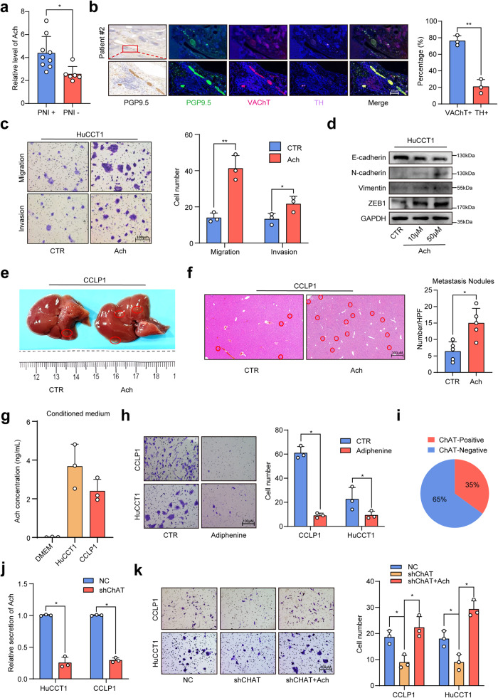
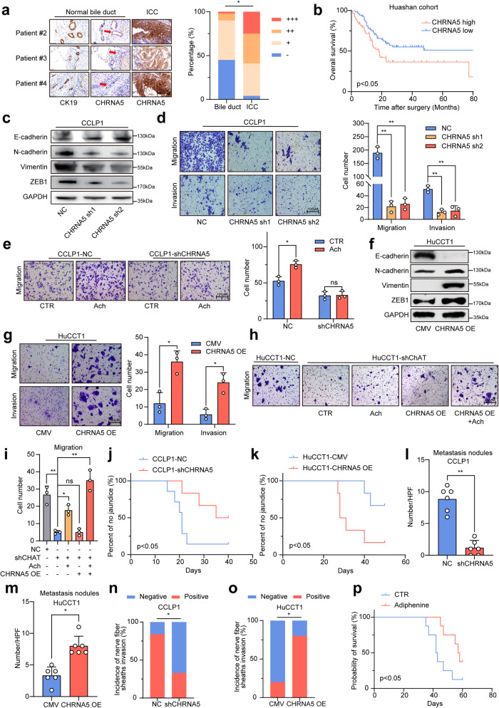
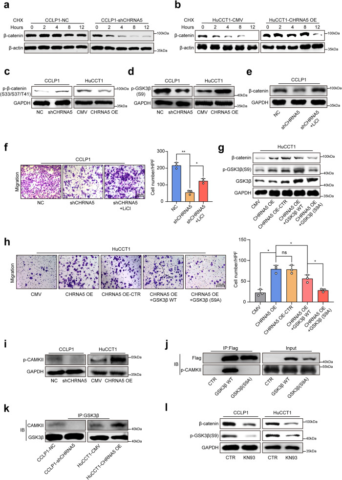
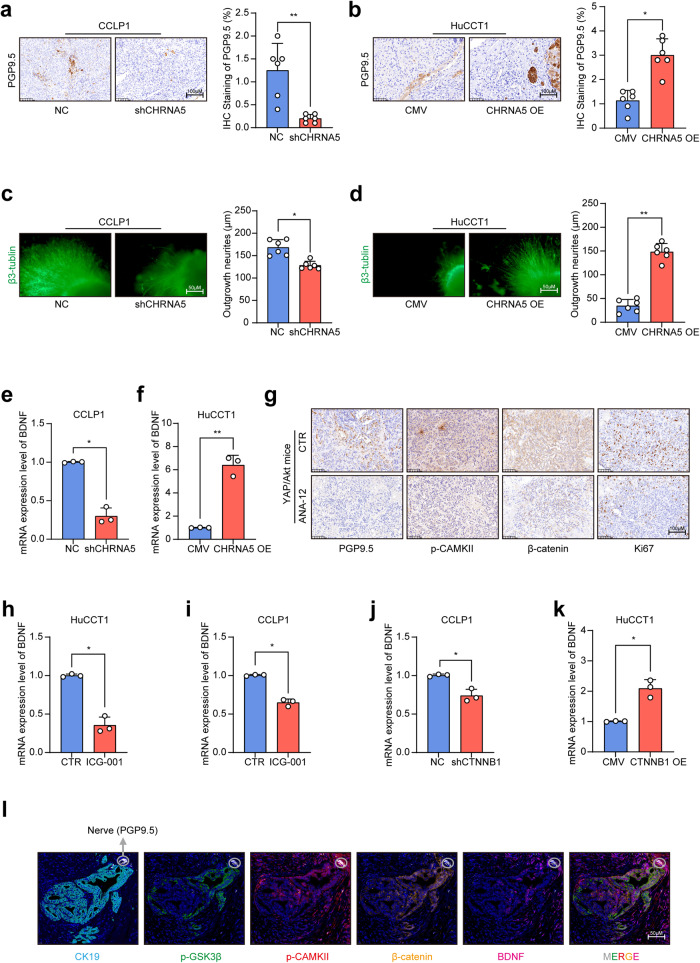
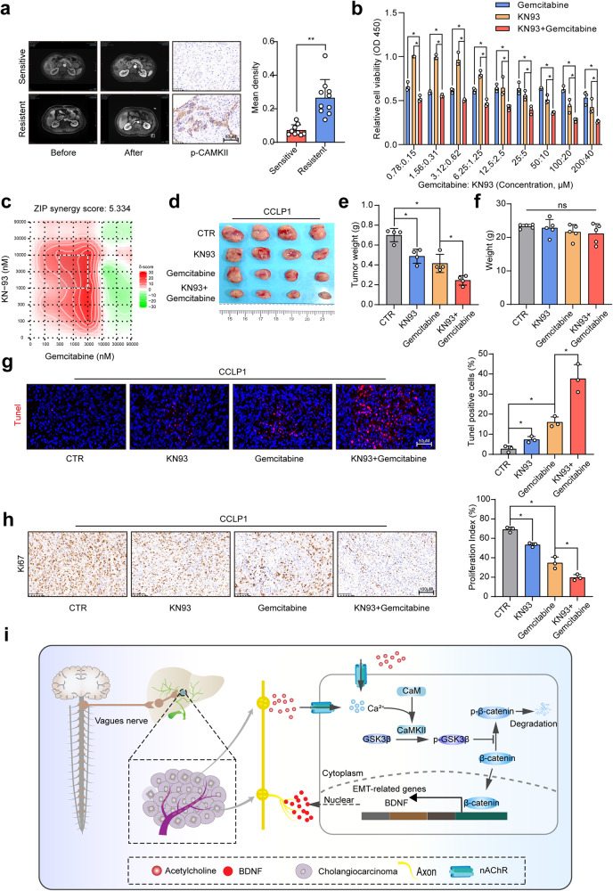
Neurotransmitter-initiated signaling pathway were reported to play an important role in regulating the malignant phenotype of tumor cells. Cancer cells could exhibit a "neural addiction" property and build up local nerve networks to achieve an enhanced neurotransmitter-initiated signaling through nerve growth factor-mediated axonogenesis. Targeting the dysregulated nervous systems might represent a novel strategy for cancer treatment. However, whether intrahepatic cholangiocarcinoma (ICC) could build its own nerve networks and the role of neurotransmitters in the progression ICC remains largely unknown. Immunofluorescence staining and Enzyme-linked immunosorbent assay suggested that ICC cells and the infiltrated nerves could generate a tumor microenvironment rich in acetylcholine that promotes ICC metastasis by inducing epithelial-mesenchymal transition (EMT). Acetylcholine promoted ICC metastasis through interacting with its receptor, alpha 5 nicotine acetylcholine receptor subunits (CHRNA5). Furthermore, acetylcholine/CHRNA5 axis activated GSK3β/β-catenin signaling pathway partially through the influx of Ca2+-mediated activation of Ca/calmodulin-dependent protein kinases (CAMKII). In addition, acetylcholine signaling activation also expanded nerve infiltration through increasing the expression of Brain-Derived Neurotrophic Factor (BDNF), which formed a feedforward acetylcholine-BDNF axis to promote ICC progression. KN93, a small-molecule inhibitor of CAMKII, significantly inhibited the migration and enhanced the sensitivity to gemcitabine of ICC cells. Above all, Acetylcholine/CHRNA5 axis increased the expression of β-catenin to promote the metastasis and resistance to gemcitabine of ICC via CAMKII/GSK3β signaling, and the CAMKII inhibitor KN93 may be an effective therapeutic strategy for combating ICC metastasis.


Conflict of interest statement

The authors declare no competing interests.

Figures

Fig. 1
Fig. 1
Acetylcholine-induced EMT to promote metastasis of ICC. a ELISA-based detection of acetylcholine in ICC tissue. b Immunofluorescence staining of PGP9.5, VAChT, and TH in ICC tissues. c Migration and invasion assays of ICC cells treated with acetylcholine. d WB-based detection of EMT-related genes in ICC cells treated with acetylcholine. e Representative images of liver metastasis of ICC cells in nude mice treated with acetylcholine. f H&E staining of liver tissues. g ELISA assay of acetylcholine in the culture supernatants of ICC cells. h Migration assays of ICC cells treated with adiphenine hydrochloride. i The results of IHC-based detection of ChAT positive rate in ICC tissues from Huashan cohort. j ELISA assays of acetylcholine in the culture supernatants of NC and sh-ChAT ICC cells. k Migration assays of NC and sh-ChAT ICC cells with or without the treatment of acetylcholine. Representative results from at least three experiments are shown. Data are shown as means ± SD. *p < 0.05, **p < 0.005
Fig. 2
Fig. 2
Blockade of acetylcholine/CHRNA5 suppressed ICC progression. a IHC staining assay of CHRNA5 in non-tumor bile duct tissues and ICC tissues. b The prognostic significance of CHRNA5 for ICC patients from the Huashan cohort. c WB of EMT-related genes in NC and sh-CHRNA5 CCLP1 cells. d Migration and invasion assays of NC and sh-CHRNA5 CCLP1 cells. e Migration assays of NC and sh-CHRNA5 CCLP1 cells treated with acetylcholine. f WB of EMT-related genes in CMV and CHRNA5-OE HuCCT1 cells. g Migration and invasion assays of CMV and CHRNA5-OE HuCCT1 cells. h, i Migration assays of NC and sh-ChAT ICC cells followed by CHRNA5 overexpression, with or without the treatment of acetylcholine. j Percentage of obstructive jaundice between NC and sh-CHRNA5 CCLP1 group. k Percentage of obstructive jaundice between HuCCT1-CMV and HuCCT1-CHRNA5 OE group. l, m Numbers of liver metastasis of ICC cells. n, o Incidence of nerve fiber sheaths invasion of ICC cells. p Overall survival of YAP/Akt-driven ICC mice treated with PBS or adiphenine. Representative results from at least three experiments are shown. Data are shown as means ± SD. *p < 0.05, **p < 0.005
Fig. 3
Fig. 3
β-catenin signaling is responsible for the malignant phenotype driven by acetylcholine/CHRNA5 axis. a WB analysis of the expression level of β-catenin in ICC cells. b IHC staining of the expression level of CHRNA5 and β-catenin in clinical ICC tissue. c IHC assay-based detection of β-catenin in YAP/Akt ICC mice model treated with acetylcholine receptor inhibitor. d, e WB analysis of the expression level of β-catenin in the nuclear and cytoplasm of ICC cells. f, g CCK8 assay-based detection of IC50 of gemcitabine in ICC cells. hk The effect of overexpressing or silencing β-catenin on the EMT phenotype in CHRNA5 silencing or overexpressing ICC cells. Representative results from at least three experiments are shown. Data are shown as means ± SD. *p < 0.05, **p < 0.005
Fig. 4
Fig. 4
Acetylcholine/CHRNA5 axis-mediated CAMKII activation suppresses GSK-3β activity to stabilize β-catenin. a, b WB-based detection of the half-life of β-catenin in CHRNA5 silencing or overexpressing ICC cells treated with CHX. c, d WB-based detection of p-β-catenin and p-s9-GSK3β in CHRNA5 silencing or overexpressing ICC cells. e WB-based detection of β-catenin expression level in CHRNA5 silencing ICC cells treated with GSK3β inhibitor LiCl. f Transwell assay-based detection of the migration ability in CHRNA5 silencing ICC cells treated with GSK3β inhibitor LiCl. g WB-based detection of β-catenin expression level in CHRNA5 overexpressing ICC cells in the condition of overexpressing WT-GSK3β or mutant-GSK3β(S9A). h Transwell assay-based detection of the migration ability in CHRNA5 overexpressing ICC cells in the condition of overexpressing WT-GSK3β or mutant-GSK3β(S9A). i WB-based detection of p-CAMKII expression level in CHRNA5 silencing or overexpressing ICC cells. j WB-based detection of CAMKII coprecipitated with GSK3β. k WB-based detection of CAMKII coprecipitated with GSK3β in CHRNA5 silencing or overexpressing ICC cells. l WB-based detection of β-catenin and p-s9-GSK3β expression level in ICC cells treated with KN93. Representative results from at least three experiments are shown. Data are shown as means ± SD. *p < 0.05, **p < 0.005
Fig. 5
Fig. 5
Acetylcholine/CHRNA5 axis induced axonogenesis through increasing the expression of BDNF in ICC. a, b IHC detection of PGP9.5 in CHRNA5 silencing or overexpressing ICC cells xenografts. c, d Immunofluorescence staining of β3-Tublin-based detection of nerve outgrowth of DRG cocultured with conditioned medium of CHRNA5 silencing or overexpressing ICC cells. e, f qRT-PCR-based detection of BDNF in CHRNA5 silencing or overexpressing ICC cells. g IHC-based detection of PGP9.5, p-CAMKII, β-catenin and Ki67 in YAP/Akt mice treated with ANA-12. h, i qRT-PCR-based detection of BDNF mRNA expression in ICC cells treated with ICG-001. j, k qRT-PCR-based detection of BDNF mRNA expression in CTNNB1 silencing or overexpressing ICC cells. l Multiplex immunofluorescence staining of CK19, PGP9.5, P-CAMK II, P-GSK3β, β-catenin and BDNF. Representative results from at least three experiments are shown. Data are shown as means ± SD. *p < 0.05; **p < 0.005
Fig. 6
Fig. 6
The combination of CAMKII inhibitor KN93 and gemcitabine exhibited an enhanced anti-tumor activity in ICC. a Representative MRI images and IHC staining of p-CAMKII in patients receiving gemcitabine-based chemotherapy. b CCK8 assay-based detection of inhibitory effect of KN93 and gemcitabine on CCLP1 cells. c Synergy score of the combination of gemcitabine and KN-93 for CCLP1. d, e In vivo detection of the therapeutic effect of KN93 and gemcitabine combined treatment. f The weight of nude mice from CTR, KN93, gemcitabine, KN93 and gemcitabine combined treatment group. g Tunel-based detection of apoptosis in CCLP1 xenograft from different groups. h IHC-based detection of Ki67 in CCLP1 xenograft from different groups. i The abstract figure of the regulatory role of acetylcholine/CHRNA5 axis in ICC: acetylcholine, derived from nerve fibers ending or cancer cells, could activate nAChR to induce the influx of Ca2+, and Ca2+-mediated CAMKII activation further phosphorylate and inactivate GSK3β, increasing the activity of β-catenin in ICC. Enhanced β-catenin activity increased the migration ability and resistance to gemcitabine in ICC. Enhanced β-catenin activity also increased the generation of BDNF to stimulate the axonogenesis, forming an Acetylcholine/CHRNA5-BDNF feedback loop to promote tumor progression in ICC. Representative results from at least three experiments are shown. Data are shown as means ± SD. *p < 0.05; **p < 0.005

Similar articles

Cited by

References

    1. Banales JM, et al. Cholangiocarcinoma 2020: the next horizon in mechanisms and management. Nat. Rev. Gastroenterol. Hepatol. 2020;17:557–588. - PMC - PubMed
    1. Ke Q, et al. The effect of adjuvant therapy for patients with intrahepatic cholangiocarcinoma after surgical resection: A systematic review and meta-analysis. PLoS ONE. 2020;15:e0229292. - PMC - PubMed
    1. Liebig C, et al. Perineural invasion in cancer: a review of the literature. Cancer. 2009;115:3379–3391. - PubMed
    1. Shi DD, et al. Therapeutic avenues for cancer neuroscience: translational frontiers and clinical opportunities. Lancet Oncol. 2022;23:e62–e74. - PMC - PubMed
    1. Guo Y, Gil Z. The Role of Extracellular Vesicles in Cancer-Nerve Crosstalk of the Peripheral Nervous System. Cells. 2022;11:1294. - PMC - PubMed

Publication types

MeSH terms

Grants and funding