

Association between patent ductus arteriosus flow and home oxygen therapy in extremely preterm infants
- PMID: 38454005
- PMCID: PMC11257949
- DOI: 10.1038/s41390-024-03120-8
Association between patent ductus arteriosus flow and home oxygen therapy in extremely preterm infants
Erratum in
-
Publisher Correction: Association between patent ductus arteriosus flow and home oxygen therapy in extremely preterm infants.Pediatr Res. 2024 Oct;96(5):1385-1386. doi: 10.1038/s41390-024-03177-5. Pediatr Res. 2024. PMID: 38693280 Free PMC article. No abstract available.
Abstract
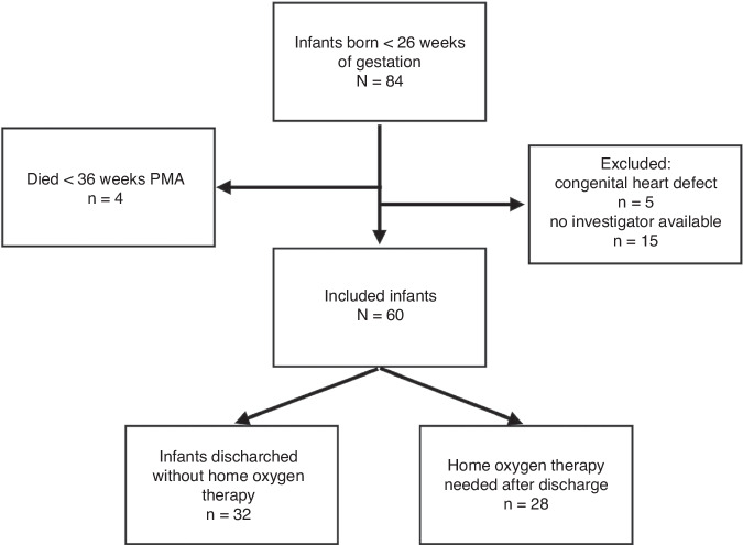
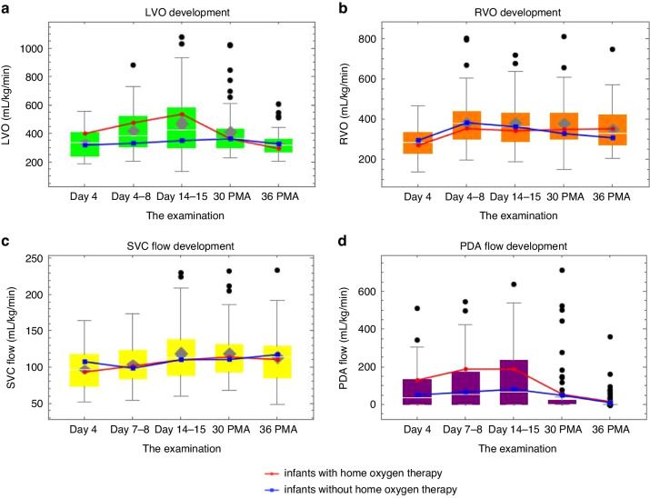
Background: Central blood flow measurements include the estimation of right and left ventricular output (RVO, LVO), superior vena cava (SVC) flow, and calculated patent ductus arteriosus (PDA) flow. We aimed to provide an overview of the maturation patterns of these values and the relationship between PDA flow and the need for home oxygen therapy.
Methods: This prospective single-center study was conducted in infants born at <26 weeks of gestation. We performed echocardiographic measurements five times during their life (from the 4th post-natal day to the 36th postmenstrual week).
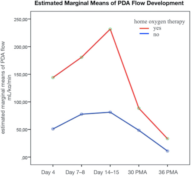
Results: Sixty patients with a mean birth weight of 680 (590, 760) g were included. Postnatal development of LVO and PDA flow peaked at the end of the second postnatal week (427 and 66 mL/kg/min, respectively). The RVO increased between days 4 and 7-8. The SVCF was most stable. The development curves of PDA flow differed between the groups with (n = 28; 47%) and without home oxygen therapy.
Conclusion: We present the central blood flow values and their postnatal development in infants <26 weeks of gestation. This study demonstrates the association between PDA flow and the future need for home oxygen therapy.
Impact: This study enriches our knowledge of the long-term development of central blood flow parameters and derived patent ductus arteriosus (PDA) flow in extremely preterm infants (<26 weeks). While pulmonary resistance decreased, PDA flow continued to increase from day 4 to the end of the second week of life. Similarly, left ventricular output increased as a marker of preload. The superior vena cava flow remained stable. The observed association between PDA flow and an unfavorable respiratory outcome is important for future studies focusing on the prevention of chronic lung disease.
© 2024. The Author(s).
Conflict of interest statement
The authors declare no competing interests.
Figures
Similar articles
-
Assessment of superior vena cava flow and cardiac output in different patterns of patent ductus arteriosus shunt.Echocardiography. 2021 Sep;38(9):1524-1533. doi: 10.1111/echo.15163. Epub 2021 Jul 26. Echocardiography. 2021. PMID: 34309068
-
Early Echocardiographic Predictors of Eventual Need for Patent Ductus Arteriosus Treatment: A Retrospective Study.Am J Perinatol. 2024 Sep;41(12):1673-1679. doi: 10.1055/a-2249-1671. Epub 2024 Jan 18. Am J Perinatol. 2024. PMID: 38237629
-
Severity of the ductal shunt: a comparison of different markers.Arch Dis Child Fetal Neonatal Ed. 2005 Sep;90(5):F419-22. doi: 10.1136/adc.2003.027698. Arch Dis Child Fetal Neonatal Ed. 2005. PMID: 16113155 Free PMC article.
-
Therapeutic strategy of patent ductus arteriosus in extremely preterm infants.Pediatr Neonatol. 2020 Apr;61(2):133-141. doi: 10.1016/j.pedneo.2019.10.002. Epub 2019 Oct 29. Pediatr Neonatol. 2020. PMID: 31740267 Review.
-
Ibuprofen for the prevention of patent ductus arteriosus in preterm and/or low birth weight infants.Cochrane Database Syst Rev. 2020 Jan 27;1(1):CD004213. doi: 10.1002/14651858.CD004213.pub5. Cochrane Database Syst Rev. 2020. PMID: 31985838 Free PMC article.
References
MeSH terms
LinkOut - more resources
Full Text Sources
Medical
