. 2024 Mar 7;14(1):5639.
doi: 10.1038/s41598-024-54903-2.

Distribution of exchangeable Ca2+ during the process of Larix decidua Mill. pollination and germination

Affiliations

Distribution of exchangeable Ca2+ during the process of Larix decidua Mill. pollination and germination

Katarzyna Rafińska et al. Sci Rep. .

Abstract

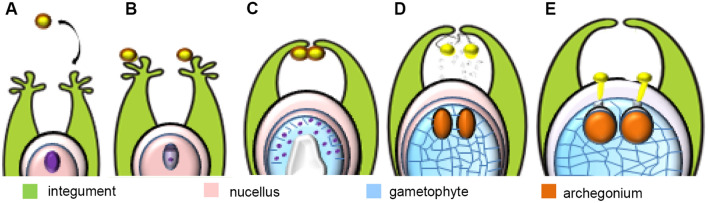
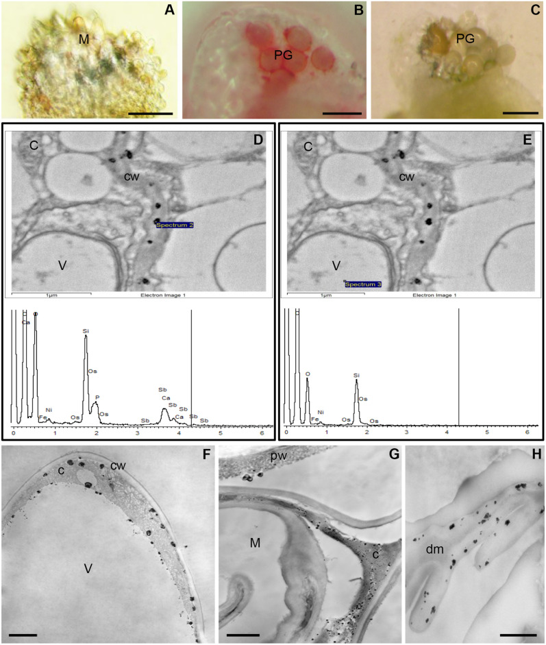
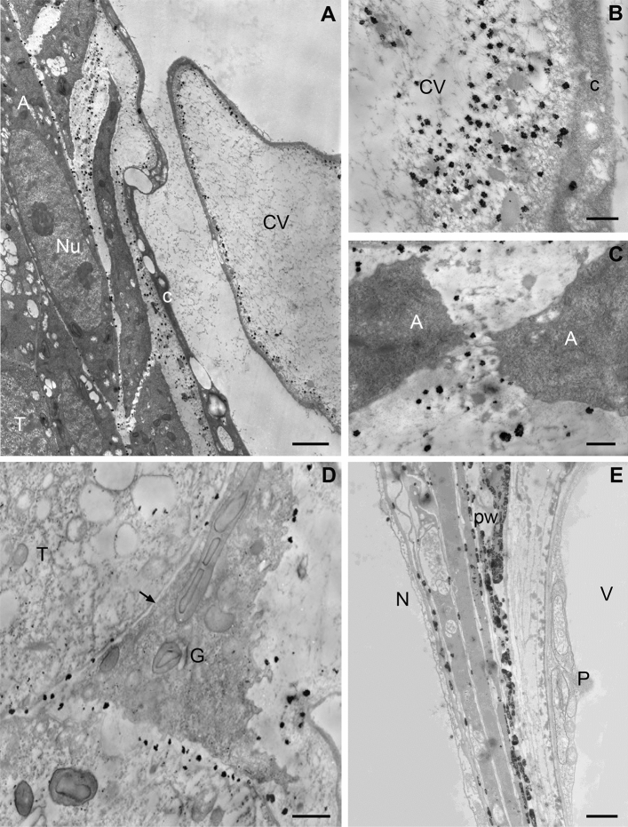
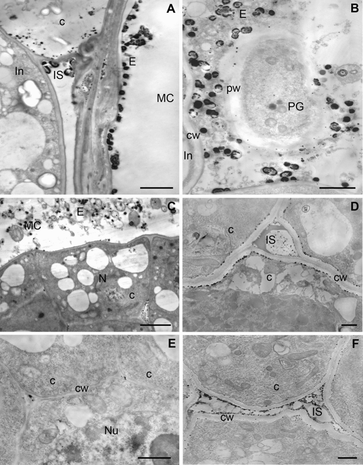
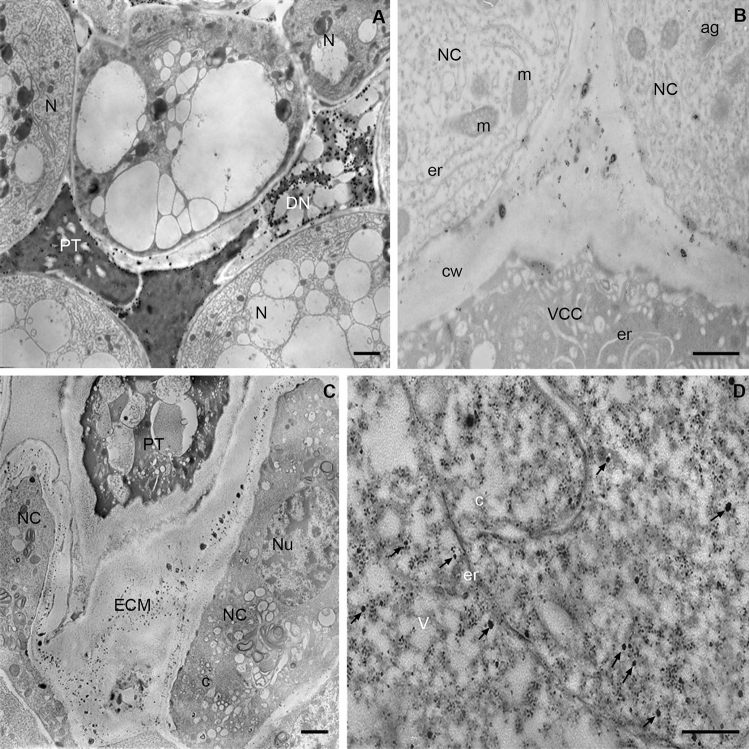
The involvement of Ca2+ ions in angiosperms sexual processes is well established, while in gymnosperms, such knowledge remains limited and is still a topic of discussion. In this study, we focused on Larix decidua, using Alizarin-red S staining and the pyroantimonate method to examine the tissue and subcellular distribution of free and loosely bound Ca2+ ions at different stages of the male gametophyte's development and its interaction with the ovule. Our findings show that in larch, both the germination of pollen grains and the growth of pollen tubes occur in an environment rich in Ca2+. These ions play a crucial role in the adhesion of the pollen grain to the stigmatic tip and its subsequent movement to the micropylar canal. There is a significant presence of free and loosely bound Ca2+ ions in both the fluid of the micropylar canal and the extracellular matrix of the nucellus. As the pollen tube extends through the nucellus, we observed a notable accumulation of Ca2+ ions just above the entry to the mature archegonium, a region likely crucial for the male gametophyte's directional growth. Meanwhile, the localized presence of free and loosely bound Ca2+ ions within the egg cell cytoplasm may inhibit the pollen tubes growth and rupture, playing an important role in fertilization.


Conflict of interest statement

The authors declare no competing interests.

Figures

Figure 1
Figure 1
Successive stages of pollen-ovule interaction in L. decidua, pollination (A), pollen grains on the stigmatic tip (B), engulfment of pollen grains into the micropylar canal (C), transfer of pollen grains to the nucellus (D), germination of pollen grains and pollen tube growth (E) (published).
Figure 2
Figure 2
Localization of Ca2+ ions in the L. decidua stigmatic tip before and after pollination. (AC) Alizarin-red S staining of the ovule. (A) Lack of staining on the surface of the unpollinated stigmatic tip is visible. (B) Intense staining is present at the adhesion of the pollen grains (PG), less staining is identified in pollen grains. (C) Control reaction (EDTA) – the lack of staining on the surface of the pollination stigmatic tip. (D, E) X-ray microanalysis of Ca/Sb precipitates present in the ovule cells. (D) X-ray spectrum of the precipitate in the cell wall (spectrum 2) is typical for spectra of calcium antimonate precipitates containing overlapping characteristic energy peaks of Ca and Sb. (E) In X-ray spectra from the area without precipitates (spectrum 3), calcium and antimony peaks are not observed. (FH). Subcellular localization of free and loosely bound Ca2+ ions. (F) Epidermal cell of the unpollinated stigmatic tip – large Ca/Sb precipitates are located mainly in the inner side of the plasma membrane and in the cytoplasm (C), much smaller in the inner side of the tonoplast (V—vacuole). No precipitates are visible in the cell wall (cw). (G) After pollination, Ca/Sb precipitates on the surface of the pollen wall (pw) and in the degenerating cytoplasm (C) of epidermal cells of the stigmatic tip (M) are present. (H) Numerous precipitates are visible in the material formed after the degeneration of the stigmatic tip cells (dm), (AC) bar 100 µm, (F, H) bar 500 nm, (G) bar 1 µm.
Figure 3
Figure 3
Localization of free and loosely bound Ca2+ ions in the developing of L. decidua female gametophyte. (AC) Free nuclear/alveoli stage. (A) Numerous Ca/Sb precipitates in the material between the alveoli (A) and in the central vacuole (CV) near the tonoplast are visible. Small precipitates are present in the wall separating the gametophyte from the tapetum (T) while single and small precipitates in the nuclei (Nu) and in the cytoplasm (C) of gametophyte and tapetum cells are localized, bar 2 µm. (B) In the central vacuole, calcium antimonate precipitates are present near the tonoplast in the area containing electron-opaque fibrillar and homogenous materials, in the cytoplasm only single precipitates are visible, bar 500 nm. (C) Gametophyte. Ca/Sb precipitates are located in the material between the forming alveoli (A), bar 500 nm. (D-E) Gametophyte-tapetum boundary. (D) Ca/Sb precipitates are present in the thin wall that separates the gametophyte (G) from tapetum cells (arrow), bar 1 µm. (E)—Prothallium stage of female gametophyte with the central cell. Accumulation of calcium antimonate precipitates are visible in the wall (PW) formed as a result of tapetum degradation; N, nucellus; P, prothallium cell; V, vacuole; bar 2 µm.
Figure 4
Figure 4
Localization of free and loosely bound Ca2+ ions in pollinated larch ovule. (AE) Nuclear gametophyte—cellular with the central cell, (F) Mature gametophyte. (A, B) micropylar canal (MC). (A) Ca/Sb precipitates are present in the secretion (E) on the surface of the micropylar canal. In the cytoplasm (C) and in the intercellular spaces (IS) between the integument cells (In), bar 2 µm. (B) numerous calcium antimonate precipitates are visible in the canal exudate (E) surrounding the pollen grain (PG), single and small Ca/Sb precipitates are present in the wall of the pollen grain (pw) and in the apoplast of the integument cell (In), bar 2 µm. (C–E) Nucellus. (C) In the surface of the nucellus Ca/Sb precipitates are localized in the secretion (E) present in the micropylar canal (MC). Numerous small precipitates in the intercellular spaces in the nucellus (N) are visible, bar 2 µm. (D) The top part of the nucellus. Numerous Ca/Sb precipitates are present in plasma membrane-cell wall (cw) boundary and in the material filling the large intracellular spaces (IS) of the tissue, bar 1 µm. (E) Only single calcium antimonate precipitates are localized in small intercellular spaces of the nucellus in the area near the prothallium, Nu-nucleus, bar 500 nm. (F) In the mature gametophyte stage, numerous small Ca/Sb precipitates are present in the intercellular space (IS) and single precipitates in the plasmalema-cell wall (cw) in the nucellus cells, bar 1 µm.
Figure 5
Figure 5
Localization of free and loosely bound Ca2+ ions in L. decidua ovule during pollen tube growth (stage of mature gametophyte). (A) Pollen tube in the nucellus. Precipitates are visible in the dense and vesicle-rich cytoplasm of the pollen tube (PT). Numerous larger Ca/Sb precipitates are present in the area of the nucellus degenerating cells (DN). The remaining cells of the nucellus (N) are devoid of Ca/Sb precipitates, bar 1 µm. (B) The top part of the archegonium. Ca/Sb precipitates are present in the extracellular matrix between neck cells (NC) and the canal-ventral cell entry (VCC), cw—cell wall, er—endoplasmic reticulum, m—mitochondrion, bar 1 µm. (C) Pollen tube (PT) after growth to the archegonium. Numerous small Ca/Sb precipitates are localized only in the extracellular matrix (ECM) between neck cells (NC), C-cytoplasm, Nu – nucleus, bar 2 µm. (D) Egg cell cytoplasm with the long cistern of the endoplasmic reticulum (er) and numerous “electron-light” vesicles (V) forming so-called inclusions. In the cytoplasm numerous larger (arrows) and smaller Ca/Sb precipitates are visible, bar 2 µm.

Similar articles

References

    1. Villar M, Knox R, Dumas C. Effective pollination period and nature of pollen-collecting apparatus in the gymnosperm, Larix leptolepis. Ann. Bot. 1984;53:279–284. doi: 10.1093/oxfordjournals.aob.a086689. - DOI
    1. Rodkiewicz, B. The Embryology of Gymnosperms (Państwowe Wydawnictwo Naukowe, 1984) [in Polish].
    1. Owens JN, Morris SJ, Catalano GL. How the pollination mechanism and prezygotic and postzygotic events affect seed production in Larix occidentalis. Can. J. For. Res. 1994;24:917–927. doi: 10.1139/x94-121. - DOI
    1. Takaso T, Owens JN. Pollen movement in the micropylar canal of Larix and its simulation. J. Plant Res. 1997;110:259–264. doi: 10.1007/BF02509314. - DOI
    1. Breygina M, Klimenko E, Schekaleva O. Pollen germination and pollen tube growth in gymnosperms. Plants. 2021;10(7):1301. doi: 10.3390/plants10071301. - DOI - PMC - PubMed