

Review
doi: 10.1038/s41556-024-01364-4.
Epub 2024 Mar 7.
Lipid droplets and cellular lipid flux
Affiliations
- PMID: 38454048
- PMCID: PMC11228001
- DOI: 10.1038/s41556-024-01364-4
Item in Clipboard
Review
Lipid droplets and cellular lipid flux
Nat Cell Biol.
2024 Mar.
Abstract
Lipid droplets are dynamic organelles that store neutral lipids, serve the metabolic needs of cells, and sequester lipids to prevent lipotoxicity and membrane damage. Here we review the current understanding of the mechanisms of lipid droplet biogenesis and turnover, the transfer of lipids and metabolites at membrane contact sites, and the role of lipid droplets in regulating fatty acid flux in lipotoxicity and cell death.
© 2024. Springer Nature Limited.
Conflict of interest statement
Competing interests
J.A.O. is a member of the scientific advisory board for Vicinitas Therapeutics and has patent applications related to ferroptosis. A.J.M. has no competing interests.
Figures
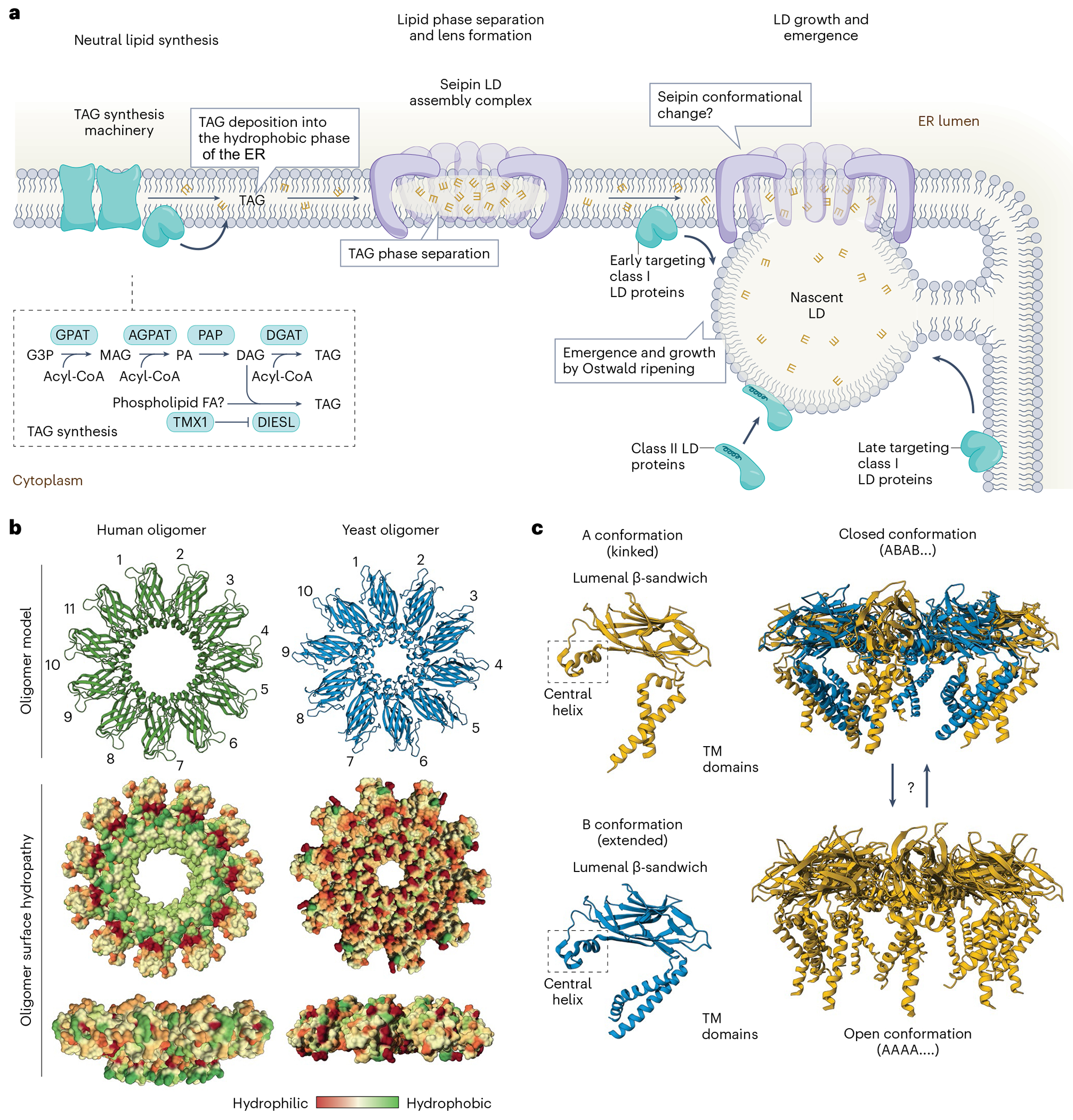
a, LD biogenesis occurs through a series of steps involving (1) the deposition of neutral lipids (for example, TAG) within the ER bilayer, (2) seipin-regulated TAG phase separation into neutral lipid lenses and (3) the emergence of the LD from the cytosolic leaflet and LD growth by Ostwald ripening. Class I LD proteins insert into the ER and traffic to LDs through lateral diffusion along membrane continuities. Early class I LD proteins traffic to nascent LDs at sites of LD biogenesis marked by the seipin LD assembly complex. Late class I LD proteins are excluded by seipin from trafficking to nascent LDs, and instead traffic across separate membrane bridges that form at specialized ER exit sites. Class II LD proteins are synthesized on cytosolic ribosomes and insert post-translationally into LDs. These proteins typically contain an amphipathic helix that is important for recognizing and inserting into unique phospholipid packing defects in the bounding LD monolayer. b, Human and yeast seipin organize into homo-oligomeric ring structures with 11 and 10 subunits, respectively. Human seipin contains a lumenal β-sandwich domain with a hydrophobic helix that extends towards the centre of the ring and likely embeds into the membrane. Molecular dynamics simulations indicate that the lumenal ring of human seipin is sufficient to promote TAG aggregation. c, Yeast seipin structures containing the transmembrane domains indicate two conformations, a kinked (A) and extended (B) conformation, and suggest that seipin functions as a flexible cage-like structure that opens towards the cytosol during LD biogenesis. The complex consisting of the alternating kinked and extended conformation (ABAB…) forms a closed conformation that may be involved in the initial phase separation of the nascent lipid lens. The complex consisting uniformly of the kinked conformation (AAAA…) forms an open conformation that may be involved in LD emergence and growth. AGPAT, acyl CoA:acylglycerol phosphate acyltransferases; G3P, glycerol-3-phosphate; PA, phosphatidic acid; PAP, phosphatidic acid phosphatase. This figure was generated using the following Protein Data Bank (PDB) entries: CryoEM human seipin (6DS5 ); CryoEM yeast seipin (7OXP ); CryoEM yeast seipin (7RSL ).
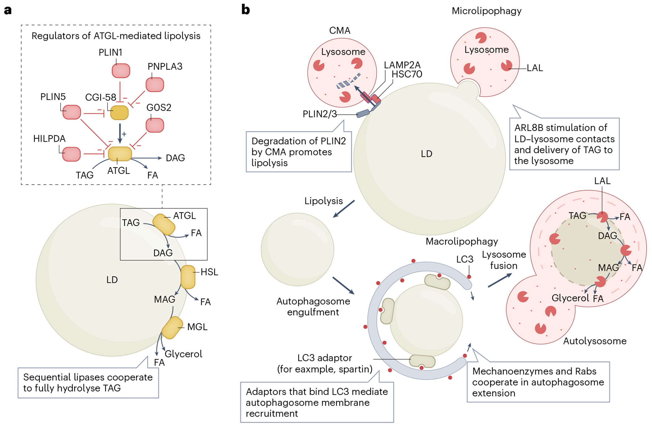
a, Lipolytic breakdown of TAGs stored in LDs occurs through the sequential actions of LD-localized lipases–ATGL, HSL and MGL. ATGL is the rate-limiting enzyme in this pathway and is subject to several mechanisms of regulation (shown in the inset box). CGI-58 is an ATGL binding partner that increases ATGL activity. The association of CGI-58 is governed by multiple regulatory proteins. The positive or negative regulatory effect is indicated, including direct ATGL inhibitors (G0S2 and HILPDA) and inhibitors that act by preventing the interaction of ATGL and CGI-58 (PLIN1, PLIN2 and PNPLA3). b, Macrolipophagy and microlipophagy provide two pathways to deliver LDs, or portions of LDs, to lysosomes for breakdown. LAL mediates the hydrolysis of lipids, including both TAGs and SEs. In macrolipophagy, CMA degradation of PLIN2 and PLIN3 promotes lipolysis, reducing the size of LDs. Autophagy receptors mediate the recruitment to LDs of LC3-decorated autophagic membranes, which are subsequently extended to fully envelope the LD. The autophagosome fuses with lysosomes to generate an autolysosome and enable the degradation of the LD. By contrast, microlipophagy mediates the direct delivery of a portion of the LD to the lysosome for degradation, independent of canonical autophagy machinery.
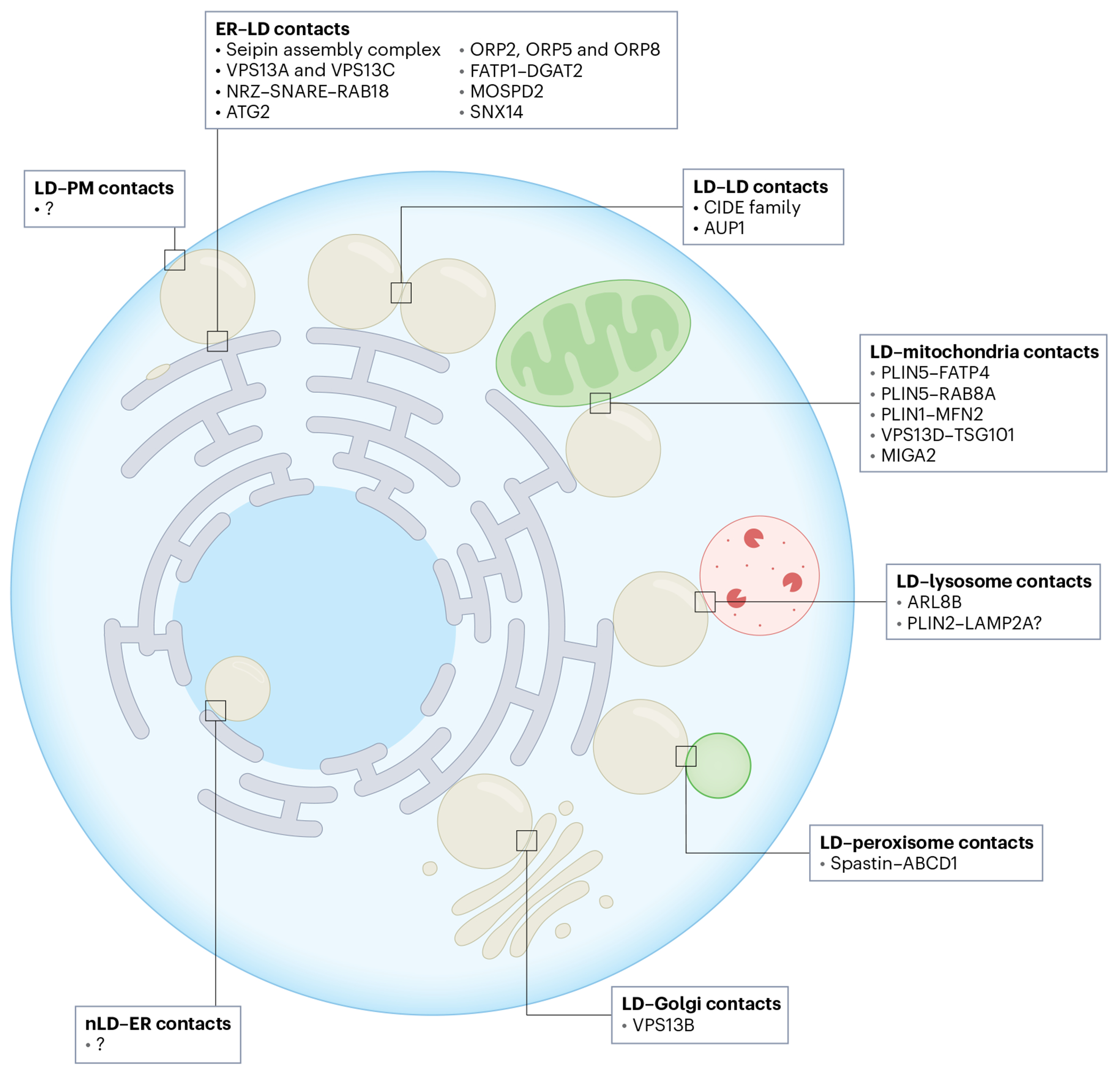
LDs form MCSs with virtually all organelles in the cell, providing sites for the organization of protein complexes and the transfer of lipids by LTPs. The membranes at these sites are held in close apposition by protein tethers. Tether and LTPs present at different LD MCSs are listed. Although LDs have been observed to make contacts with the plasma membrane (PM), the proteins present in these contact sites are not known. In Drosophila, snazarus localizes to LD–plasma membrane contacts and it is possible that snazarus orthologues may have similar roles in mammalian cells. Nuclear LDs (nLDs) are present in some cell types, but little is known about their membrane contact sites.
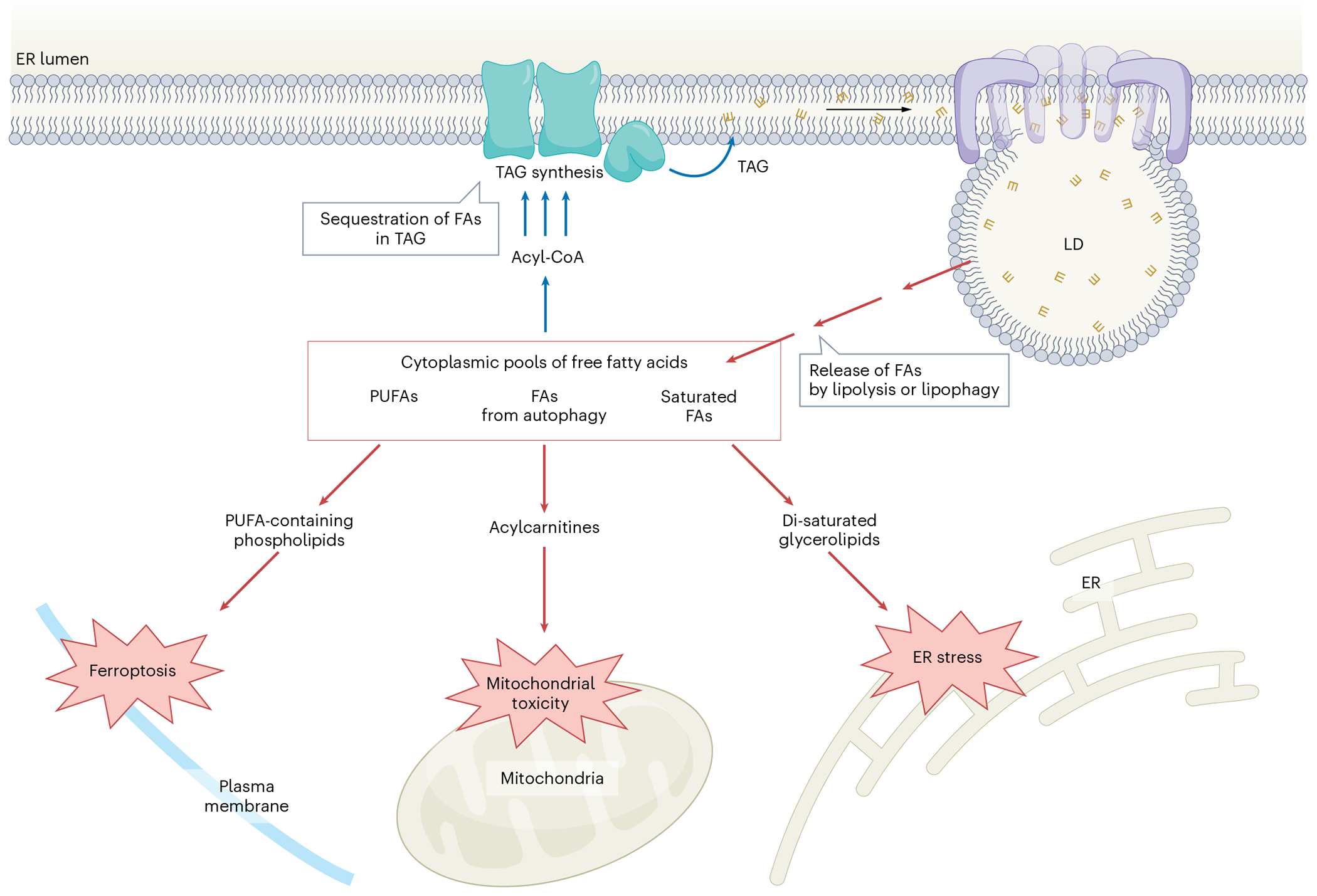
In addition to their canonical role In cellular energy homeostasis, LDs act as a lipid buffering system that prevents lipotoxicity. Various types of FA can be esterified and safely stored as TAGs within LDs. Excessively high amounts of FAs can elicit different manifestations of lipotoxicity, either by acting as direct membrane detergents or by being incorporated into specific lipids that cause dysfunction at high amounts. PUFA-containing phospholipids are prone to oxidation and sensitize cells to lipid peroxidation and ferroptosis. The incorporation of saturated FAs (such as palmitate) into glycerolipids can lead to high amounts of di-saturated glycerolipids that may alter membrane fluidity, inducing ER stress and apoptosis. FAs released during the autophagic breakdown of membranous organelles, such as during nutrient deprivation, can accumulate as acylcarnitine and induce mitochondrial dysfunction. Under certain conditions, lipolysis or lipophagy could lead to excessive free FAs that contribute to lipotoxicity.
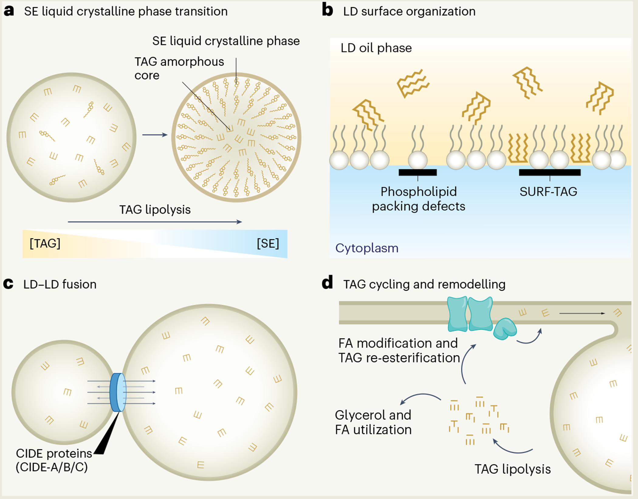
a, SE lipid phase transition under high SE to TAG ratios. b, Phospholipid packing defects and SURF-TAGs. c, LD–LD fusion via CIDE-mediated TAG transfer driven by Ostwald ripening. d, FA modifications and remodelling of TAG composition during cycles of TAG breakdown and FA re-esterification.
References
-
- Petan T. Lipid droplets in cancer. Rev. Physiol. Biochem Pharm 185, 53–86 (2023). - PubMed
