. 2024 Jul;29(7):2185-2198.
doi: 10.1038/s41380-024-02490-z. Epub 2024 Mar 7.

Glial-restricted precursors stimulate endogenous cytogenesis and effectively recover emotional deficits in a model of cytogenesis ablation

Affiliations

Glial-restricted precursors stimulate endogenous cytogenesis and effectively recover emotional deficits in a model of cytogenesis ablation

Joana Martins-Macedo et al. Mol Psychiatry. 2024 Jul.

Abstract

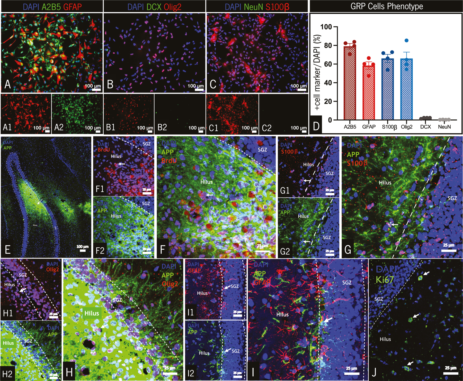
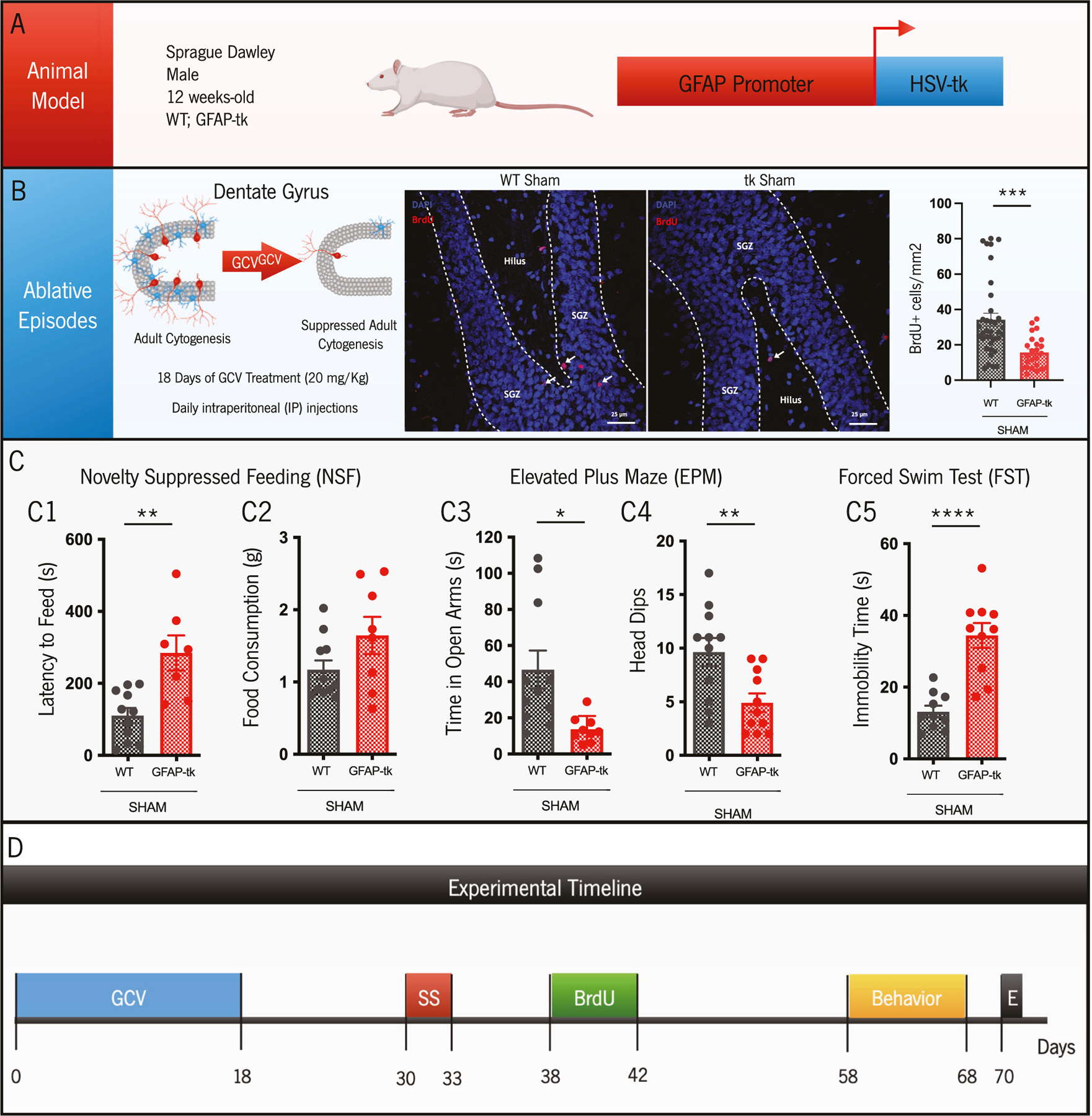
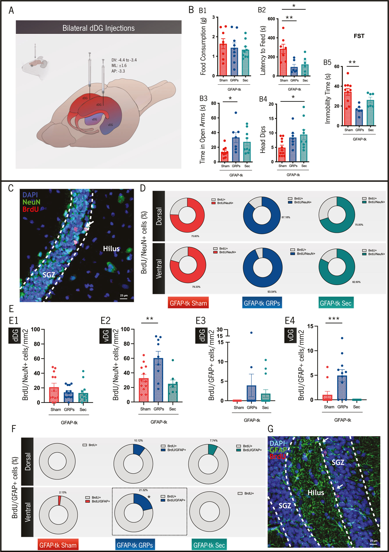
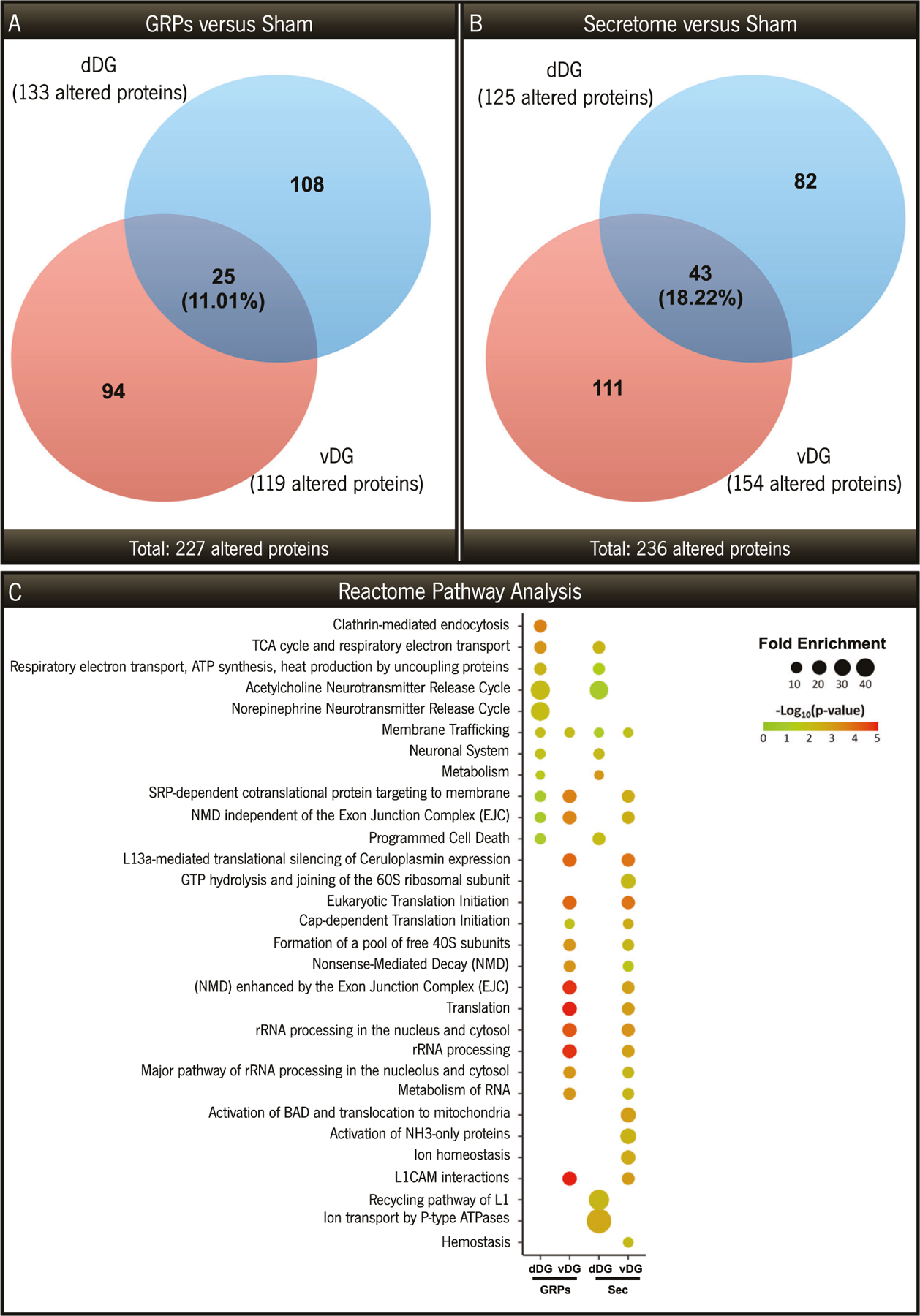
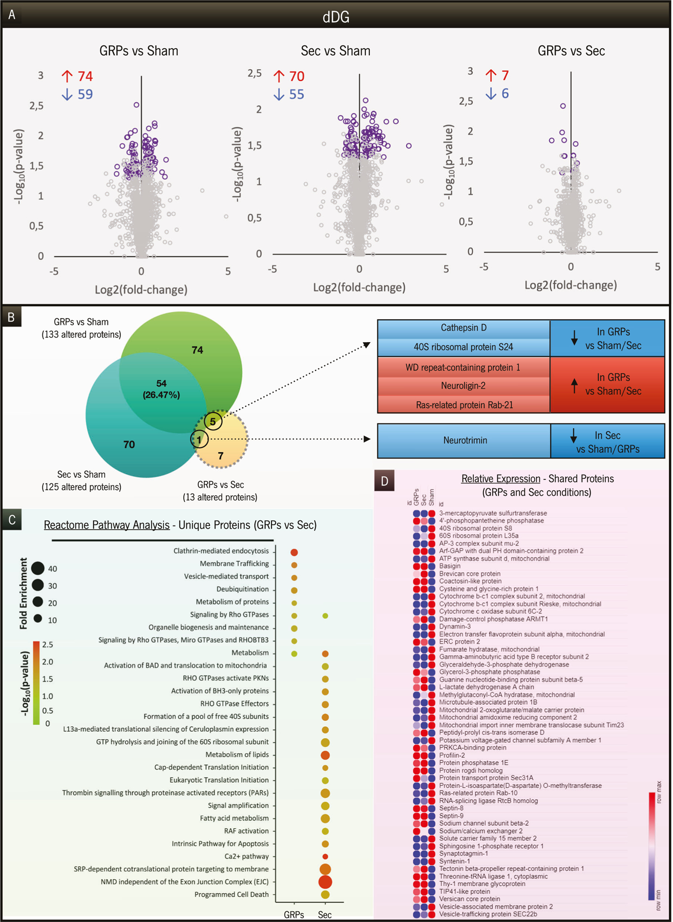
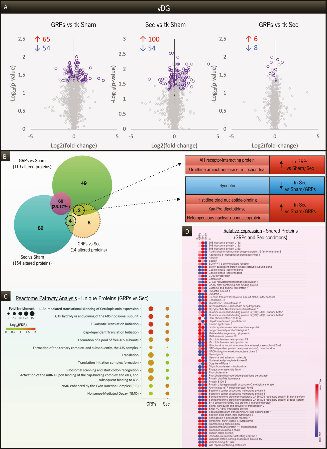
Adult cytogenesis, the continuous generation of newly-born neurons (neurogenesis) and glial cells (gliogenesis) throughout life, is highly impaired in several neuropsychiatric disorders, such as Major Depressive Disorder (MDD), impacting negatively on cognitive and emotional domains. Despite playing a critical role in brain homeostasis, the importance of gliogenesis has been overlooked, both in healthy and diseased states. To examine the role of newly formed glia, we transplanted Glial Restricted Precursors (GRPs) into the adult hippocampal dentate gyrus (DG), or injected their secreted factors (secretome), into a previously validated transgenic GFAP-tk rat line, in which cytogenesis is transiently compromised. We explored the long-term effects of both treatments on physiological and behavioral outcomes. Grafted GRPs reversed anxiety-like deficits and demonstrated an antidepressant-like effect, while the secretome promoted recovery of only anxiety-like behavior. Furthermore, GRPs elicited a recovery of neurogenic and gliogenic levels in the ventral DG, highlighting the unique involvement of these cells in the regulation of brain cytogenesis. Both GRPs and their secretome induced significant alterations in the DG proteome, directly influencing proteins and pathways related to cytogenesis, regulation of neural plasticity and neuronal development. With this work, we demonstrate a valuable and specific contribution of glial progenitors to normalizing gliogenic levels, rescuing neurogenesis and, importantly, promoting recovery of emotional deficits characteristic of disorders such as MDD.


Conflict of interest statement

COMPETING INTERESTS

The authors declare no competing interests.

Figures

Fig. 1
Fig. 1. Characterization of GRPs at P5, both in vitro and in vivo (post transplantation).
Immunocytochemistry for GFAP (A1)/A2B5 (A2), Olig2 (B1)/DCX (B2) and S100β (C1)/ NeuN (C2) markers, and their respective quantification (D). GRPs maintained an overall progenitor phenotype, mainly differentiating into glial cells. E Representative brain section of a WT rat transplanted with GRPs, with most of the cells primarily concentrated in the dDG. F BrdU+ cells, within the hilar region of the dDG, co-localized (white arrows) with APP+ cells (GRPs), confirming that transplanted cells were proliferating up to 12 days post grafting. Approximately 40 days after transplantation, GRPs differentiated into mature astrocytes (S100β + ), white arrows in (G), most likely cells from the oligodendroglial lineage (Olig2 + ), white arrows in (H) and immature astrocytes (GFAP + ), white arrows in (I), highlighting the glial differentiation pattern of these cells. J Proliferating cells (Ki67 + , white arrows) were still found within the dDG, 40 days after GRPs transplantation. Data is presented as Mean ± SEM. Scale (A–E): 100 μm; scale (F–J): 25 μm.
Fig. 2
Fig. 2. GFAP-tk rat model characterization and experimental timeline.
A In this study, 12 weeks-old Sprague-Dawley transgenic male rats were used, expressing the viral HSV-tk under the GFAP promoter, with their WT littermates being used as controls. B Both groups were treated with the anti-viral GCV for 18 days, leading to suppression of adult cytogenesis in GFAP-tk animals (fewer BrdU+ cells in the DG). Afterwards, due to this impaired cytogenic process, transgenic animals revealed anxiety-like behavior in the NSF (C1) and EPM tests (C3-C4), while maintaining a normal food consumption pattern (C2). GFAP-tk rats further presented increased immobility time in the FST (C5). D GFAP-tk and WT male Sprague-Dawley rats were submitted to a protocol of GCV injections for 18 days. Twelve days after the end of GCV treatment, animals were submitted to stereotaxic surgeries, for GRPs and secretome transplantation. One week after surgeries, BrdU injections were performed for five consecutive days. Forty-days post-GCV treatment, behavioral tests were conducted to assess different emotional domains. At the end of the in vivo experiments, animals were euthanized for subsequent molecular and cellular analyses. GCV Ganciclovir, SS stereotaxic surgeries, BrdU 5’-Bromo-2’-Deoxyuridine, Behavior: EPM, NSF and FST, E Euthanasia. Data is presented as Mean ± SEM. *p < 0.05, **p < 0.01, ***p < 0.001, ****p < 0.0001.
Fig. 3
Fig. 3. Effects of GRP and secretome treatments on behavior and generation of new cells in GFAP-tk rats.
A Schematic representation of bilateral injections delivered at the dDG, at specific coordinates. B Anxiety-like and antidepressant-like effect behavioral characterization in GFAP-tk animals through the NSF (B1-B2), EPM (B3-B4), and FST tests (B5). Data regarding GFAP-tk sham group was intentionally replicated from Fig. 2C. Both treatments with GRPs and their secretome induced a recovery in anxiety-like behavior, while GRPs were also capable of presenting an antidepressant-like effect. C–G Evaluation of the cytogenic potential in GFAP-tk animals. C Representative microphotograph of BrdU+ cells co-localizing with NeuN+ cells in the Subgranular Zone (SGZ) of the dDG, in a GFAP-tk rat treated with GRPs. D Percentage of cells co-localizing BrdU/NeuN markers, from the total BrdU+ cells, for all groups in the study and for the dDG and vDG regions. E Quantification of the total number of cells co-localizing BrdU/NeuN and BrdU/GFAP markers, for all groups in the study, regarding dDG (E1, E3) and vDG (E2, E4) regions, respectively. F Percentage of cells co-localizing BrdU/GFAP markers, from the total BrdU+ cells, for all groups in the study and for the dDG and vDG regions. Statistical differences between GFAP-tk GRPs and the other two groups (GFAP-tk Sham and GFAP-tk Sec) were highlighted within a square. G Representative microphotograph of BrdU+ cells co-localizing with GFAP+ cells in the SGZ of the dDG, in a GFAP-tk rat treated with GRPs. Data is presented as Mean ± SEM. *p < 0.05, **p < 0.01, ***p < 0.001.
Fig. 4
Fig. 4. Analysis of the proteins significantly altered between GRPs or Secretome treatments and GFAP-tk sham animals.
Venn diagrams representing the total number of proteins significantly altered between GRPs-treated (A) or Secretome-treated rats (B) and GFAP-tk sham animals, both for the dDG (blue) and vDG regions (red). C Comparative pathway analysis covering the most significant and enriched Reactome pathways, taking into consideration the proteins uniquely altered in each region (dDG and vDG) between GRP-treated or secretome-treated animals and GFAP-tk sham rats.
Fig. 5
Fig. 5. Analysis of the proteins altered between groups in the dDG region.
A Volcano plots presenting the significantly altered proteins (highlighted in purple) in each comparison under study: GRPs vs. Sham, Sec vs. Sham, and GRPs vs. Sec. The number of increased (red) or decreased (blue) proteins is indicated for each comparison. B Venn diagram representing the specific number of proteins altered between GRPs vs. Sham, Sec vs. Sham, and GRPs vs. Sec. Six of the 13 proteins altered between treatments were also significantly altered compared to Sham animals, herein highlighted. C Reactome pathway analysis covering the most significant and enriched pathways, only considering the unique proteins in the comparisons of GRPs vs. Sham and Sec vs. Sham. D Heatmap of the relative expression levels of the 54 proteins commonly altered between GRP and secretome groups, in comparison to GFAP-tk animals.
Fig. 6
Fig. 6. Analysis of the proteins altered between groups in the vDG region.
A Significantly altered proteins (highlighted in purple), among the ones that were unique to each comparison under study: GRPs vs. Sham, Sec vs. Sham, and GRPs vs. Sec. The number of increased (red) or decreased (blue) proteins is indicated for each comparison. B Venn diagram representing the specific number of proteins altered between GRPs vs. Sham, Sec vs. Sham, and GRPs vs. Sec. Six of the 14 proteins altered between treatments were also significantly altered compared to Sham animals, herein highlighted. C Reactome pathway analysis covering the most significant and enriched pathways, only considering the unique proteins in the comparisons of GRPs vs. Sham and Sec vs. Sham. D Heatmap of the relative expression levels of the 68 proteins commonly altered between GRPs and secretome groups, in comparison to GFAP-tk animals.

Update of

Similar articles

Cited by

References

    1. Eriksson PS, Perfilieva E, Björk-Eriksson T, Alborn AM, Nordborg C, Peterson DA, et al. Neurogenesis in the adult human hippocampus. Nat Med 1998;4:1313–7. - PubMed
    1. Sorrells SF, Paredes MF, Cebrian-Silla A, Sandoval K, Qi D, Kelley KW, et al. Human hippocampal neurogenesis drops sharply in children to undetectable levels in adults. Nature. 2018;555:377–81. - PMC - PubMed
    1. Lucassen PJ, Fitzsimons CP, Salta E, Maletic-Savatic M. Adult neurogenesis, human after all (again): Classic, optimized, and future approaches. Behav Brain Res 2020;381:112458. - PubMed
    1. Kempermann G, Gage FH, Aigner L, Song H, Curtis MA, Thuret S, et al. Human adult neurogenesis: evidence and remaining questions. Cell Stem Cell. 2018;23:25–30. - PMC - PubMed
    1. Boldrini M, Fulmore CA, Tartt AN, Simeon LR, Pavlova I, Poposka V, et al. Human Hippocampal neurogenesis persists throughout aging. Cell Stem Cell. 2018;22:589–599.e5. - PMC - PubMed

LinkOut - more resources