. 2024 Mar 7;24(1):76.
doi: 10.1186/s12866-024-03203-w.

Exploring Aeromonas dhakensis in Aldabra giant tortoises: a debut report and genetic characterization

Affiliations

Exploring Aeromonas dhakensis in Aldabra giant tortoises: a debut report and genetic characterization

Chenxu Zhao et al. BMC Microbiol. .

Abstract

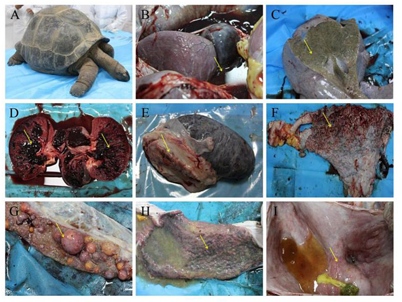
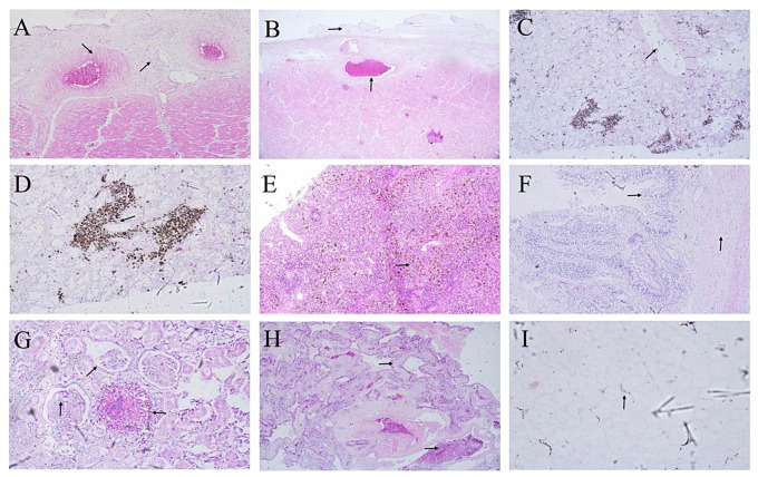
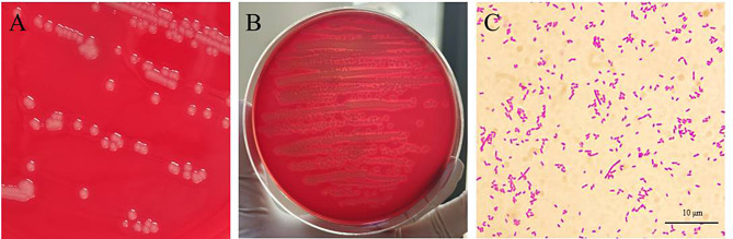
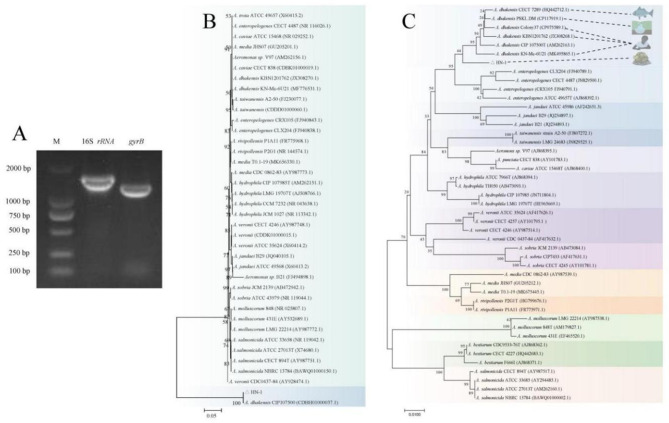
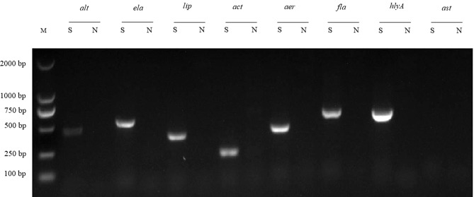
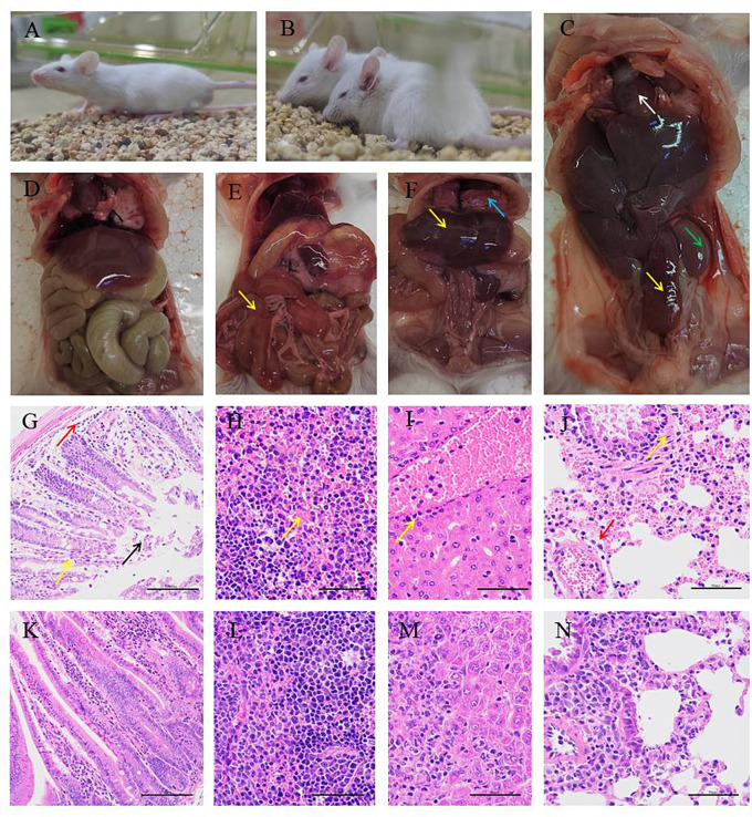
Aeromonas dhakensis (A. dhakensis) is becoming an emerging pathogen worldwide, with an increasingly significant role in animals and human health. It is a ubiquitous bacteria found in terrestrial and aquatic milieus. However, there have been few reports of reptile infections. In this study, a bacterial strain isolated from a dead Aldabra giant tortoise was identified as A. dhakensis HN-1 through clinical observation, matrix-assisted laser desorption ionization-time of flight mass spectrometry (MALDI-TOF/MS), and gene sequencing analysis. Subsequently, to evaluate its pathogenicity, the detection of virulence genes and mice infection experiments were performed. A. dhakensis HN-1 was found to contain seven virulence genes, including alt, ela, lip, act, aerA, fla, and hlyA. Mice infected with A. dhakensis HN-1 exhibited hemorrhage of varying degrees in multiple organs. The half-maximal lethal dose (LD50) value of A. dhakensis HN-1 for mice was estimated to be 2.05 × 10⁷ colony forming units (CFU)/mL. The antimicrobial susceptibility test revealed that A. dhakensis HN-1 was resistant to amoxicillin, penicillin, ampicillin and erythromycin. This is the first report of A. dhakensis in Aldabra giant tortoises, expanding the currently known host spectrum. Our findings emphasize the need for One Health surveillance and extensive research to reduce the spread of A. dhakensis across the environment, humans, and animals.

Keywords: Aeromonas dhakensis; Aldabra giant tortoise; Multidrug resistance; Pathogenicity; Public health.


Conflict of interest statement

The authors declare no competing interests.

Figures

Fig. 1
Fig. 1
Gross lesions of the dead Aldabra giant tortoise in Henan, China. (A) Effusion pleural effusion. (B) Fibrinous exudation and hemorrhage in heart. (C) Swelling in the greyish-yellow liver. (D) Hemorrhage with multiple papillae in kidneys. (E) Edema and necrosis in spleen. (F) Congestion in lung. (G) Hemorrhage on the surface of the follicle. (H) duodenal ulcer with necrotic foci in cecum. (I) hemorrhage in bladder
Fig. 2
Fig. 2
Histological lesions of the dead Aldabra giant tortoise. HE. (A) Vessel walls hyperplasia and thickened with infiltration of heterophile leukocytes (200×). (B) Verrucous hyperplasia in the epicardium. hyperemia and protein exudation in the blood vessel (100×). (C) Fatty degeneration and vacuolar degeneration of hepatocytes, and fused into large vacuoles in the live (400×). (D) Interstitial collagen fibroplasia with infiltration of a great quantity of heterophil leukocyte in the liver (400×). (E) Spleen trabecular loosing, White pulp necrosis, a great quantity of scattered golden clumps or particles in the spleen (100×). (F) Intestinal villus fusion, villous epithelial cell degeneration and necrosis. A large number of neutrophils infiltrated in the intestinal lamina propria (200×). (G) Infiltration of a great quantity of neutrophils around the purulent foci. Fibrin exudation in the renal capsule cavity. Necrosis of renal tubular epithelia in the kidney (400×). (H) Infiltration of red blood cells and protein-like exudates in the alveoli pulmonum matrix and alveolar cavity (100×). (I) Fatty degeneration in the bladder (400×)
Fig. 3
Fig. 3
Colony morphology and microscopic findings of bacterial isolate HN-1. (A, B) The bacteria grew well and produced a β-hemolytic zone on sheep blood agar plates. (C) Gram-negative bacilli were observed under an oil immersion lens, with different sizes and staining depths.
Fig. 4
Fig. 4
Molecular analysis of HN-1 gene amplicons. (A) Agarose gel electrophoresis of PCR products from bacterial isolate HN-1 for 16S rRNA and gyrB. Lane M: DL2000 DNA marker. (B-C) Unrooted neighbor-joining phylogenetic trees of the bacterial isolate HN-1 sequence (in bold) and sequences with highest identity of Aeromonas species, for the 16S rRNA gene (B), and gyrB gene (C). Numbers at nodes indicated bootstrap percentages obtained after 1000 replicates
Fig. 5
Fig. 5
Agarose gel electrophoresis of PCR products from A. dhakensis HN-1 for detecting eight virulence genes. Lane M: DL2000 DNA marker. S indicates the nucleic acid samples from A. dhakensis HN-1. N indicates negative control samples
Fig. 6
Fig. 6
The pathological changes of A. dhakensis HN-1 infected mice. (A, D) No obvious clinic symptoms and visible lesions in the negative control group; (B) Mental malaise, lethargy and tremor in the infected groups; (C) Swelling and hemorrhage in the spleen (yellow arrow) and kidney (green arrow); (E) Oedema with acute hemorrhage in the intestine wall (yellow arrow); (F) Hepatic oedema (yellow arrow), severe hemorrhage in the lung (blue arrow); (G) Intestine mucosal epithelial cells shedding (black arrow), capillary congestion expansion (yellow arrow), mucosal layer and submucosa infiltrated with a small amount of inflammatory cell (red arrow). (H) Spleen red pulp congestion; (I) Inflammatory cell infiltration around the hepatic portal area; (J) Mucosal epithelial cells arranged irregularly (yellow arrow), perivascular inflammatory cell infiltration (red arrow); (K-N) PBS control

References

    1. World Health Organization. Zoonoses. 2022. Available from: https://www.who.int/news-room/fact-sheets/detail/zoonoses.
    1. Guerra RM, Maleno FD. Potential Pathogenicity of Aeromonas spp. Recovered in River Water, Soil, and Vegetation from a Natural Recreational Area, Pathogens, 11 (11) (2022) 1382. 10.3390/pathogens11111382. - PMC - PubMed
    1. Rusiñol M, Hundesa A, Cárdenas-Youngs Y, Fernández-Bravo A, Pérez-Cataluña A, Moreno-Mesonero L, Girones R, et al. Microbiological contamination of conventional and reclaimed irrigation water: evaluation and management measures. Sci Total Environ. 2020;710:136298. doi: 10.1016/j.scitotenv.2019.136298. - DOI - PubMed
    1. Pablos M, Remacha MA, Rodríguez-Calleja JM, Santos JA, Otero A, García-López ML. Identity, virulence genes, and clonal relatedness of Aeromonas isolates from patients with diarrhea and drinking water. Eur J Clin Microbiol Infect Dis. 2010;29(9):1163–72. doi: 10.1007/s10096-010-0982-3. - DOI - PubMed
    1. Aravena-Román M, Beaz-Hidalgo R, Inglis TJJ, Riley TV, Martínez-Murcia AJ, Chang BJ, Figueras MJ. Aeromonas australiensis sp. nov., isolated from irrigation water. Int J Syst Evol Microbiol. 2013;63:2270–6. doi: 10.1099/ijs.0.040162-0. - DOI - PubMed

Supplementary concepts