. 2024 Mar 7;24(1):311.
doi: 10.1186/s12903-024-04062-7.

Periodontal ligament stem cell-derived exosome-loaded Emodin mediated antimicrobial photodynamic therapy against cariogenic bacteria

Affiliations

Periodontal ligament stem cell-derived exosome-loaded Emodin mediated antimicrobial photodynamic therapy against cariogenic bacteria

Maryam Pourhajibagher et al. BMC Oral Health. .

Abstract

Background: This study was conducted to investigate the efficiency of periodontal ligament (PDL) stem cell-derived exosome-loaded Emodin (Emo@PDL-Exo) in antimicrobial photodynamic therapy (aPDT) on Streptococcus mutans and Lactobacillus acidophilus as the cariogenic bacteria.

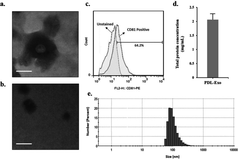
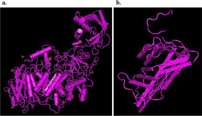
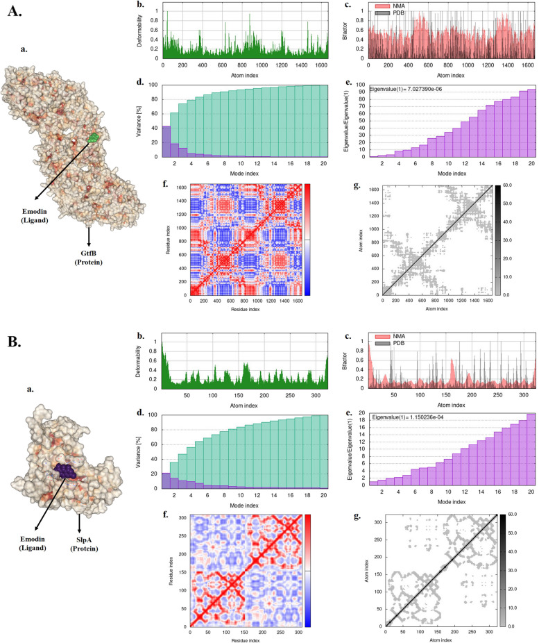
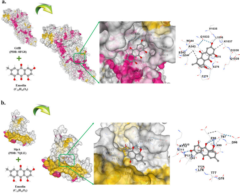
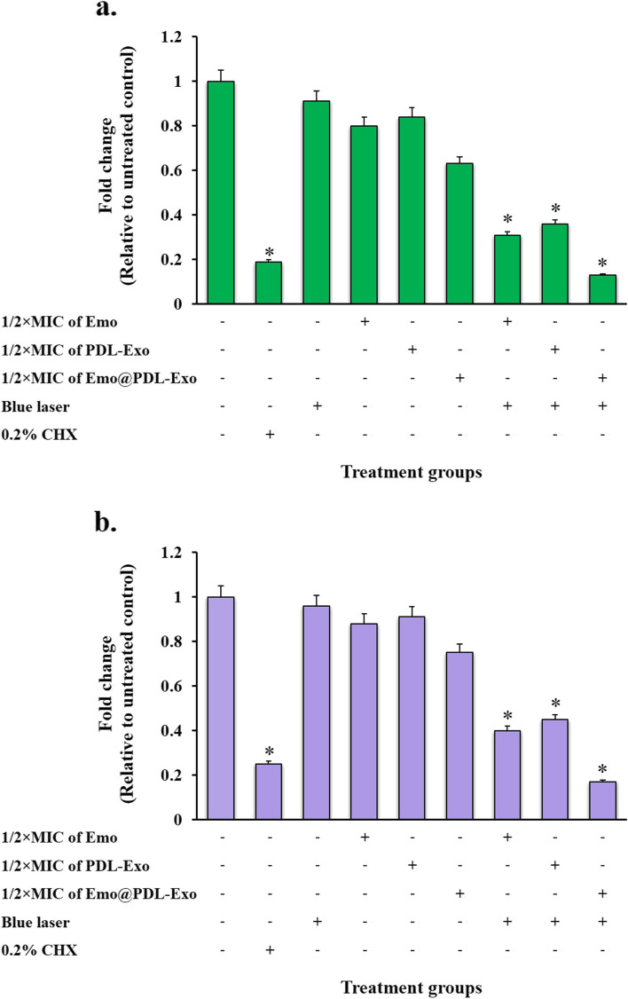
Materials and methods: After isolating and characterizing PDL-Exo, the study proceeded to prepare and verify the presence of Emo@PDL-Exo. The antimicrobial effect, anti-biofilm activity, and anti-metabolic potency of Emo, PDL-Exo, and Emo@PDL-Exo were then evaluated with and without irradiation of blue laser at a wavelength of 405 ± 10 nm with an output intensity of 150 mW/cm2 for a duration of 60 s. In addition, the study assessed the binding affinity of Emodin with GtfB and SlpA proteins using in silico molecular docking. Eventually, the study examined the generation of endogenous reactive oxygen species (ROS) and changes in the gene expression levels of gelE and sprE.

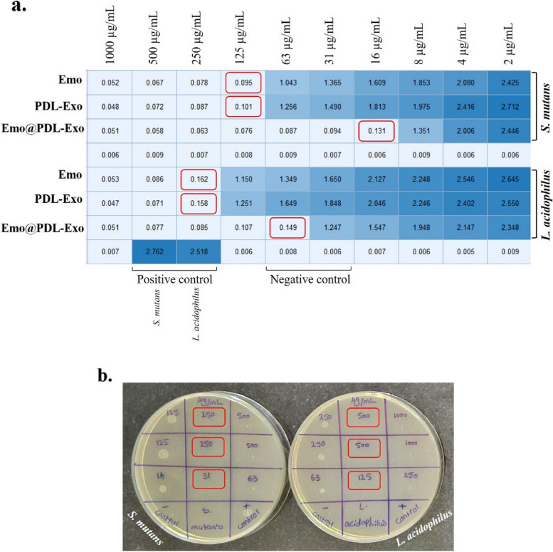
Results: The study found that using Emo@PDL-Exo-mediated aPDT resulted in a significant decrease in L. acidophilus and S. mutans by 4.90 ± 0.36 and 5.07 log10 CFU/mL, respectively (P < 0.05). The study found that using Emo@PDL-Exo for aPDT significantly reduced L. acidophilus and S. mutans biofilms by 44.7% and 50.4%, respectively, compared to untreated biofilms in the control group (P < 0.05). Additionally, the metabolic activity of L. acidophilus and S. mutans decreased by 58.3% and 71.2%, respectively (P < 0.05). The molecular docking analysis showed strong binding affinities of Emodin with SlpA and GtfB proteins, with docking scores of -7.4 and -8.2 kcal/mol, respectively. The study also found that the aPDT using Emo@PDL-Exo group resulted in the most significant reduction in gene expression of slpA and gtfB, with a decrease of 4.2- and 5.6-folds, respectively, compared to the control group (P < 0.05), likely due to the increased generation of endogenous ROS.

Discussion: The study showed that aPDT using Emo@PDL-Exo can effectively reduce the cell viability, biofilm activity, and metabolic potency of S. mutans and L. acidophilus. aPDT also significantly reduced the expression levels of gtfB and slpA mRNA due to the increased endogenous ROS generation. The findings suggest that Emo@PDL-Exo-mediated aPDT could be a promising antimicrobial approach against cariogenic microorganisms.

Keywords: Antimicrobial photodynamic therapy; Bioinformatics tools; Emodin; Exosome.


Conflict of interest statement

The authors declare no competing interests.

Figures

Fig. 1
Fig. 1
Characterization of PDL-Exo and Emo@PDL-Exo: a TEM micrograph of PDL-Exo (Scale bar = 100 nm), b TEM micrograph of Emo@PDL-Exo (Scale bar = 50 nm), c Flow cytometry analysis of Exo-specific marker CD81, d Total protein concentration of PDL-Exo, e Average particle size distribution of Emo@PDL-Exo
Fig. 2
Fig. 2
a Determination of minimum inhibitory concentration (MIC) after taking optical density using a spectrometer at 600 nm for the various dilutions of Emo, PDL-Exo, and Emo@PDL-Exo incubated with S. mutans and L. acidophilus; Red rectangle = MIC, b Determination of minimum bactericidal concentration (MBC) of Emo, PDL-Exo, and Emo@PDL-Exo against S. mutans and L. acidophilus; Red rectangle = MBC
Fig. 3
Fig. 3
Histograms of endogenous reactive oxygen species (ROS) in: a L. acidophilus cells treated with normal saline (control group), b L. acidophilus cells treated with Emo@PDL-Exo, c L. acidophilus cells treated with aPDT using Emo@PDL-Exo, d S. mutans cells treated with normal saline (control group), e S. mutans cells treated with Emo@PDL-Exo, and f S. mutans cells treated with aPDT using Emo@PDL-Exo
Fig. 4
Fig. 4
Effect of different treatment groups on cell viability of: a S. mutans; b L. acidophilus. *Significantly different from the control group (no treatment), P < 0.05
Fig. 5
Fig. 5
Effect of different treatment groups on biofilm of: a S. mutans; b L. acidophilus. *Significantly different from the control group (no treatment), P < 0.05
Fig. 6
Fig. 6
Effect of different treatment groups on metabolic activity of: a S. mutans; b L. acidophilus. *Significantly different from the control group (no treatment), P < 0.05
Fig. 7
Fig. 7
Three-dimensional structure of proteins: a GtfB (PDB ID: 8FG8), b SlpA (PDB ID: 7QLE)
Fig. 8
Fig. 8
Molecular dynamic simulation: A GtfB, B. SlpA. a Protein–ligand complex, b Deformability, c B-factor values, d Variance (violet: individual variances, green: cumulative variances), e Eigenvalues, f Co-variance map (residues with correlated motions in red, uncorrelated motions in white, and anti-correlated motions in blue), and g Elastic network (darker grays indicate stiffer springs) of the complex
Fig. 9
Fig. 9
Depiction of docked ligand–protein complex along with interaction of the amino acid residues of the protein with ligand: a GtfB-Emodin, b SlpA-Emodin
Fig. 10
Fig. 10
Effect of natural photosensitizers-mediated aPDT on gene expression of: a gtfb, b slpA. *Significantly different from the control group (no treatment), P < 0.05

Similar articles

Cited by

References

    1. Pitts NB, Zero DT, Marsh PD, Ekstrand K, Weintraub JA, Ramos-Gomez F, Tagami J, Twetman S, Tsakos G, Ismail A. Dental caries Nat Rev Dis Primers. 2017;3:17030. doi: 10.1038/nrdp.2017.30. - DOI - PubMed
    1. Cavallari T, Arima LY, Ferrasa A, Moysés SJ, Tetu Moysés S, Hirochi Herai R, Iani WR. Dental caries: Genetic and protein interactions. Arch Oral Biol. 2019;108:104522. doi: 10.1016/j.archoralbio.2019.104522. - DOI - PubMed
    1. Cogulu D, Saglam C. Genetic aspects of dental caries. Frontiers in Dental Medicine. 2022;3:1060177. doi: 10.3389/fdmed.2022.1060177. - DOI
    1. Zhang Q, Ma Q, Wang Y, Wu H, Zou J. Molecular mechanisms of inhibiting glucosyltransferases for biofilm formation in Streptococcus mutans. Int J Oral Sci. 2021;13(1):30. doi: 10.1038/s41368-021-00137-1. - DOI - PMC - PubMed
    1. Schormann N, Patel M, Thannickal L, Purushotham S, Wu R, Mieher JL, Wu H, Deivanayagam C. The catalytic domains of Streptococcus mutans glucosyltransferases: a structural analysis. Acta Crystallogr F Struct Biol Commun. 2023;79(Pt 5):119–127. doi: 10.1107/S2053230X23003199. - DOI - PMC - PubMed

LinkOut - more resources