. 2024 Mar 7;24(1):319.
doi: 10.1186/s12885-024-12085-0.

Regulation of cancer stem cells by CXCL1, a chemokine whose secretion is controlled by MCM2

Affiliations

Regulation of cancer stem cells by CXCL1, a chemokine whose secretion is controlled by MCM2

Yeon-Jee Kahm et al. BMC Cancer. .

Abstract

Background: A high expression pattern of minichromosome maintenance 2 (MCM2) has been observed in various cancers. MCM2 is a protein involved in the cell cycle and plays a role in cancer growth and differentiation by binding to six members of the MCM subfamily. The MCM protein family includes MCM2 through MCM7.

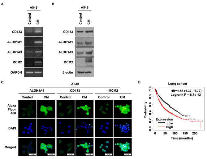
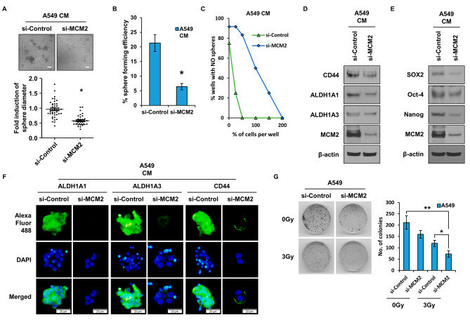
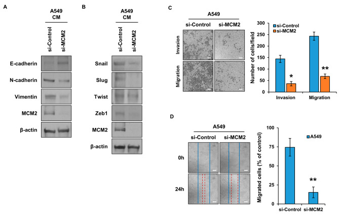
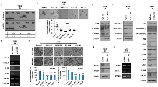
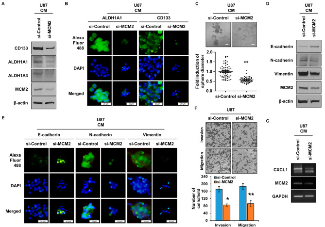
Methods: MCM2 has shown high expression in both lung cancer stem cells (LCSCs) and glioma stem cells (GSCs). We investigated the characteristics of CSCs and the regulation of the epithelial-to-mesenchymal transition (EMT) phenomenon in LCSCs and GSCs by MCM2. Additionally, we explored secreted factors regulated by MCM2.

Results: There was a significant difference in survival rates between lung cancer patients and brain cancer patients based on MCM2 expression. MCM2 was found to regulate both markers and regulatory proteins in LCSCs. Moreover, MCM2 is thought to be involved in cancer metastasis by regulating cell migration and invasion, not limited to lung cancer but also identified in glioma. Among chemokines, chemokine (C-X-C motif) ligand 1 (CXCL1) was found to be regulated by MCM2.

Conclusions: MCM2 not only participates in the cell cycle but also affects cancer cell growth by regulating the external microenvironment to create a favorable environment for cells. MCM2 is highly expressed in malignant carcinomas, including CSCs, and contributes to the malignancy of various cancers. Therefore, MCM2 may represent a crucial target for cancer therapeutics.

Keywords: CXCL1; Cancer stem cells; Chemokine; EMT; MCM2.


Conflict of interest statement

The authors declare no competing interests.

Figures

Fig. 1
Fig. 1
MCM2 Expression in Cancer Stem Cells and Prognostic Implications. (A) Comparison of RNA expression levels of CSC markers and MCM2 in A549 cells cultured in conditioned medium (CM). GAPDH was used as a loading control. (B) Comparison of protein expression of CSC markers and MCM2 in A549 cells cultured with CM. β-actin was used as a loading control. Full-length blots are presented in Supplementary Fig. 1A. (C) Comparison of expression levels of CSC marker protein and MCM2 using immunocytochemistry (ICC). (D) Kaplan-Meier survival curves for lung cancer patients according to MCM2 gene expression levels. Sample derive from the same experiment and that gels/blots were processed in parallel
Fig. 2
Fig. 2
Impact of MCM2 Expression on Characteristics of LCSCs. (A) Comparative analysis of differences in sphere formation ability according to MCM2 expression. Comparison of sphere formation after 96 h of treatment of A549 cells with CM. (B) Single cell analysis of lung cancer stem cells (LCSCs) based on MCM2 expression. A549 cells were cultured in a 96-well plate to assess sphere-forming ability over a period of 10 days. (C) Limiting dilution assay to analyze LCSCs ability. Varying numbers of cells (1, 50, 100, 150, and 200) were plated in each well of a 96-well plate to compare sphere formation capacity. (D) Analysis of the expression amount of CSC marker proteins (CD44, ALDH1A1, ALDH1A3) according to MCM2 expression. Full-length blots are presented in Supplementary Fig. 1B. (E) Confirmation of the expression level of CSC regulatory proteins (SOX2, Oct-4, Nanog) according to MCM2 expression. Full-length blots are presented in Supplementary Fig. 1C. (F) Comparative analysis of the expression level of CSC marker proteins by ICC. (G) Colony formation assay to measure radioresistance based on MCM2 expression. To this end, 1 × 10 3 cells were planted in each cell and irradiated with 3 Gy of radiation the next day. Sample derive from the same experiment and that blots were processed in parallel. Data represent the mean ± standard deviation of three replicates. Scale bar, 50 μm. *p˂0.005, **p˂0.05
Fig. 3
Fig. 3
Influence of MCM2 Expression on EMT Marker Proteins and Cell Migration in LCSCs. (A) Confirmation of the expression levels of EMT marker proteins E-cadherin, N-cadherin, and vimentin according to the expression levels of MCM2. Full-length blots are presented in Supplementary Fig. 1D. (B) Expression levels of Snail, Slug, Twist, and Zeb1, which are EMT regulators. Full-length blots are presented in Supplementary Fig. 1E. (C) Confirmation of cell migration and invasion ability regulated by MCM2. (D) Measurement of cell migration ability according to the expression level of MCM2. Sample derive from the same experiment and that blots were processed in parallel. Data are presented as the mean ± standard deviation of three replicates. Scale bar, 50 μm. *p˂0.001, **p˂0.0005
Fig. 4
Fig. 4
Regulation of Secreted Factors by MCM2 in LCSC. (A) Identification of secreted factors regulated by MCM2 using cytokine array. (B) Gene expression of secreted factors (CCL5, CXCL1, IL-18, IL-21) according to the expression level of MCM2. (C) Comparative analysis after conducting sphere-forming assay by treating with neutralizing antibodies of each secreted factor. (D) Comparative analysis of cell migration and invasion ability by treatment with neutralizing antibodies of each secreted factor. (E) Expression levels of CSC marker proteins after treatment with CXCL1 neutralizing antibody. Full-length blots are presented in Supplementary Fig. 1F. (F) Expression levels of EMT marker proteins after treatment with CXCL1 neutralizing antibody. Full-length blots are presented in Supplementary Fig. 1G. (G) Confirmation of the protein expression levels of MCM2 and CXCL1 after treatment with CXCL1 neutralizing antibody. Full-length blots are presented in Supplementary Fig. 1H. (H) Confirmation of gene expression levels of MCM2 and CXCL1 after treatment with CXCL1 neutralizing antibody. (I) Investigation of the CXCL1 signaling mechanism after inhibiting the action of CXCL1. Full-length blots are presented in Supplementary Fig. 1I. Sample derive from the same experiment and that gels/blots were processed in parallel. Data are presented as the mean ± standard deviation of three replicates. Scale bar, 50 μm. *p˂0.05, **p˂0.005, ***p˂0.0005
Fig. 5
Fig. 5
Regulation of GSC and EMT Properties by MCM2 Inhibition. (A) Expression of GSC marker proteins by MCM2 gene suppression in GSCs. Full-length blots are presented in Supplementary Fig. 1J. (B) Confirmation of marker protein expression in GSCs using ICC. (C) Comparison of differences in sphere formation according to the expression level of MCM2. (D) Comparison of expression of EMT marker proteins by MCM2 gene suppression. Full-length blots are presented in Supplementary Fig. 1K. (E) Confirmation of EMT marker proteins expression using ICC. (F) Analysis of cell migration and invasion ability after inhibiting the expression of MCM2. (G) Analysis of the regulation of CXCL1 expression by MCM2 in GSCs. Sample derive from the same experiment and that gels/blots were processed in parallel. Data are presented as the mean ± standard deviation of three replicates. Scale bar, 50 μm. *p˂0.001, **p˂0.005

Similar articles

Cited by

References

    1. Zhai Y, Li N, Jiang H, Huang X, Gao N, Tye BK. Unique roles of the non-identical MCM subunits in DNA replication licensing. Mol Cell. 2017;67:168–79. doi: 10.1016/j.molcel.2017.06.016. - DOI - PubMed
    1. Ishimi Y. Regulation of MCM2-7 function. Genes Genet Syst. 2018;93(4):125–33. doi: 10.1266/ggs.18-00026. - DOI - PubMed
    1. Tye BK. The MCM2-3-5 proteins: are they replication licensing factors? Trends Cell Biol. 1994;4(5):160–6. doi: 10.1016/0962-8924(94)90200-3. - DOI - PubMed
    1. Nishitani H, Lygerou Z. Control of DNA replication licensing in a cell cycle. Genes Cells. 2002;7(6):523–34. doi: 10.1046/j.1365-2443.2002.00544.x. - DOI - PubMed
    1. Evrin C, Clarke P, Zech J, Lurz R, Sun J, Uhle S, Li H, Stillman B, Speck C. A double-hexameric MCM2-7 complex is loaded onto origin DNA during licensing of eukaryotic DNA replication. Proc Natl Acad Sci U S A. 2009;106(48):20240–5. doi: 10.1073/pnas.0911500106. - DOI - PMC - PubMed

MeSH terms