. 2024 Mar 7;21(1):13.
doi: 10.1186/s12989-024-00574-w.

Exposure to high dose of polystyrene nanoplastics causes trophoblast cell apoptosis and induces miscarriage

Affiliations

Exposure to high dose of polystyrene nanoplastics causes trophoblast cell apoptosis and induces miscarriage

Shukun Wan et al. Part Fibre Toxicol. .

Abstract

Background: With rapid increase in the global use of various plastics, microplastics (MPs) and nanoplastics (NPs) pollution and their adverse health effects have attracted global attention. MPs have been detected out in human body and both MPs and NPs showed female reproductive toxicological effects in animal models. Miscarriage (abnormal early embryo loss), accounting for 15-25% pregnant women worldwide, greatly harms human reproduction. However, the adverse effects of NPs on miscarriage have never been explored.

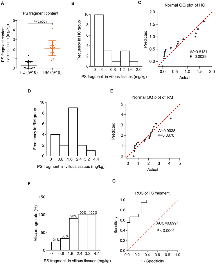
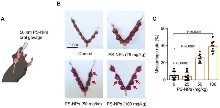
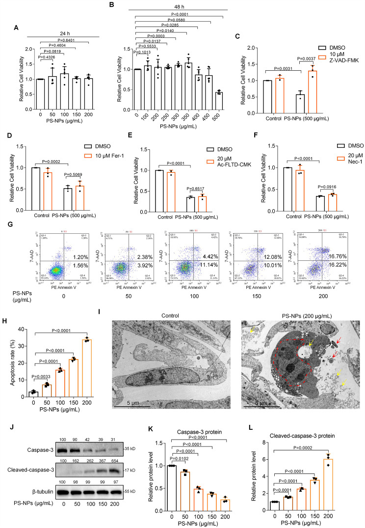
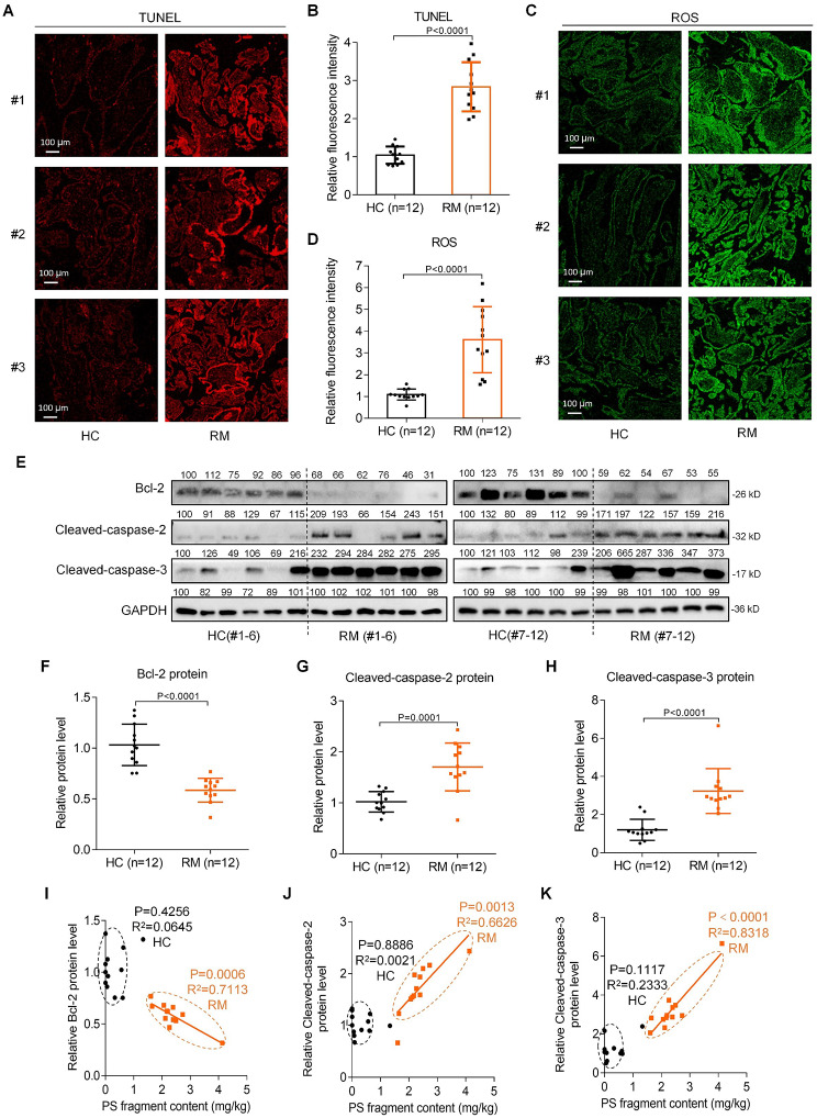
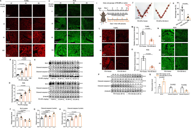
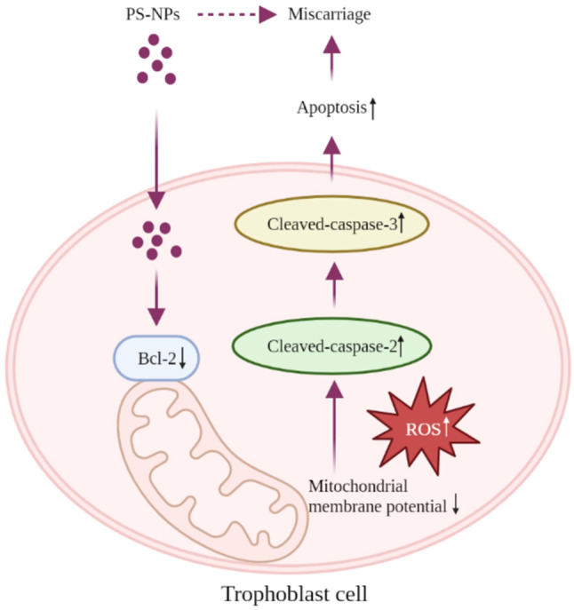
Results: In this study, we identified that polystyrene (PS) plastics particles were present in women villous tissues. Their levels were higher in villous tissues of unexplained recurrent miscarriage (RM) patients vs. healthy control (HC) group. Furthermore, mouse assays further confirmed that exposure to polystyrene nanoplastics (PS-NPs, 50 nm in diameter, 50 or 100 mg/kg) indeed induced miscarriage. In mechanism, PS-NPs exposure (50, 100, 150, or 200 µg/mL) increased oxidative stress, decreased mitochondrial membrane potential, and increased apoptosis in human trophoblast cells by activating Bcl-2/Cleaved-caspase-2/Cleaved-caspase-3 signaling through mitochondrial pathway. The alteration in this signaling was consistent in placental tissues of PS-NPs-exposed mouse model and in villous tissues of unexplained RM patients. Supplement with Bcl-2 could efficiently suppress apoptosis in PS-NPs-exposed trophoblast cells and reduce apoptosis and alleviate miscarriage in PS-NPs-exposed pregnant mouse model.

Conclusions: Exposure to PS-NPs activated Bcl-2/Cleaved-caspase-2/Cleaved-caspase-3, leading to excessive apoptosis in human trophoblast cells and in mice placental tissues, further inducing miscarriage.

Keywords: Apoptosis; Miscarriage; Polystyrene nanoplastics; Trophoblast.


Conflict of interest statement

The authors declare no competing interests.

Figures

Fig. 1
Fig. 1
PS plastic fragments were detected out in human villous tissues and their high contents were positively associated with miscarriage. (A) The contents of PS plastic fragment in HC and RM villous tissues analyzed by Py-GC/MS (each n = 18). (B) The histogram of PS plastic fragment frequency in HC villous tissues. (C) The normal QQ plot of PS fragment content in HC villous tissues. (D) The histogram of PS fragment frequency in RM villous tissues. (E) The normal QQ plot of PS fragment content in RM villous tissues. (F) The ratios of RM women in total women in each range of the contents of PS plastic fragments. (G) The ROC curve of the diagnostic value of PS fragment contents for RM
Fig. 2
Fig. 2
PS-NPs exposure induced mouse miscarriage. (A) Schematic diagram of mouse model exposed to 50 nm PS-NPs by oral gavage. (B, C) The embryo adsorption and miscarriage rates in 0, 25, 50, or 100 mg/kg PS-NPs-exposed pregnant mice (each n = 9, scale bar, 1 cm)
Fig. 3
Fig. 3
The characterization and internalization of PS-NPs in human trophoblast cells. (A, B) TEM image of PS-NPs (scale bar, 200 nm). (C) Size analysis of PS-NPs in TEM images. (D, E) Internalization of FITC-PS-NPs into Swan 71 cells analyzed by flow cytometry. (F) Distribution of FITC-PS-NPs in Swan71 cells detected by confocal microscopy. Nucleus (blue), cytoplasm (red), and FITC-PS-NPs (green, indicated by yellow arrows). Scale bar, 5 μm for all images. (G) The average number of FITC-PS-NPs were counted in the nucleus and cytoplasm of Swan 71 cells
Fig. 4
Fig. 4
PS-NPs exposure induced human trophoblast cell apoptosis. (A) Cell viability of Swan 71 cells exposed to 0-200 µg/mL PS-NPs for 24 h. (B) Cell viability of Swan 71 cells exposed to 0-500 µg/mL PS-NPs for 48 h. (C-F) Cell viability of 500 µg/mL PS-NPs-exposed Swan 71 cells treated with apoptosis inhibitor cystatin Z-VAD-FMK (C), ferroptosis inhibitor Fer-1 (D), pyroptosis inhibitor Ac-FLTD-CMK (E), or necrosis inhibitor Nec-1 (F) for 48 h. (G, H) Flow cytometry analysis and their quantification of apoptosis rates (total early and late apoptosis) in 0-200 µg/mL PS-NPs-exposed Swan 71 cells for 24 h. (I) TEM image of 0 or 200 µg/mL PS-NPs-exposed Swan 71 cells (scale bar, 5 μm; PS-NPs were indicated by yellow arrows; cell pyknosis, nuclear membrane wrinkling, chromatin aggregation, division, and edge shift were indicated by red dashed line; apoptotic bodies were indicated by red arrows). (J-L) Western blot analysis and the relative quantification of the protein levels of Caspase-3 and Cleaved-caspase-3 in 0-200 µg/mL PS-NPs-exposed Swan 71 cells for 24 h
Fig. 5
Fig. 5
PS-NPs induced trophoblast cell apoptosis through mitochondrial Bcl-2/Caspase-2/Caspase-3 pathway. (A, B) MMP levels in Swan 71 cells exposed to 0, 100, or 200 µg/mL PS-NPs for 0, 1, or 2 h (scale bar, 20 μm). (C, D) ROS levels in Swan 71 cells exposed to 0, 100, or 200 µg/mL PS-NPs for 0, 1, or 2 h (scale bar, 20 μm). (E-H) The protein levels of Bcl-2, Cleaved-caspase-2, and Cleaved-caspase-3 in Swan 71 cells exposed to 0, 50, 100, 150, or 200 µg/mL PS-NPs for 24 h. (I, J) The mRNA and protein levels of Bcl-2 in trophoblast cells with Bcl-2 knockdown. (K-N) The protein levels of Cleaved-caspase-2, Caspase-3, and Cleaved-caspase-3 in trophoblast cells with Bcl-2 knockdown. (O, P) Flow cytometry analysis and their quantification of apoptosis rates (total early and late apoptosis) of trophoblast cells with Bcl-2 knockdown
Fig. 6
Fig. 6
Supplement with Bcl-2 alleviated apoptosis in PS-NPs-exposed human trophoblast cells. (A-C) The mRNA and protein levels of Bcl-2 in Swan 71 cells with Bcl-2 overexpression. (D-G) The protein levels of Cleaved-caspase-2, Caspase-3, and Cleaved-caspase-3 in trophoblast cells exposed to 0 or 100 µg/mL PS-NPs and transfected with or without Bcl-2 overexpression plasmid for 24 h. (H, I) MMP levels in trophoblast cells exposed to 0 or 100 µg/mL PS-NPs and transfected with or without Bcl-2 overexpression plasmid for 1 h (scale bar, 20 μm). (J, K) Flow cytometry analysis and quantification of apoptosis rates (total early and late apoptosis) in trophoblast cells exposed to 0 or 100 µg/mL PS-NPs and transfected with or without Bcl-2 overexpression plasmid for 24 h
Fig. 7
Fig. 7
Higher levels of apoptosis and ROS in RM vs. HC villous tissues. (A-B) Representative images of TUNEL analysis of apoptosis levels in HC and RM villous tissues (scale bar, 100 μm) and their relative quantification of the fluorescence intensity (each n = 12). (C-D) Representative images of ROS assays in HC and RM villous tissues (scale bar, 100 μm) and their relative quantification of fluorescence intensity (each n = 12). (E-H) The protein levels of Bcl-2, Cleaved-caspase-2, and Cleaved-caspase-3 in HC and RM villous tissues (each n = 12). (I-K) The correlation analysis between the protein levels of Bcl-2 (I), Cleaved-caspase-2 (J), or Cleaved-caspase-3 (K) and PS fragment content in HC (round) and RM (square) groups (each n = 12)
Fig. 8
Fig. 8
PS-NPs exposure increased placental apoptosis and thus induced mouse miscarriage. (A-B) Representative images of TUNEL analysis of apoptosis levels in PS-NPs-exposed mouse placental tissues (scale bar, 100 μm) and their relative quantification of fluorescence intensity (each n = 9). (C-D) Representative images of ROS assays in PS-NPs-exposed mouse placental tissues (scale bar, 100 μm) and their relative quantification of fluorescence intensity (each n = 9). (E-H) The protein levels of murine Bcl-2, Cleaved-caspase-2, and Cleaved-caspase-3 in placental tissues of PS-NPs-exposed mice (each n = 6). (I) Schematic diagram of mouse intervention assays. (J-K) The embryo adsorption and miscarriage rates in pregnant mice treated with PS-NPs + Vector or PS-NPs + OE-Bcl-2 (each n = 9, scale bar, 1 cm). (L-M) Representative images of TUNEL analysis of apoptosis levels in placental tissues of PS-NPs-exposed mice treated with PS-NPs + Vector or PS-NPs + OE-Bcl-2 (scale bar, 100 μm) and their relative quantification of fluorescence intensity (each n = 9). (N-O) Representative images of ROS assays in placental tissues of PS-NPs-exposed mice treated with PS-NPs + Vector or PS-NPs + OE-Bcl-2 (scale bar, 100 μm) and their relative quantification of fluorescence intensity (each n = 9). (P, Q) The protein levels of Bcl-2, Cleaved-caspase-2, and Cleaved-caspase-3 in placental tissues of mice treated with PS-NPs + Vector or PS-NPs + OE-Bcl-2 (each n = 6)
Fig. 9
Fig. 9
The proposed regulatory mechanism. PS-NPs can readily enter trophoblast cells and distribute primarily in cytoplasm, where PS-NPs activate Bcl-2/Cleaved-caspase-2/Cleaved-caspase-3 signaling through the classical mitochondrial pathway, leading to trophoblast cell apoptosis and further inducing miscarriage

References

    1. Plastics-the. Facts 2021—An Analysis of European Plastics Production, Demand and Waste Data.
    1. Lamba P, Kaur DP, Raj S, Sorout J. Recycling/reuse of plastic waste as construction material for sustainable development: a review. Environ Sci Pollut Res Int. 2022;29(57):86156–79. doi: 10.1007/s11356-021-16980-y. - DOI - PMC - PubMed
    1. de Souza Machado AA, Kloas W, Zarfl C, Hempel S, Rillig MC. Microplastics as an emerging threat to terrestrial ecosystems. Glob Change Biol. 2018;24(4):1405–16. doi: 10.1111/gcb.14020. - DOI - PMC - PubMed
    1. Jiang B, Kauffman AE, Li L, McFee W, Cai B, Weinstein J, Lead JR, Chatterjee S, Scott GI, Xiao S. Health impacts of environmental contamination of micro- and nanoplastics: a review. Environ Health Prev Med. 2020;25(1):29. doi: 10.1186/s12199-020-00870-9. - DOI - PMC - PubMed
    1. Hartmann NB, Hüffer T, Thompson RC, Hassellöv M, Verschoor A, Daugaard AE, Rist S, Karlsson T, Brennholt N, Cole M, et al. Are we speaking the same Language? Recommendations for a definition and categorization Framework for plastic debris. Environ Sci Technol. 2019;53(3):1039–47. doi: 10.1021/acs.est.8b05297. - DOI - PubMed

Publication types