

Does Magnetic Resonance Imaging Predict Neurological Deficit in Patients with Traumatic Lower Lumbar Fractures?
- PMID: 38454754
- PMCID: PMC11065513
- DOI: 10.31616/asj.2023.0311
Does Magnetic Resonance Imaging Predict Neurological Deficit in Patients with Traumatic Lower Lumbar Fractures?
Abstract
Study design: A retrospective cohort study.
Purpose: This study aimed to understand the role of magnetic resonance imaging (MRI) in predicting neurological deficits in traumatic lower lumbar fractures (LLFs; L3-L5).
Overview of literature: Despite studies on the radiological risk factors for neurological deficits in thoracolumbar fractures, very few have focused on LLFs. Moreover, the potential utility of MRI in LLFs has not been evaluated.
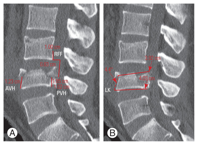
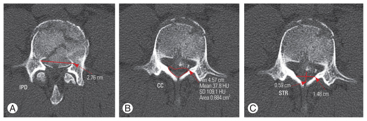
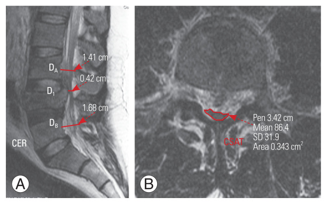
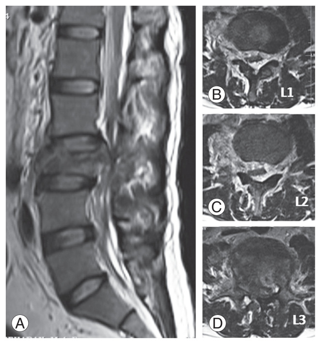
Methods: In total, 108 patients who underwent surgery for traumatic LLFs between January 2010 and January 2020 were reviewed to obtain their demographic details, injury level, and neurology status at the time of presentation (American Spinal Injury Association [ASIA] grade). Preoperative computed tomography scans were used to measure parameters such as anterior vertebral body height, posterior vertebral body height, loss of vertebral body height, local kyphosis, retropulsion of fracture fragment, interpedicular distance, canal compromise, sagittal transverse ratio, and presence of vertical lamina fracture. MRI was used to measure the canal encroachment ratio (CER), cross-sectional area of the thecal sac (CSAT), and presence of an epidural hematoma.
Results: Of the 108 patients, 9 (8.3%) had ASIA A, 4 (3.7%) had ASIA B, 17 (15.7%) had ASIA C, 21 (19.4%) had ASIA D, and 57 (52.9%) had ASIA E neurology upon admission. The Thoracolumbar Injury Classification and Severity score (p =0.000), CER (p =0.050), and CSAT (p =0.019) were found to be independently associated with neurological deficits on the multivariate analysis. The receiver operating characteristic curves showed that only CER (area under the curve [AUC], 0.926; 95% confidence interval [CI], 0.860-0.968) and CSAT (AUC, 0.963; 95% CI, 0.908-0.990) had good discriminatory ability, with the optimal cutoff of 50% and 65.3 mm2, respectively.
Conclusions: Based on the results, the optimal cutoff values of CER >50% and CSAT >65.3 mm2 can predict the incidence of neurological deficits in LLFs.
Keywords: Canal encroachment ratio; Epidural hematoma; Lower lumbar fractures; Magnetic resonance imaging; Neurological deficit.
Conflict of interest statement
No potential conflict of interest relevant to this article was reported.
Figures
References
-
- Seybold EA, Sweeney CA, Fredrickson BE, Warhold LG, Bernini PM. Functional outcome of low lumbar burst fractures: a multicenter review of operative and nonoperative treatment of L3–L5. Spine (Phila Pa 1976) 1999;24:2154–61. - PubMed
-
- Reinhold M, Knop C, Beisse R, et al. Operative treatment of 733 patients with acute thoracolumbar spinal injuries: comprehensive results from the second, prospective, Internet-based multicenter study of the Spine Study Group of the German Association of Trauma Surgery. Eur Spine J. 2010;19:1657–76. - PMC - PubMed
-
- Moore TA, Bransford RJ, France JC, et al. Low lumbar fractures: does thoracolumbar injury classification and severity score work? Spine (Phila Pa 1976) 2014;39:E1021–5. - PubMed
-
- Sansur CA, Shaffrey CI. Diagnosis and management of low lumbar burst fractures. Semin Spine Surg. 2010;22:33–7.
LinkOut - more resources
Full Text Sources
Research Materials
