

A Nomogram to Predict Benign/Malignant Mediastinal Lymph Nodes Based on EBUS Sonographic Features
- PMID: 38454935
- PMCID: PMC10919979
- DOI: 10.1155/2024/3711123
A Nomogram to Predict Benign/Malignant Mediastinal Lymph Nodes Based on EBUS Sonographic Features
Abstract
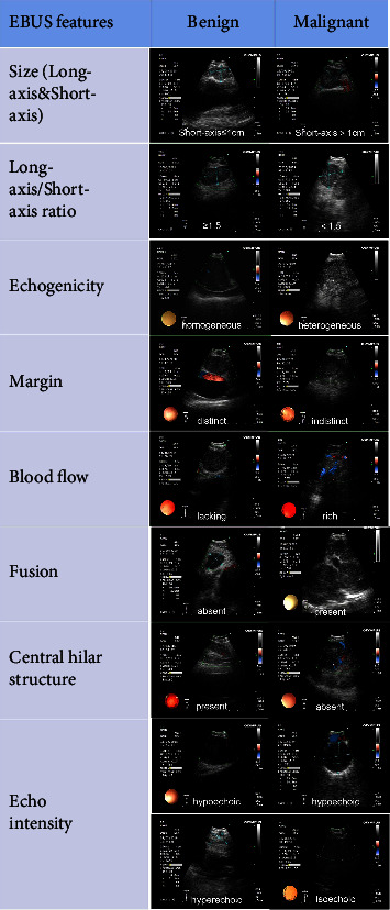
Background: Endobronchial ultrasound (EBUS) sonographic features help identify benign/malignant lymph nodes while conducting transbronchial needle aspiration (TBNA). This study aims to identify risk factors for malignancy based on EBUS sonographic features and to estimate the risk of malignancy in lymph nodes by constructing a nomogram.
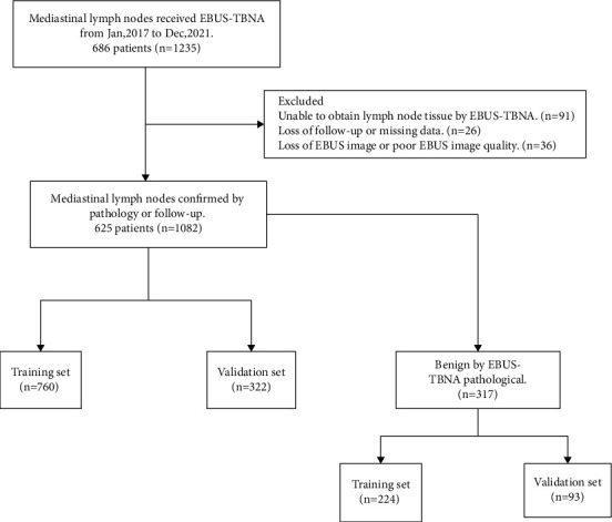
Methods: 1082 lymph nodes from 625 patients were randomly enrolled in training (n = 760) and validation (n = 322) sets. The subgroup of EBUS-TBNA postoperative negative lymph nodes (n = 317) was randomly enrolled in a training (n = 224) set and a validation (n = 93) set. Logistic regression analysis was used to identify the EBUS features of malignant lymph nodes. A nomogram was formulated using the EBUS features in the training set and later validated in the validation set.
Results: Multivariate analysis revealed that long-axis, short-axis, echogenicity, fusion, and central hilar structure (CHS) were the independent predictors of malignant lymph nodes. Based on these risk factors, a nomogram was constructed. Both the training and validation sets of 5 EBUS features nomogram showed good discrimination, with area under the curve values of 0.880 (sensitivity = 0.829 and specificity = 0.807) and 0.905 (sensitivity = 0.819 and specificity = 0.857). Subgroup multivariate analysis revealed that long-axis, echogenicity, and CHS were the independent predictors of malignancy outcomes of EBUS-TBNA postoperative negative lymph nodes. Based on these risk factors, a nomogram was constructed. Both the training and validation sets of 3 EBUS features nomogram showed good discrimination, with the area under the curve values of 0.890 (sensitivity = 0.882 and specificity = 0.786) and 0.834 (sensitivity = 0.930 and specificity = 0.636).
Conclusions: Our novel scoring system based on two nomograms can be utilized to predict malignant lymph nodes.
Copyright © 2024 Bingchao Ling et al.
Conflict of interest statement
The authors declare that they have no conflicts of interest.
Figures


Similar articles
-
Identification of specific EBUS sonographic characteristics for predicting benign mediastinal lymph nodes.Clin Respir J. 2018 Feb;12(2):681-690. doi: 10.1111/crj.12579. Epub 2016 Nov 15. Clin Respir J. 2018. PMID: 27805323
-
The combination of endobronchial elastography and sonographic findings during endobronchial ultrasound-guided transbronchial needle aspiration for predicting nodal metastasis.Thorac Cancer. 2019 Oct;10(10):2000-2005. doi: 10.1111/1759-7714.13186. Epub 2019 Sep 1. Thorac Cancer. 2019. PMID: 31474004 Free PMC article.
-
The value of endobronchial ultrasound-guided transbronchial needle aspiration, 18-fluorodeoxyglucose positron emission tomography/computed tomography, and ultrasonography imaging techniques in the diagnosis of mediastinal and/or hilar malignant, anthracotic, and other benign lymph nodes.Medicine (Baltimore). 2021 Feb 19;100(7):e24728. doi: 10.1097/MD.0000000000024728. Medicine (Baltimore). 2021. PMID: 33607816 Free PMC article.
-
Comparing diagnostic sensitivity of different needle sizes for lymph nodes suspected of lung cancer in endobronchial ultrasound transbronchial needle aspiration: Systematic review and meta-analysis.Clin Respir J. 2021 Dec;15(12):1328-1336. doi: 10.1111/crj.13436. Epub 2021 Aug 31. Clin Respir J. 2021. PMID: 34402194
-
Ultrasonographic characteristics of lymph nodes as predictors of malignancy during endobronchial ultrasound (EBUS): A systematic review.Lung Cancer. 2018 Dec;126:97-105. doi: 10.1016/j.lungcan.2018.10.020. Epub 2018 Oct 30. Lung Cancer. 2018. PMID: 30527199
References
Publication types
MeSH terms
LinkOut - more resources
Full Text Sources
Medical