

Automated volumetry of meningiomas in contrast-enhanced T1-Weighted MRI using deep learning
- PMID: 38455247
- PMCID: PMC10918322
- DOI: 10.1016/j.wnsx.2024.100353
Automated volumetry of meningiomas in contrast-enhanced T1-Weighted MRI using deep learning
Abstract
Background: Meningiomas are among the most common intracranial tumors. In these tumors, volumetric assessment is not only important for planning therapeutic intervention but also for follow-up examination.However, a highly accurate automated volumetric method for meningiomas using single-modality magnetic resonance imaging (MRI) has not yet been reported. Here, we aimed to develop a deep learning-based automated volumetry method for meningiomas in MRI and investigate its accuracy and potential clinical applications.
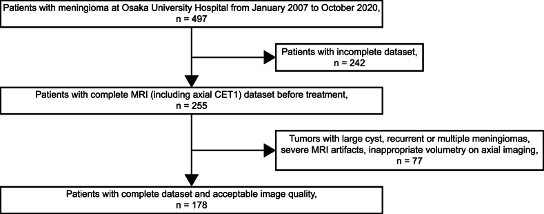
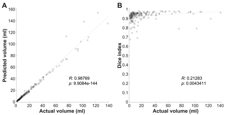
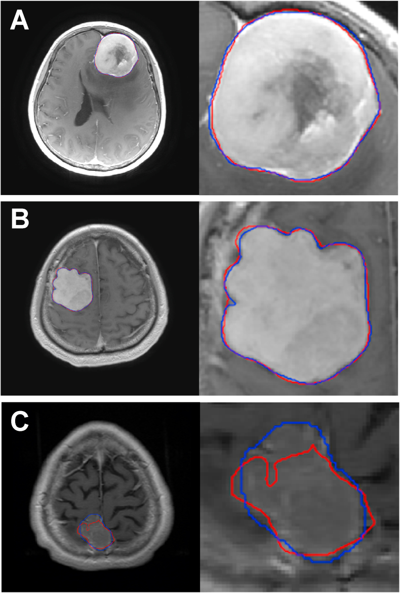
Methods: For deep learning, we used MRI images of patients with meningioma who were referred to Osaka University Hospital between January 2007 and October 2020. Imaging data of eligible patients were divided into three non-overlapping groups: training, validation, and testing. The model was trained and tested using the leave-oneout cross-validation method. Dice index (DI) and root mean squared percentage error (RMSPE) were measured to evaluate the model accuracy. Result: A total of 178 patients (64.6 ± 12.3 years [standard deviation]; 147 women) were evaluated. Comparison of the deep learning model and manual segmentation revealed a mean DI of 0.923 ± 0.051 for tumor lesions. For total tumor volume, RMSPE was 9.5 ± 1.2%, and Mann-Whitney U test did not show a significant difference between manual and algorithm-based measurement of the tumor volume (p = 0.96).
Conclusion: The automatic tumor volumetry algorithm developed in this study provides a potential volume-based imaging biomarker for tumor evaluation in the field of neuroradiological imaging, which will contribute to the optimization and personalization of treatment for central nervous system tumors in the near future.
Keywords: Algorithm; Biomarkers; Brain neoplasms; Deep learning; Magnetic resonance imaging; Meningeal neoplasms; Meningioma.
© 2024 The Authors.
Conflict of interest statement
The authors declare that they have no known competing financial interests or personal relationships that could have appeared to influence the work reported in this paper.
Figures
Similar articles
-
Automated Meningioma Segmentation in Multiparametric MRI : Comparable Effectiveness of a Deep Learning Model and Manual Segmentation.Clin Neuroradiol. 2021 Jun;31(2):357-366. doi: 10.1007/s00062-020-00884-4. Epub 2020 Feb 14. Clin Neuroradiol. 2021. PMID: 32060575
-
Fully Automated MRI Segmentation and Volumetric Measurement of Intracranial Meningioma Using Deep Learning.J Magn Reson Imaging. 2023 Mar;57(3):871-881. doi: 10.1002/jmri.28332. Epub 2022 Jul 1. J Magn Reson Imaging. 2023. PMID: 35775971
-
Fully automated detection and segmentation of meningiomas using deep learning on routine multiparametric MRI.Eur Radiol. 2019 Jan;29(1):124-132. doi: 10.1007/s00330-018-5595-8. Epub 2018 Jun 25. Eur Radiol. 2019. PMID: 29943184 Free PMC article.
-
Deep learning-based automatic segmentation of meningioma from multiparametric MRI for preoperative meningioma differentiation using radiomic features: a multicentre study.Eur Radiol. 2022 Oct;32(10):7248-7259. doi: 10.1007/s00330-022-08749-9. Epub 2022 Apr 14. Eur Radiol. 2022. PMID: 35420299
-
Primary Central Nervous System Lymphoma: Clinical Evaluation of Automated Segmentation on Multiparametric MRI Using Deep Learning.J Magn Reson Imaging. 2021 Jan;53(1):259-268. doi: 10.1002/jmri.27288. Epub 2020 Jul 13. J Magn Reson Imaging. 2021. PMID: 32662130
References
LinkOut - more resources
Full Text Sources
