. 2024 Feb 25;14(2):917-930.
doi: 10.62347/TYNU2341. eCollection 2024.

PPIB/Cyclophilin B expression associates with tumor progression and unfavorable survival in patients with pulmonary adenocarcinoma

Affiliations

PPIB/Cyclophilin B expression associates with tumor progression and unfavorable survival in patients with pulmonary adenocarcinoma

Ilseon Hwang et al. Am J Cancer Res. .

Abstract

Cyclophilin B (CypB), encoded by peptidylprolyl isomerase B (PPIB), is involved in cellular transcriptional regulation, immune responses, chemotaxis, and proliferation. Recent studies have shown that PPIB/CypB is associated with tumor progression and chemoresistance in various cancers. However, the clinicopathologic significance and mechanism of action of PPIB/CypB in non-small cell lung cancer (NSCLC) remain unclear. In this study, we used RNA in situ hybridization to examine PPIB expression in 431 NSCLC tissue microarrays consisting of 295 adenocarcinomas (ADCs) and 136 squamous cell carcinomas (SCCs). Additionally, Ki-67 expression was evaluated using immunohistochemistry. The role of PPIB/CypB was assessed in five human NSCLC cell lines. There was a significant correlation between PPIB/CypB expression and Ki-67 expression in ADC (Spearman correlation r=0.374, P<0.001) and a weak correlation in SCC (r=0.229, P=0.007). In ADCs, high PPIB expression (PPIBhigh) was associated with lymph node metastasis (P=0.023), advanced disease stage (P=0.014), disease recurrence (P=0.013), and patient mortality (P=0.015). Meanwhile, high Ki-67 expression (Ki-67high) was correlated with male sex, smoking history, high pT stage, lymph node metastasis, advanced stage, disease recurrence, and patient mortality in ADC (all P<0.001). However, there was no association between either marker or clinicopathological factors, except for old age and PPIBhigh (P=0.038) in SCC. Survival analyses revealed that the combined expression of PPIBhigh/Ki-67high was an independent prognosis factor for poor disease-free survival (HR 1.424, 95% CI 1.177-1.723, P<0.001) and overall survival (HR 1.266, 95% CI 1.036-1.548, P=0.021) in ADC, but not in SCC. Furthermore, PPIB/CypB promoted the proliferation, colony formation, and migration of NSCLC cells. We also observed the oncogenic properties of PPIB/CypB expression in human bronchial epithelial cells. In conclusion, PPIB/CypB contributes to tumor growth in NSCLC, and elevated PPIB/Ki-67 levels are linked to unfavorable survival, especially in ADC.

Keywords: Ki-67; Peptidylprolyl isomerase B; adenocarcinoma; non-small cell lung cancer; prognosis.


Conflict of interest statement

None.

Figures
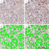
Figure 1
Figure 1
Assessment of PPIB expression in human NSCLC tissues. Digital images were automatically analyzed using Visiopharm software (v6.9.1) for quantification of PPIB expression. A captured RNAscope image of poorly differentiated adenocarcinoma (A) showed outlined tumor nuclei (green) (B), followed by identification of positive PPIB signals (red dots) (C). The mean value of DAB dots per tumor cells was calculated (D).
Figure 2
Figure 2
Correlation analysis between PPIB and Ki-67 expression in NSCLC (A), adenocarcinoma (ADC) group (B), and squamous cell carcinoma (SCC) group (C). PPIB expression showed a significant positive correlation with Ki-67 in ADC (Spearman correlation r=0.374, P<0.001), but a weak correlation in all NSCLC (Spearman correlation r=0.191, P<0.001) and SCC (Spearman correlation r=0.229, P=0.007).
Figure 3
Figure 3
Kaplan-Meier survival analysis for PPIB and Ki-67 expression in adenocarcinoma patients. Patients with high PPIB (A, D) and high Ki-67 (B, E) expression showed poor disease-free (log rank P=0.008 and P<0.001, respectively) and overall survival rates (log rank P=0.014 and P<0.001, respectively) compared to patients with low expressions of both markers, respectively. A significant difference in survival rate was found among four groups (C, F), classified based on combined PPIB and Ki-67 expression (log rank P<0.001) and patients with PPIBhigh/Ki-67high had worst disease-free (C) and overall survival rates (F).
Figure 4
Figure 4
The impact of suppressing PPIB on the proliferation, colony formation, and migration of human NSCLC cells. (A) Protein level of PPIB in HBE cells (1799 and BEAS-2B) and NSCLC cells (A549, PC-9, Calu-3, H460 and H2009) was determined by Western blot. β-ACTIN was included as an internal loading control. Numbers below blot images indicate the expression as measured by fold change. Graph depicts the experimental quantitation based on at least three independent experiments. (B-F) H460 and PC-9 cells were transfected with specific siRNA for GFP (siGFP) or PPIB (siPPIB-#3). (B) PPIB protein level in these cells was determined by Western blot. β-ACTIN was included as an internal loading control. Numbers below blot images indicate the expression as measured by fold change. (C) The proliferation index of these cells, as measured by the mean fluorescence intensity of Ki-67 staining. Graphs represent three independent experiments performed in triplicate (n=3). (D) Proliferation rate of these cells. Cells were harvested at the indicated times and counted after trypan blue staining to exclude dead cells. (E) Colony formation assay. 500 cells were plated in 12 well plates and cultured for 1 week and formed colonies were stained with crystal violet. (F) The migration ability of these cells was measured using the wound healing assays. After scratching the confluent cell layer by using pipette tips, photographs were taken at 24 h. All experiments were performed in triplicate. *P<0.05, **P<0.01 and ***P<0.001 by student’s t-test (two-tailed, unpaired) (C-F). Data represent the mean ± SD.
Figure 5
Figure 5
The effect of overexpressing PPIB on the proliferation, colony formation, and migration of NSCLC and HBE cells. H2009 and 1799 cells were stably transfected with empty vector (no insert) or PPIB expression vector. (A) FLAG-PPIB protein level in these cells was determined by Western blot. β-ACTIN was included as an internal loading control. The numbers below blot images indicate the expression as measured by fold change. (B) The proliferation index of these cells, as measured by the mean fluorescence intensity of Ki-67 staining. Graphs represent three independent experiments performed in triplicate (n=3). (C) Proliferation rate of these cells. Cells were harvested at the indicated times and counted after trypan blue staining to exclude dead cells. (D) Colony formation assay. 500 cells were plated in 12 well plates and cultured for 1 week and formed colonies were stained with crystal violet. (E) The migration ability of these cells was measured using the wound healing assays. After scratching the confluent cell layer by using pipette tips, photographs were taken at 24 h. All experiments were performed in triplicate. *P<0.05, **P<0.01 and ***P<0.001 by student’s t-test (two-tailed, unpaired) (B-E). Data represent the mean ± SD.

References

    1. Siegel RL, Miller KD, Wagle NS, Jemal A. Cancer statistics, 2023. CA Cancer J Clin. 2023;73:17–48. - PubMed
    1. Ganti AK, Klein AB, Cotarla I, Seal B, Chou E. Update of incidence, prevalence, survival, and initial treatment in patients with non-small cell lung cancer in the US. JAMA Oncol. 2021;7:1824–1832. - PMC - PubMed
    1. Rami-Porta R, Asamura H, Travis WD, Rusch VW. Lung cancer - major changes in the American Joint Committee on Cancer eighth edition cancer staging manual. CA Cancer J Clin. 2017;67:138–155. - PubMed
    1. Hou S, Zhou S, Qin Z, Yang L, Han X, Yao S, Ji H. Evidence, mechanism, and clinical relevance of the transdifferentiation from lung adenocarcinoma to squamous cell carcinoma. Am J Pathol. 2017;187:954–962. - PubMed
    1. Yun JK, Kwon Y, Kim J, Lee GD, Choi S, Kim HR, Kim YH, Kim DK, Park SI. Clinical impact of histologic type on survival and recurrence in patients with surgically resected stage II and III non-small cell lung cancer. Lung Cancer. 2023;176:24–30. - PubMed

LinkOut - more resources