

Doxazosin inhibits vasculogenic mimicry in human non‑small cell lung cancer through inhibition of the VEGF‑A/VE‑cadherin/mTOR/MMP pathway
- PMID: 38455663
- PMCID: PMC10918514
- DOI: 10.3892/ol.2024.14303
Doxazosin inhibits vasculogenic mimicry in human non‑small cell lung cancer through inhibition of the VEGF‑A/VE‑cadherin/mTOR/MMP pathway
Abstract
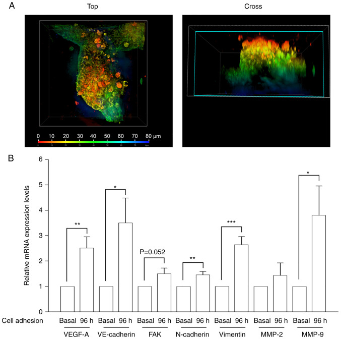
Lung cancer is the leading cause of cancer-related death worldwide, and ~85% of lung cancers are non-small cell lung cancer (NSCLC), which has a low 5-year overall survival rate and high mortality. Several therapeutic strategies have been developed, such as targeted therapy, immuno-oncotherapy and combination therapy. However, the low survival rate indicates the urgent need for new NSCLC treatments. Vasculogenic mimicry (VM) is an endothelial cell-free tumor blood supply system of aggressive and metastatic tumor cells present during tumor neovascularization. VM is clinically responsible for tumor metastasis and resistance, and is correlated with poor prognosis in NSCLC, making it a potential therapeutic target. In the present study, A549 cells formed glycoprotein-rich lined tubular structures, and transcript levels of VM-related genes were markedly upregulated in VM-forming cells. Based on a drug repurposing strategy, it was demonstrated that doxazosin (an antihypertensive drug) displayed inhibitory activity on VM formation at non-cytotoxic concentrations. Doxazosin significantly reduced the levels of vascular endothelial growth factor A (VEGF-A) and matrix metalloproteinase-2 (MMP-2) in the cell media during VM formation. Further experiments revealed that the protein expression levels of VEGF-A and vascular endothelial-cadherin (VE-cadherin), which contribute to tumor aggressiveness and VM formation, were downregulated following doxazosin treatment. Moreover, the downstream signaling Ephrin type-A receptor 2 (EphA2)/AKT/mTOR/MMP/Laminin-5γ2 network was inhibited in response to doxazosin treatment. In conclusion, the present study demonstrated that doxazosin displayed anti-VM activity in an NSCLC cell model through the downregulation of VEGF-A and VE-cadherin levels, and the suppression of signaling pathways related to the receptor tyrosine kinase, EphA2, protein kinases, AKT and mTOR, and proteases, MMP-2 and MMP-9. These results support the add-on anti-VM effect of doxazosin as a potential agent against NSCLC.
Keywords: doxazosin; drug repurposing; non-small cell lung cancer; vascular endothelial growth factor A/vascular endothelial-cadherin pathway; vasculogenic mimicry.
Copyright: © 2024 Hsu et al.
Conflict of interest statement
The authors declare that they have no competing interests.
Figures


Similar articles
-
Ginsenoside Rg3 inhibition of vasculogenic mimicry in pancreatic cancer through downregulation of VE‑cadherin/EphA2/MMP9/MMP2 expression.Int J Oncol. 2014 Sep;45(3):1065-72. doi: 10.3892/ijo.2014.2500. Epub 2014 Jun 16. Int J Oncol. 2014. PMID: 24938458
-
Serum promotes vasculogenic mimicry through the EphA2/VE-cadherin/AKT pathway in PC-3 human prostate cancer cells.Life Sci. 2019 Mar 15;221:267-273. doi: 10.1016/j.lfs.2019.02.043. Epub 2019 Feb 21. Life Sci. 2019. PMID: 30797819
-
Contribution of the PI3K/MMPs/Ln-5γ2 and EphA2/FAK/Paxillin signaling pathways to tumor growth and vasculogenic mimicry of gallbladder carcinomas.Int J Oncol. 2013 Jun;42(6):2103-15. doi: 10.3892/ijo.2013.1897. Epub 2013 Apr 15. Int J Oncol. 2013. PMID: 23588386
-
Molecular mechanisms of Thrombospondin-2 modulates tumor vasculogenic mimicry by PI3K/AKT/mTOR signaling pathway.Biomed Pharmacother. 2023 Nov;167:115455. doi: 10.1016/j.biopha.2023.115455. Epub 2023 Sep 9. Biomed Pharmacother. 2023. PMID: 37696083 Review.
-
Vasculogenic mimicry: current status and future prospects.Cancer Lett. 2007 Sep 8;254(2):157-64. doi: 10.1016/j.canlet.2006.12.036. Epub 2007 Feb 15. Cancer Lett. 2007. PMID: 17306454 Review.
Cited by
-
Vasculogenic mimicry in non-small cell lung cancer: a systematic review.Front Oncol. 2025 Jul 25;15:1481726. doi: 10.3389/fonc.2025.1481726. eCollection 2025. Front Oncol. 2025. PMID: 40786517 Free PMC article.
-
Microbial Metabolite Effects on Vasculogenic Mimicry in Metastatic Cancers.Cells. 2025 May 30;14(11):811. doi: 10.3390/cells14110811. Cells. 2025. PMID: 40497987 Free PMC article. Review.
References
LinkOut - more resources
Full Text Sources
Miscellaneous