. 2024 Mar 1;32(1):76-82.
doi: 10.53854/liim-3201-10. eCollection 2024.

Histopathologic and transmission electron microscopic findings in monkeypox cutaneous lesions

Affiliations

Histopathologic and transmission electron microscopic findings in monkeypox cutaneous lesions

Consuelo Venturi et al. Infez Med. .

Abstract

Background: a few pathologic and ultrastructural findings of monkeypox skin lesions are available in the literature. To integrate such evidence, we aimed to describe the pathologic features of monkeypox skin lesions and to show monkeypox virions by transmission electron microscopy (TEM).

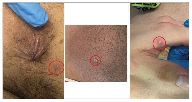
Methods: we studied the cutaneous biopsies of three patients affected by monkeypox during the 2022 monkeypox outbreak. Skin biopsies have been collected only from body sites with a recent laboratory-confirmed mpox virus infection, defined by a polymerase chain reaction (PCR) positive result in specimens taken through skin swabs.

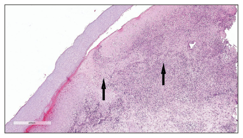
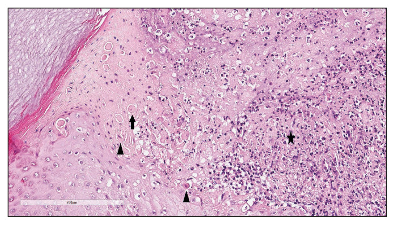
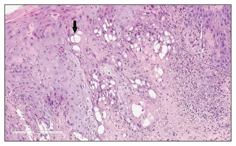
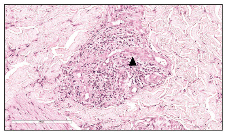
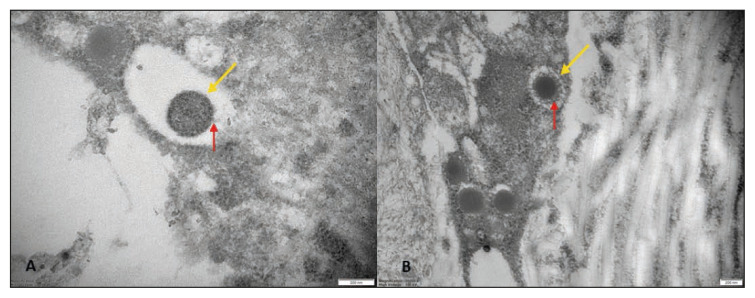
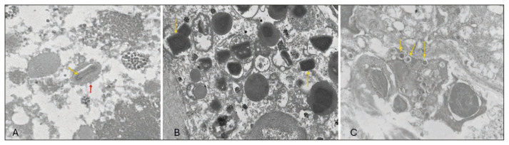
Results: in all the samples the epidermis showed keratinocytes ballooning degeneration; perivascular/periadnexal infiltrates composed of neutrophils and lymphocytes were observed in the deep dermis. Immunohistochemistry showed that the infiltrate was mostly composed of CD3+ T-cells. TEM revealed monkeypox virus-like particles in various stages of morphogenesis in the dermis and epidermis; virions were interspersed among keratinocytes and within their cytoplasm. At the intracellular level, virions showed a biconcaveshaped central core, surrounded by lateral bodies and an external membrane; they also appeared as rectangular, brick-shaped, or oval particles with eccentric nucleoids. The histologic features of our skin samples confirmed the few other studies on this topic, except for the eosinophilic inclusions of the cytoplasm of keratinocytes (Guarnieri's bodies).

Conclusion: the role of molecular biology is crucial for monkeypox diagnosis but when it is not disposable and/or in doubtful cases, skin biopsy and TEM may be helpful to establish the diagnosis.

Keywords: histopathology; monkeypox virus infection; transmission electron microscopy.


Conflict of interest statement

Conflict of interest: None to declare.

Figures

Figure 1
Figure 1
The skin biopsies have been performed on lesions (highlighted by the red circles) located in the following body sites: perianal skin (erythematous eroded papule), abdomen (whitish pustule), and hand (erythematous pustule).
Figure 2
Figure 2
Haematoxylin and eosin (H&E) stain. At scanning magnification (10x) the histopathology showed a broad dermo-epidermal ulceration with an underlying purulent base (arrows).
Figure 3
Figure 3
Haematoxylin and eosin (H&E) stain. At higher magnification (40x), the epidermis was characterized by necrotic keratinocytes with pycnotic nuclei and dense eosinophilic cytoplasm (arrowhead) and by scattered keratinocytes with a “shadow cell” appearance (enlarged cells with eosinophilic cytoplasm without well-defined nuclei) (arrow); suppurative changes were detected in the papillary dermis characterized by marked neutrophilic exocytosis (asterisk).
Figure 4
Figure 4
Haematoxylin and eosin (H&E) stain. The keratinocytes showed ballooning degeneration (arrow): keratinocytes appear swollen, pale, and round due to intra-cellular oedema and loss of intercellular bridges (magnification 40x).
Figure 5
Figure 5
Haematoxylin and eosin (H&E) stain. An interstitial and perivascular/peri-adnexal inflammatory infiltrate composed of neutrophils and lymphocytes was observed in the deep dermis, associated with endothelial swelling (arrowhead) without fibrinoid necrosis of the vascular wall (magnification 40x).
Figure 6
Figure 6
Transmission electron microscopy. Epidermal-dermal extracellular environment: viral particles with an electrondense core (yellow arrow) and a surface characterized by inhomogeneously (A, red arrow) or evenly distributed protrusions “threads” (B, red arrow) near the collagen fibers (magnification: 50000× [A], 30000× [B]).
Figure 7
Figure 7
A) Transmission electron microscopy. Epidermal intracellular environment (keratinocytes): virions showed a biconcave-shaped central core (yellow arrow), surrounded by lateral bodies and an external membrane with typical threads (red arrow), magnification 30000x; virions also appeared as rectangular, brick-shaped particles (B, yellow arrow), magnification 15000x, or as oval particles with eccentric nucleoids like “frog spawns” (C, yellow arrow), magnification 10000x.

Similar articles

Cited by

References

    1. Hughes AL, Irausquin S, Friedman R. The evolutionary biology of poxviruses. Infect Genet Evol. 2010;10(1):50–59. - PMC - PubMed
    1. World Health Organization. Mpox (monkeypox) [accessed January 10, 2024]. Available at: https://www.who.int/news-room/fact-sheets/detail/monkeypox.
    1. Farahat RA, Sah R, El-Sakka AA, et al. Human monkeypox disease (MPX) Infez Med. 2022;30(3):372–391. - PMC - PubMed
    1. Bunge EM, Hoet B, Chen L, et al. The changing epidemiology of human monkeypox-a potential threat? A systematic review. PLoS Neglected Trop Dis. 2022;16(2):e0010141. - PMC - PubMed
    1. Ciccarese G, Di Biagio A, Bruzzone B, et al. Monkeypox outbreak in Genoa, Italy: clinical, laboratory, histopathologic features, management, and outcome of the infected patients. J Med Virol. 2023;95(2):e28560. - PubMed

LinkOut - more resources