

Seeing beneath the surface: Harnessing point-of-care ultrasound for internal jugular vein evaluation
- PMID: 38456073
- PMCID: PMC10915892
- DOI: 10.4330/wjc.v16.i2.73
Seeing beneath the surface: Harnessing point-of-care ultrasound for internal jugular vein evaluation
Abstract
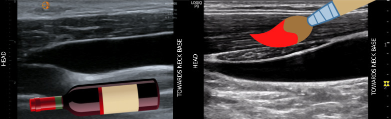
Point-of-care ultrasound (POCUS) of the internal jugular vein (IJV) offers a non-invasive means of estimating right atrial pressure (RAP), especially in cases where the inferior vena cava is inaccessible or unreliable due to conditions such as liver disease or abdominal surgery. While many clinicians are familiar with visually assessing jugular venous pressure through the internal jugular vein, this method lacks sensitivity. The utilization of POCUS significantly enhances the visualization of the vein, leading to a more accurate identification. It has been demonstrated that combining IJV POCUS with physical examination enhances the specificity of RAP estimation. This review aims to provide a comprehensive summary of the various sonographic techniques available for estimating RAP from the internal jugular vein, drawing upon existing data.
Keywords: Bedside ultrasound; Central venous pressure; Internal jugular vein; Point-of-care ultrasound; Right atrial pressure.
©The Author(s) 2024. Published by Baishideng Publishing Group Inc. All rights reserved.
Conflict of interest statement
Conflict-of-interest statement: All authors have no conflicts of interest to declare.
Figures


Similar articles
-
Evaluating the Role of Point-of-Care Ultrasound in Central Venous Pressure Monitoring for Critically Ill Patients. A Comprehensive Systematic Review and Meta-analysis.Ultrasound Med Biol. 2025 May 21:S0301-5629(25)00125-5. doi: 10.1016/j.ultrasmedbio.2025.04.008. Online ahead of print. Ultrasound Med Biol. 2025. PMID: 40404525 Review.
-
A Novel Method for Estimating Right Atrial Pressure With Point-of-Care Ultrasound.J Am Soc Echocardiogr. 2023 Mar;36(3):278-283. doi: 10.1016/j.echo.2022.12.008. Epub 2022 Dec 13. J Am Soc Echocardiogr. 2023. PMID: 36521834
-
Ultrasound imaging and central venous pressure in spontaneously breathing patients: a comparison of ultrasound-based measures of internal jugular vein and inferior vena cava.Anaesthesiol Intensive Ther. 2022;54(2):150-155. doi: 10.5114/ait.2022.114469. Anaesthesiol Intensive Ther. 2022. PMID: 35416439 Free PMC article.
-
Correlation of internal jugular and subclavian vein diameter variation on bedside ultrasound with invasive right heart catheterization.Indian Heart J. 2021 Mar-Apr;73(2):231-235. doi: 10.1016/j.ihj.2021.01.024. Epub 2021 Feb 2. Indian Heart J. 2021. PMID: 33865526 Free PMC article.
-
Noninvasive evaluation of right atrial pressure.J Am Soc Echocardiogr. 2013 Sep;26(9):1033-42. doi: 10.1016/j.echo.2013.06.004. Epub 2013 Jul 13. J Am Soc Echocardiogr. 2013. PMID: 23860098 Review.
Cited by
-
Ultrasound based estimate of central venous pressure: Are we any closer?World J Cardiol. 2024 Jun 26;16(6):310-313. doi: 10.4330/wjc.v16.i6.310. World J Cardiol. 2024. PMID: 38993581 Free PMC article.
-
Contemporary Perspectives on Congestion in Heart Failure: Bridging Classic Signs with Evolving Diagnostic and Therapeutic Strategies.Diagnostics (Basel). 2025 Apr 24;15(9):1083. doi: 10.3390/diagnostics15091083. Diagnostics (Basel). 2025. PMID: 40361901 Free PMC article. Review.
-
Point-of-care ultrasonography in cirrhosis-related acute kidney injury: How I do it.World J Crit Care Med. 2024 Jun 9;13(2):93812. doi: 10.5492/wjccm.v13.i2.93812. eCollection 2024 Jun 9. World J Crit Care Med. 2024. PMID: 38855271 Free PMC article. Review.
-
Echocardiography in the Ventilated Patient: What the Clinician Has to Know.J Clin Med. 2024 Dec 27;14(1):77. doi: 10.3390/jcm14010077. J Clin Med. 2024. PMID: 39797158 Free PMC article. Review.
References
-
- Assavapokee T, Thadanipon K. Examination of the Neck Veins. N Engl J Med. 2020;383:e132. - PubMed
-
- Jang T, Aubin C, Naunheim R, Char D. Ultrasonography of the internal jugular vein in patients with dyspnea without jugular venous distention on physical examination. Ann Emerg Med. 2004;44:160–168. - PubMed
-
- Donahue SP, Wood JP, Patel BM, Quinn JV. Correlation of sonographic measurements of the internal jugular vein with central venous pressure. Am J Emerg Med. 2009;27:851–855. - PubMed
-
- Baumann UA, Marquis C, Stoupis C, Willenberg TA, Takala J, Jakob SM. Estimation of central venous pressure by ultrasound. Resuscitation. 2005;64:193–199. - PubMed
-
- Parienti JJ, Mongardon N, Mégarbane B, Mira JP, Kalfon P, Gros A, Marqué S, Thuong M, Pottier V, Ramakers M, Savary B, Seguin A, Valette X, Terzi N, Sauneuf B, Cattoir V, Mermel LA, du Cheyron D 3SITES Study Group. Intravascular Complications of Central Venous Catheterization by Insertion Site. N Engl J Med. 2015;373:1220–1229. - PubMed
Publication types
LinkOut - more resources
Full Text Sources
