. 2024 Mar 19;13(6):e030460.
doi: 10.1161/JAHA.123.030460. Epub 2024 Mar 8.

Stroke Causes DNA Methylation at Ncx1 Heart Promoter in the Brain Via DNMT1/MeCP2/REST Epigenetic Complex

Affiliations

Stroke Causes DNA Methylation at Ncx1 Heart Promoter in the Brain Via DNMT1/MeCP2/REST Epigenetic Complex

Natascia Guida et al. J Am Heart Assoc. .

Abstract

Background: REST (Repressor-Element 1 [RE1]-silencing transcription factor) inhibits Na+/Ca2+exchanger-1 (Ncx1) transcription in neurons through the binding of RE1 site on brain promoter (Br) after stroke. We identified a new putative RE1 site in Ncx1 heart promoter (Ht) sequence (Ht-RE1) that participates in neuronal Ncx1 transcription. Because REST recruits DNA-methyltransferase-1 (DNMT1) and MeCP2 (methyl-CpG binding protein 2) on different neuronal genes, we investigated the role of this complex in Ncx1 transcriptional regulation after stroke.

Methods and results: Luciferase experiments performed in SH-SY5Y cells demonstrated that Br activity was selectively decreased by REST, whereas Ht activity was reduced by DNMT1, MeCP2, and REST. Notably, site-direct mutagenesis of Ht-RE1 prevented REST-dependent downregulation of Ncx1. Furthermore, in temporoparietal cortex of 8-week-old male wild-type mice (C57BL/6) subjected to transient middle cerebral artery occlusion, DNMT1, MeCP2, and REST binding to Ht promoter was increased, with a consequent DNA promoter hypermethylation. Intracerebroventricular injection of siREST prevented DNMT1/MeCP2 binding to Ht and Ncx1 downregulation, thus causing a reduction in stroke-induced damage. Consistently, in cortical neurons subjected to oxygen and glucose deprivation plus reoxygenation Ncx1 knockdown counteracted neuronal protection induced by the demethylating agent 5-azacytidine. For comparisons between 2 experimental groups, Student's t test was used, whereas for more than 2 experimental groups, 1-way ANOVA was used, followed by Tukey or Newman Keuls. Statistical significance was set at P<0.05.

Conclusions: If the results of this study are confirmed in humans, it could be asserted that DNMT1/MeCP2/REST complex disruption could be a new pharmacological strategy to reduce DNA methylation of Ht in the brain, ameliorating stroke damage.

Keywords: DNA methylation; DNMT1; MeCP2; NCX1; REST; stroke.


Figures

Figure 1
Figure 1. The epigenetic factors DNMT1 and MeCP2 and the transcriptional factor REST are involved in the negative modulation of Ncx1 mRNA and protein expression.
A and B, qRT‐PCR for DNMT1 and MECP2 transcripts in SH‐SY5Y cells transfected for 48 hours with siDNMT1 or siMeCP2 at a final concentration of 50 nM. An siCTL was used as scrambled. *P≤0.05 vs siCTL by Student's t test (n=3). C, Ncx1 mRNA and protein expression in SH‐SY5Y cells transfected for 48 hours with siCTL, siDNMT1, siMeCP2, and siREST. *P≤0.05 vs siCTL by 1‐way ANOVA analysis followed by Tukey's post hoc test (n=3/4). D and E, qRT‐PCR for DNMT1 and MECP2 transcripts in SH‐SY5Y transfected for 48 hours with vector overexpressing DNMT1 or MeCP2. pcDNA3.1 vector was used as EV. *P≤0.05 vs EV by Student's t test (n=3). F, Ncx1 mRNA and protein expression in SH‐SY5Y cells transfected for 48 hour with EV, DNMT1, MeCP2, or REST plasmids. *P≤0.05 vs EV by 1‐way ANOVA analysis followed by Tukey's post hoc test (n=3/4). CTL indicates control; DNMT1, DNA‐methyltransferase‐1; EV, empty vector; MeCP2, methyl‐CpG binding protein 2; NCX1, sodium/calcium exchanger 1; qRT‐PCR, quantitative real time polymerase chain reaction; REST, repressor element 1‐silencing transcription factor; si, small interfering; and siCTL, control siRNA.
Figure 2
Figure 2. The epigenetic factors DNMT1 and MeCP2 and the transcriptional factor REST are involved in the negative modulation of Ncx1 promoter activity.
A, Alignment of the human, mouse, and rat Ht sequences is shown. Conserved nucleotides are marked with asterisk. The arrow indicates the TSS. B through E, Luciferase assay in SH‐SY5Y cells transfected for 48 hour with pGL3‐Br (B and D) or pGL3‐Ht (C and E) and cotransfected with siCTL, siDNMT1, siMeCP2, or siREST (B, C), or with EV, DNMT1, MeCP2, or REST (D and E). *P≤0.05 vs siCTL or EV by 1‐way ANOVA analysis followed by Tukey's post hoc test (n=4). CTL indicates control; DNMT1, DNA‐methyltransferase‐1; EV, empty vector; Ht, heart promoter, MeCP2, methyl‐CpG binding protein 2; NCX1, sodium/calcium exchanger 1; pGL3‐Br, Ncx1 brain promoter construct inserted in pGL3basic; pGL3‐Ht, Ncx1 heart promoter construct inserted in pGL3basic; REST, repressor element 1‐silencing transcription factor; si, small interfering; and TSS, transcription start site.
Figure 3
Figure 3. REST represses Ncx1 transcription through a direct binding of a RE1 site on Ht promoter region.
A, Top: JASPAR matrix representation (MA0138.1) of the consensus REST binding site on Ht gene (Ht‐RE1). Ht‐RE1 sequence is represented in International Union of Pure and Applied Chemistry code. Bottom: Partial human genomic Ht sequence containing the predicted REST binding site (underlined). B, Luciferase assay in SH‐SY5Y cells under the following experimental conditions: (1) pGL3basic, (2) pGL3‐Ht+EV, (3) pGL3‐Ht+REST, (4) pGL3‐Ht‐RE1mut+EV, (5) pGL3‐Ht‐RE1mut+REST. *P≤0.05 vs pGL3Ht+EV by 1‐way ANOVA analysis followed by Tukey's post hoc test (n=4). C, ChIP with anti‐REST antibody followed by qPCR of the promoter region containing the RE1 site on the Ht gene in SH‐SY5Y cells transiently transfected with (1) pGL3‐Ht+EV, (2) pGL3‐Ht+REST, (3) pGL3‐Ht+RE1mut+REST. *P≤0.05 vs pGL3‐Ht+EV by 1‐way ANOVA analysis followed by Tukey's post hoc test (n=4). ChIP indicates chromatin immunoprecipitation; CTL, control; DNMT1, DNA‐methyltransferase‐1; EV, empty vector; Ht, heart promoter, MeCP2, methyl‐CpG binding protein 2; NCX1, sodium/calcium exchanger 1; pGL3‐Br, Ncx1 brain promoter construct inserted in pGL3basic; pGL3‐Ht, Ncx1 heart promoter construct inserted in pGL3basic; qRT‐PCR, quantitative real time polymerase chain reaction; RE1, repressor element 1; and REST, repressor element 1‐silencing transcription factor.
Figure 4
Figure 4. DNMT1 and MeCP2 bind Ht, but not Br promoter sequence, in the temporoparietal cortex 24 hours after tMCAO.
Chromatin immunoprecipitation with anti‐DNMT1 (A), anti‐MeCP2 (B) anti‐REST (C), anti‐HDAC1 (D), anti‐HDAC2 (E), antibodies followed by qPCR of Ncx1 brain promoter (Br) (white columns) and Ncx1 heart promoter (Ht) (gray columns) in peri‐ischemic cortex of mice euthanized 12 or 24 hours after tMCAO. IgG was used as negative control. *P≤0.05 vs Sham immunopreciptated with IgG antibody by 1‐way ANOVA analysis followed by Tukey's post hoc test (n=3). DNMT1 indicates DNA‐methyltransferase‐1; HDAC, histone deacetylase; IP, immunoprecipitation; MeCP2, methyl‐CpG binding protein 2; NCX1, sodium/calcium exchanger 1; qRT‐PCR, quantitative real time polymerase chain reaction; REST, repressor element 1‐silencing transcription factor; and tMCAO, transient middle cerebral artery occlusion.
Figure 5
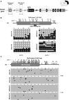
Figure 5. CpG group 1 containing Ht promoter is hypermethylated in peri‐ischemic cortex of mice after tMCAO, whereas CpG island 2 remains hypomethylated.
A, Schematic representation of the 5′‐region of murine Slc8a1 gene encoding for Ncx1. Exons are represented as boxes, introns as thin connecting lines. Black filled boxes correspond to genomic regions encoding for mature protein, whereas gray regions correspond to exons transcribing for untranslated regions. Open boxes represent regions frequently excluded from mature Ncx1 mRNA by alternative splicing in neurons. Heart, kidney, and brain promoters are represented as 1Ht, 1Kc, and 1Br boxes, respectively. CpG island 1 and 2 are represented above Ht and Br promoter regions. (B and C) top: schematic representation of CpG sites on CpG group 1 region in panel B and on CpG island 2 in (C). Ht and Br promoters are represented by a gray box, whereas black box represents the relative position of Ht‐RE1 sequence. Vertical black lines indicate locations of each CpG site. Bottom: Bisulfite sequencing patterns of the “CpG group 1” region (B) and CpG island 2 (C). Black filled circles and open circles represent methylated and nonmethylated CpG sites, respectively. Gray filled circles represent no CpG at the expected site. Each row represents a single clone, whereas each column represents a single CpG site. Percentage of methylated CpG sites in each group was calculated as mean percentage of methylation on 20 clones. *P≤0.05 vs sham‐operated animals, by Mann–Whitney U test. Br indicates brain promoter; Ht, heart promoter; NCX1, sodium/calcium exchanger 1; RE1, repressor element 1; and tMCAO, transient middle cerebral artery occlusion.
Figure 6
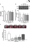
Figure 6. The knockdown of DNMT1, MeCP2, and REST counteracts the stroke‐mediated downregulation of Ncx1 mRNA and protein levels and ameliorates the ischemic damage in mice.
A and B, Effect of intracerebroventricular injections of siRNA for DNMT1, MeCP2, and REST on Ncx1 mRNA and protein expression levels in mice subjected to tMCAO and euthanized after 24 hours. *P≤0.05 vs Sham, # P≤0.05 vs tMCAO 24 h+siCTL by 1‐way ANOVA analysis followed by Tukey's post hoc test (n=4). C, % of brain infarct volume in mice subjected to tMCAO after intracerebroventricular administration of siCTL, siDNMT1, siMeCP2, siREST, and siNCX1. Mice were euthanized 24 hours after tMCAO. Representative brain slices from each experimental group are shown on the top. # P≤0.05 vs tMCAO 24 h+siCTL, by 1‐way ANOVA, followed by Newman–Keuls test (n=4). CTL indicates control; DNMT1, DNA‐methyltransferase‐1; MeCP2, methyl‐CpG binding protein 2; NCX1, sodium/calcium exchanger 1; REST, repressor element 1‐silencing transcription factor; si, small interfering; and tMCAO, transient middle cerebral artery occlusion.
Figure 7
Figure 7. Schematic Illustration of transcriptional and epigenetic complexes controlling Ncx1 during OGD/reoxygenation phase and stroke.
The transcriptional factor REST modulates Ncx1 through 2 different mechanisms involving Br and Ht at different time points after stroke. AZA indicates azacytidine; Br, brain promoter; DNMT1, DNA‐methyltransferase‐1; HDAC, histone deacetylase; Ht, heart promoter; MeCP2, methyl‐CpG binding protein 2; NCX1, sodium/calcium exchanger 1; OGD, oxygen and glucose deprivation; RE1, repressor element 1; REST, repressor element 1‐silencing transcription factor; and tMCAO, transient middle cerebral artery occlusion.

Similar articles

Cited by

References

    1. Molinaro P, Sirabella R, Pignataro G, Petrozziello T, Secondo A, Boscia F, Vinciguerra A, Cuomo O, Philipson KD, De Felice M, et al. Neuronal NCX1 overexpression induces stroke resistance while knockout induces vulnerability via Akt. J Cereb Blood Flow Metab. 2016;36:1790–1803. doi: 10.1177/0271678X15611913 - DOI - PMC - PubMed
    1. Annunziato L, Pignataro G, Di Renzo GF. Pharmacology of brain Na+/Ca2+ exchanger: from molecular biology to therapeutic perspectives. Pharmacol Rev. 2004;56:633–654. doi: 10.1124/pr.56.4.5 - DOI - PubMed
    1. Formisano L, Guida N, Valsecchi V, Cantile M, Cuomo O, Vinciguerra A, Laudati G, Pignataro G, Sirabella R, Di Renzo G, et al. Sp3/REST/HDAC1/HDAC2 complex represses and Sp1/HIF‐1/p300 complex activates ncx1 gene transcription, in brain ischemia and in ischemic brain preconditioning, by epigenetic mechanism. J Neurosci. 2015;35:7332–7348. doi: 10.1523/JNEUROSCI.2174-14.2015 - DOI - PMC - PubMed
    1. Ng GY, Lim YA, Sobey CG, Dheen T, Fann DY, Arumugam TV. Epigenetic regulation of inflammation in stroke. Ther Adv Neurol Disord. 2018;11:1–30. doi: 10.1177/1756286418771815 - DOI - PMC - PubMed
    1. Rhee I, Bachman KE, Park BH, Jair KW, Yen RW, Schuebel KE, Cui H, Feinberg AP, Lengauer C, Kinzler KW, et al. DNMT1 and DNMT3b cooperate to silence genes in human cancer cells. Nature. 2002;416:552–556. doi: 10.1038/416552a - DOI - PubMed