

Patient Screening for Self-Expanding Percutaneous Pulmonary Valves Using Virtual Reality
- PMID: 38456473
- PMCID: PMC11009987
- DOI: 10.1161/JAHA.123.033239
Patient Screening for Self-Expanding Percutaneous Pulmonary Valves Using Virtual Reality
Abstract
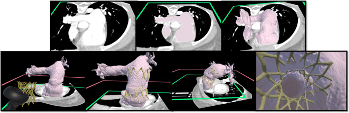
Background: In recent years, self-expanding technology to treat pulmonary regurgitation in the native right ventricular outflow tract became Food and Drug Administration approved in the United States and is now routinely used. The current practice for selection of patients who are candidates for these devices includes screening for "anatomic fit," performed by each of the manufacturing companies. Our study aims to validate the use of virtual reality (VR) as a tool for local physician-led screening of patients.
Methods and results: This retrospective study from Children's Hospital Colorado included patients who underwent pulmonary valve replacement and had screening for a Harmony TPV or Alterra Prestent performed between September 2020 and January 2022. The data from the commercial companies' dedicated analysis for self-expanding transcatheter pulmonary valve frames evaluation with perimeter analysis were collected. VR simulation was performed blinded by 2 congenital interventional cardiologists using Elucis VR software and an Oculus Quest 2 headset. Among the 27 evaluated cases, the use of a self-expandable valve was recommended by companies' dedicated analysis in 23 cases (85.2%), by VR assessment in 26 cases (96.3), and finally implanted in 25 cases (92.6%). Regarding the level of agreement, both modalities (manufacturer and VR) were good at screening-in patients who received a self-expanding valve (100% versus 96.1%). When it came to screening-out the patients, VR presented good capacity to accurately classify nonsuitable patients (50% versus 100%).
Conclusions: Our institutional experience with VR transcatheter pulmonary valve implantation planning accurately predicted clinical outcomes. This paves the way for routine use of VR in patient selection for self-expanding valve technologies.
Keywords: Alterra Prestent; anatomic fit; harmony TPV; self‐expanding technology; transcatheter pulmonary valve implantation; virtual reality.
Figures

References
-
- Zablah JE, Morgan GJ. Current treatment options for catheter‐based pulmonary valve replacement in children. Curr Treat Options Pediatr. 2020;6:274–282. doi: 10.1007/s40746-020-00209-0 - DOI
-
- Ghosh RM, Jolley MA, Mascio CE, Chen JM, Fuller S, Rome JJ, Silvestro E, Whitehead KK. Clinical 3D modeling to guide pediatric cardiothoracic surgery and intervention using 3D printed anatomic models, computer aided design and virtual reality. 3D Print Med. 2022;8(1):11. doi: 10.1186/s41205-022-00137-9 - DOI - PMC - PubMed
MeSH terms
LinkOut - more resources
Full Text Sources
