. 2024 Mar 8;26(1):4.
doi: 10.1007/s12017-023-08771-0.

Effect of 2-Week Naringin Supplementation on Neurogenesis and BDNF Levels in Ischemia-Reperfusion Model of Rats

Affiliations

Effect of 2-Week Naringin Supplementation on Neurogenesis and BDNF Levels in Ischemia-Reperfusion Model of Rats

Esen Yilmaz et al. Neuromolecular Med. .

Abstract

Background: Ischemic stroke is the leading cause of mortality and disability worldwide with more than half of survivors living with serious neurological sequelae; thus, it has recently attracted a lot of attention in the field of medical study.

Purpose: The aim of this study was to determine the effect of naringin supplementation on neurogenesis and brain-derived neurotrophic factor (BDNF) levels in the brain in experimental brain ischemia-reperfusion.

Study design: The research was carried out on 40 male Wistar-type rats (10-12 weeks old) obtained from the Experimental Animals Research and Application Center of Selçuk University. Experimental groups were as follows: (1) Control group, (2) Sham group, (3) Brain ischemia-reperfusion group, (4) Brain ischemia-reperfusion + vehicle group (administered for 14 days), and (5) Brain ischemia-reperfusion + Naringin group (100 mg/kg/day administered for 14 days).

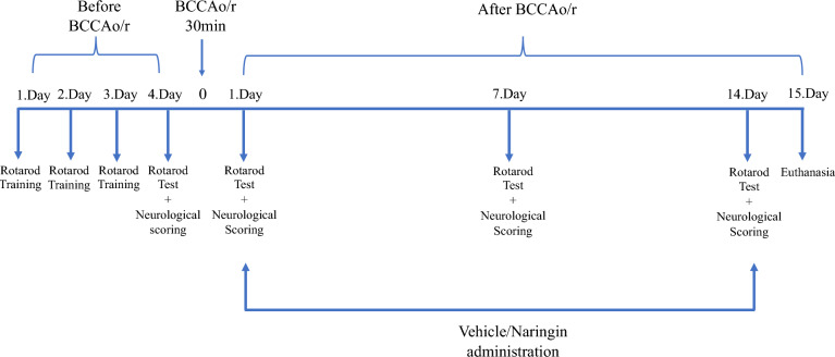
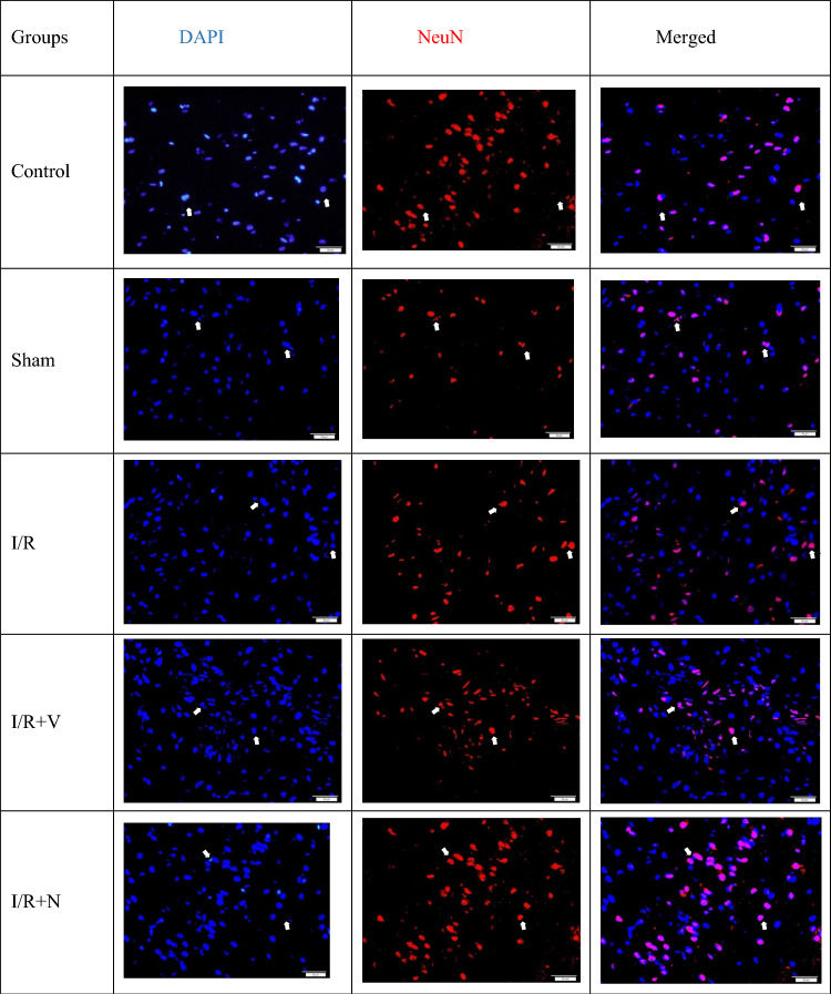
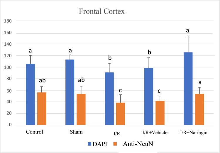
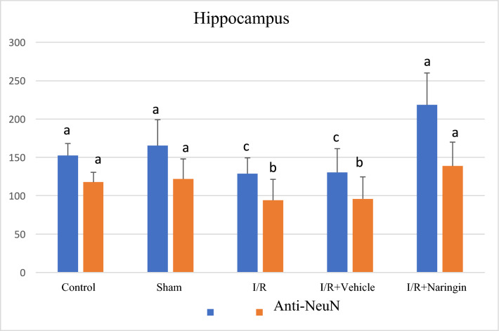
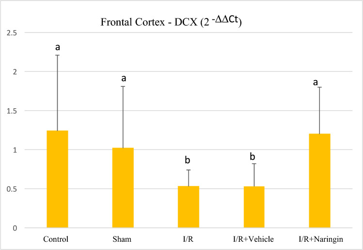
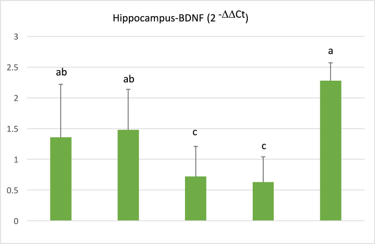
Methods: In the ischemia-reperfusion groups, global ischemia was performed in the brain by ligation of the right and left carotid arteries for 30 min. Naringin was administered to experimental animals by intragastric route for 14 days following reperfusion. The training phase of the rotarod test was started 4 days before ischemia-reperfusion, and the test phase together with neurological scoring was performed the day before and 1, 7, and 14 days after the operation. At the end of the experiment, animals were sacrificed, and then hippocampus and frontal cortex tissues were taken from the brain. Double cortin marker (DCX), neuronal nuclear antigen marker (NeuN), and BDNF were evaluated in hippocampus and frontal cortex tissues by Real-Time qPCR analysis and immunohistochemistry methods.

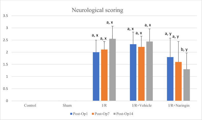
Results: While ischemia-reperfusion increased the neurological score values, DCX, NeuN, and BDNF levels decreased significantly after ischemia in the hippocampus and frontal cortex tissues. However, naringin supplementation restored the deterioration to a certain extent.

Conclusion: The results of the study show that 2 weeks of naringin supplementation may have protective effects on impaired neurogenesis and BDNF levels after brain ischemia and reperfusion in rats.

Keywords: BDNF; DCX; Flavonoid; Naringin; NeuN; Neurogenesis.


Conflict of interest statement

The authors declare no competing interests.

Figures

Fig. 1
Fig. 1
Timeline of the study. Plan of performing rotarod tests and neurological scoring before and after bilateral common carotid artery occlusion/reperfusion (BCCAo/r)
Fig. 2
Fig. 2
*a,b,c shows the statistical difference within the same group (a > b > c) (P < 0.001); x,y,z shows the statistical difference in groups (x > y > z) (P < 0.001). I/R led to increasing neurologic score levels, however, naringin supplementation improved neurologic score levels especially on days 7th and 14th after BCCAo/r
Fig. 3
Fig. 3
*a,b,c shows the statistical difference within the same group; x,y,z shows the statistical difference in the groups (a > b > c; x > y >) (P < 0.001). I/R led to increasing rotarod test values, however, naringin supplementation increased rotarod test values back to control levels on the 7th and 14th days after BCCAo/r
Fig. 4
Fig. 4
Fluorescent images with immunolabeling to demonstrate neurogenesis in the frontal cortex. DAPI (blue) column shows all nucleated cells (neurons and glia) in frontal cortex tissue, NeuN (red) column shows mature neurons in frontal cortex tissue, DAPI and NeuN images combined (pink) are shown in merged column. Arrows indicate mature neurons as examples (X40 Magnification; 1 bar: 20 µm)
Fig. 5
Fig. 5
Levels of DAPI and anti-NeuN antibodies in the frontal cortex in experimental groups. While the number of cells stained with DAPI and NeuN was unchanged in the control and sham groups, it decreased in the I/R and I/R + solvent groups, but increased significantly with naringin supplementation (a > b > c; P < 0.001)
Fig. 6
Fig. 6
Fluorescent images with immunolabeling to demonstrate neurogenesis in the hippocampus. The DAPI (blue) column shows all nucleated cells (neurons and glia) in the hippocampus tissue, the NeuN (red) column shows mature neurons in the hippocampus tissue, the merged (pink) version of the DAPI and NeuN images is shown in the merged column. Arrows indicate mature neurons as examples (X40 Magnification; 1 bar: 50 µm)
Fig. 7
Fig. 7
Levels of DAPI and anti-NeuN antibodies in the hippocampus in experimental groups. While the number of cells stained with DAPI and NeuN did not change in the control and sham groups, it decreased in the I/R and I/R + solvent groups, but increased significantly with naringin supplementation (a > b > c; P < 0.001)
Fig. 8
Fig. 8
Expression level of DCX gene in the frontal cortex in experimental groups. Expression levels of the DCX gene did not change in the control and sham groups, but decreased in the I/R and I/R + solvent groups. With naringin supplementation, it again reached control group levels (a > b; P < 0.03)
Fig. 9
Fig. 9
Expression level of DCX gene in the hippocampus in experimental groups. Expression levels of the DCX gene did not change in the control and sham groups, but decreased in the I/R and I/R + solvent groups. With naringin supplementation, it increased back to the control group levels (a > b > c; P < 0.001)
Fig. 10
Fig. 10
Expression level of BDNF gene in the frontal cortex in experimental groups. While the BDNF gene expression level was close in the control and sham groups, it decreased in the I/R and I/R + solvent groups, but this decrease was restored with naringin supplementation and increased to the control level (a > b; P < 0.01)
Fig. 11
Fig. 11
Expression level of BDNF gene in the hippocampus in experimental groups. BDNF gene expression levels were close in the control and sham groups. The decreased BDNF gene expression in the I/R and I/R + solvent groups reached control level again with naringin supplementation (a > b > c; P < 0.01)

Similar articles

Cited by

References

    1. Ojaghihaghighi, S., Vahdati, S. S., Mikaeilpour, A., & Ramouz, A. (2017). Comparison of neurological clinical manifestation in patients with hemorrhagic and ischemic stroke. World Journal of Emergency Medicine,8, 34–38. - PMC - PubMed
    1. Prabhakaran, S., Ruff, I., & Bernstein, R. A. (2015). Acute stroke intervention: A systematic review. JAMA,313, 1451–1462. - PubMed
    1. Dorado, L., Millan, M., & Davalos, A. (2014). Reperfusion therapies for acute ischemic stroke: An update. Current Cardiology Reviews,10, 327–335. - PMC - PubMed
    1. Rahman, A. A., Amruta, N., Pinteaux, E., & Bix, G. J. (2021). Neurogenesis after stroke: A therapeutic perspective. Translational Stroke Research,12, 1–14. - PMC - PubMed
    1. Marques, B. L., Carvalho, G. A., Freitas, E. M. M., Chiareli, R. A., Barbosa, T. G., Di Araujo, A. G. P., Nogueira, Y. L., Ribeiro, R. I., Parreira, R. C., Vieira, M. S., Resende, R. R., Gomez, R. S., Oliveira-Lima, O. C., & Pinto, M. C. X. (2019). The role of neurogenesis in neurorepair after ischemic stroke. Seminars in Cell & Developmental Biology,95, 98–110. - PubMed

LinkOut - more resources