. 2024 May 2;143(18):1816-1824.
doi: 10.1182/blood.2023023370.

T-cell help in the tumor microenvironment enhances rituximab-mediated NK-cell ADCC

Affiliations

T-cell help in the tumor microenvironment enhances rituximab-mediated NK-cell ADCC

Jyoti Arora et al. Blood. .

Abstract

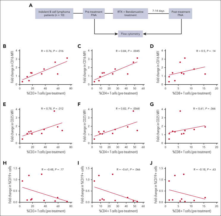
Rituximab (RTX) and other monoclonal antibodies (mAbs) that bind directly to malignant cells are of great clinical value but are not effective for all patients. A major mechanism of action of RTX is antibody-dependent cellular cytotoxicity (ADCC) mediated by natural killer (NK) cells. Prior in vitro studies in our laboratory demonstrated that T cells contribute to maintaining the viability and cytotoxic potential of NK cells activated by anti-CD20-coated target B cells. Here, we conducted studies using a novel mouse model and clinical correlative analysis to assess whether T-cell help contribute to RTX-mediated NK-cell ADCC in the tumor microenvironment (TME) in vivo. A humanized mouse model was developed using Raji lymphoma cells and normal donor peripheral blood mononuclear cells that allows for control of T-cell numbers in the lymphoma TME. In this model, NK-cell viability and CD16 and CD25 expression dropped after RTX in the absence of T cells but increased in the presence of T cells. RTX therapy was more effective when T cells were present and was ineffective when NK cells were depleted. In patients with indolent lymphoma, fine needle aspirates were obtained before and ∼1 week after treatment with a RTX-containing regimen. There was a strong correlation between CD4+ T cells as well as total T cells in the pretherapy TME and an increase in NK-cell CD16 and CD25 expression after RTX. We conclude that T-cell help in the TME enhances RTX-mediated NK-cell viability and ADCC.


Conflict of interest statement

Conflict-of-interest disclosure: The authors declare no competing financial interests.

Figures

None
Graphical abstract
Figure 1.
Figure 1.
Emerging tumors exhibit uniform distribution of T cells and NK cells in the TME. Humanized mouse xenograft tumors were harvested 27 days after tumor inoculation. IHC staining for hematoxylin and eosin, along with specific markers for CD3 (A), CD16 (B), and CD56 (C), was used to assess the distribution of immune cells within the TME.
Figure 2.
Figure 2.
Adjusting T-cell numbers in the inoculum results in varying T-cell numbers in the emerging tumors but does not have a detectable impact on fractions of malignant cells or NK cells. (A) Schematic diagram of a humanized mouse model in which mice were inoculated on day 0 with consistent numbers of Raji tumor cells and T-cell–depleted PBMCs but varying numbers of T cells. (B) Flow cytometric analysis of FNAs obtained from mouse xenografts 20 days after inoculation and evaluated for percent of cells that are CD19+, CD56+, or CD3+ (n = 12-15 mice per group).
Figure 3.
Figure 3.
Humanized mouse model: T cells in the pretherapy TME increase NK-cell viability and CD16 and CD25 expression after RTX therapy. Flow cytometric analysis of intratumoral NK cells before and after RTX therapy. Mice were inoculated on day 0 with Raji tumor cells mixed with PBMCs containing varying percentages of T cells. After tumors developed on day 20, FNAs were obtained, and mice were treated systemically with RTX (or TRA as control). On day 24 (4 days after mAb dose), a second FNAs were obtained and before to after therapy changes in NK-cell numbers (A), NK-cell CD16 expression (B), and NK-cell CD25 expression (C) were determined.
Figure 4.
Figure 4.
Intratumoral T cells enhance NK-cell–mediated RTX efficacy. (A-C) Mice were inoculated with luciferase-expressing Raji-Luc cells mixed with PBMCs containing varying percentages of T cells, on day 0. Weekly treatment with RTX or TRA was started on day 7. (A) Spider plot of total flux determined using bioluminescent imaging. (B) Changes in tumor flux after treatment compared with that of before treatment (day 21 vs day 4). (C) Kaplan-Meier curve of survival. Mice were euthanized when tumors exceeded 20 mm in any dimension. (D) In a separate experiment, mice were inoculated with Raji-Luc mixed with either PBMCs or CD56-depleted PBMCs on day 0, followed by weekly RTX treatment starting on day 7. Bioluminescent imaging was used to determine changes in tumor flux after treatment compared with before treatment (day 21 vs day 4).
Figure 5.
Figure 5.
Correlative clinical trial results: CD4+ T cells in the pretherapy TME correlate with an increase in NK-cell CD16 and CD25 expression after RTX-containing therapy. (A) Clinical trial schema: patients (n = 10) with indolent B-cell lymphoma were treated with RTX and bendamustine based on clinical indications. FNAs from involved nodes were obtained before and 7 to 14 days after the first course of treatment. Pretherapy frequency of CD3+, CD4+, and CD8+ cells in the TME was correlated with pretherapy to posttherapy changes in expression intensity by NK cells of CD16 (B-D) and CD25 (E-G) as well as CD19 cell frequency (H-J).

Comment in

References

    1. Rezvani AR, Maloney DG. Rituximab resistance. Best Pract Res Clin Haematol. 2011;24(2):203–216. - PMC - PubMed
    1. Freeman CL, Sehn LH. A tale of two antibodies: obinutuzumab versus rituximab. Br J Haematol. 2018;182(1):29–45. - PubMed
    1. Minard-Colin V, Xiu Y, Poe JC, et al. Lymphoma depletion during CD20 immunotherapy in mice is mediated by macrophage FcgammaRI, FcgammaRIII, and FcgammaRIV. Blood. 2008;112(4):1205–1213. - PMC - PubMed
    1. Di Gaetano N, Cittera E, Nota R, et al. Complement activation determines the therapeutic activity of rituximab in vivo. J Immunol. 2003;171(3):1581–1587. - PubMed
    1. Weiner LM, Dhodapkar MV, Ferrone S. Monoclonal antibodies for cancer immunotherapy. Lancet. 2009;373(9668):1033–1040. - PMC - PubMed

Publication types

MeSH terms

Substances