. 2024 Mar 8;10(10):eadg7380.
doi: 10.1126/sciadv.adg7380. Epub 2024 Mar 8.

Activation of CGRP receptor-mediated signaling promotes tendon-bone healing

Affiliations

Activation of CGRP receptor-mediated signaling promotes tendon-bone healing

Xibang Zhao et al. Sci Adv. .

Abstract

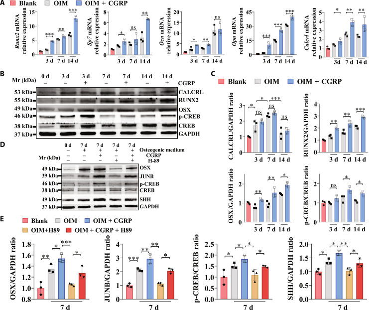
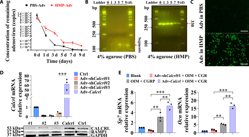
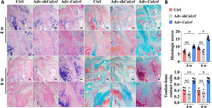
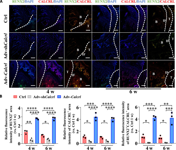
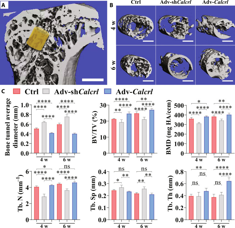
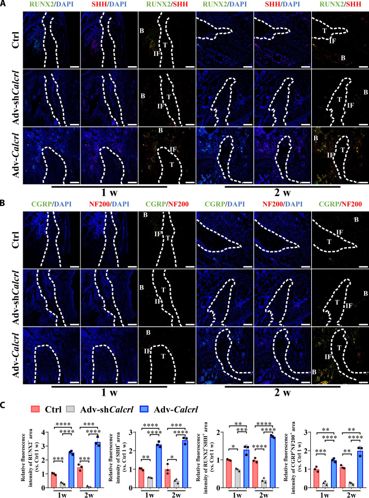
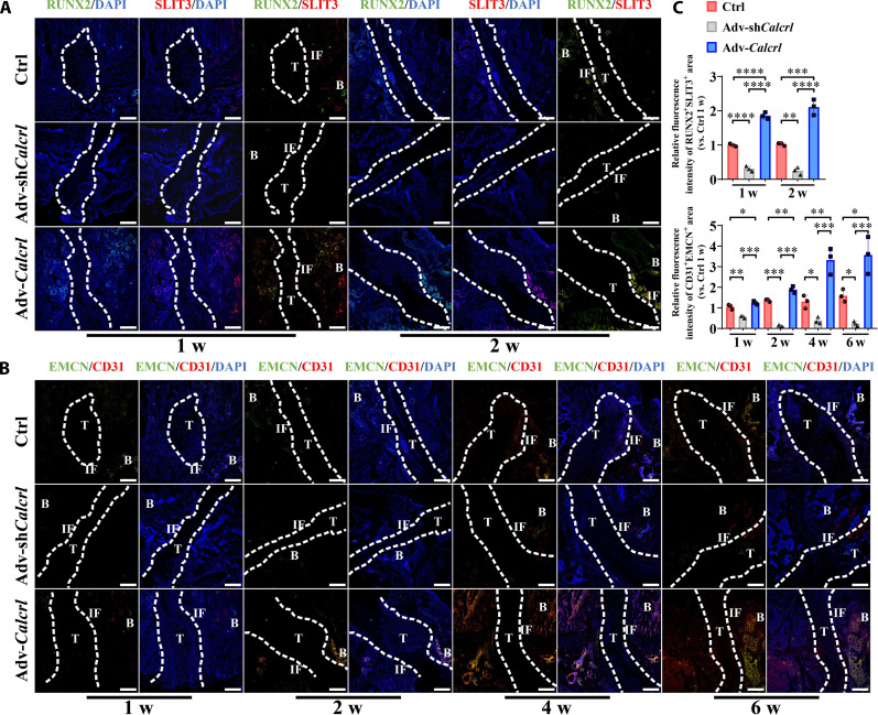
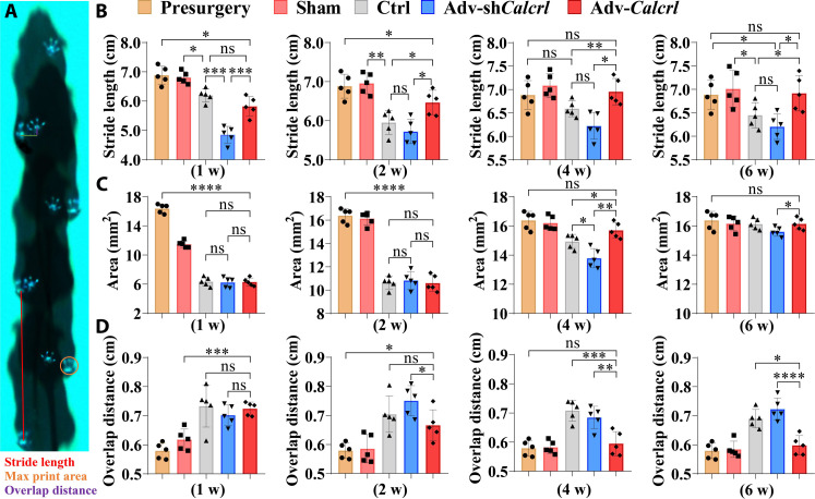
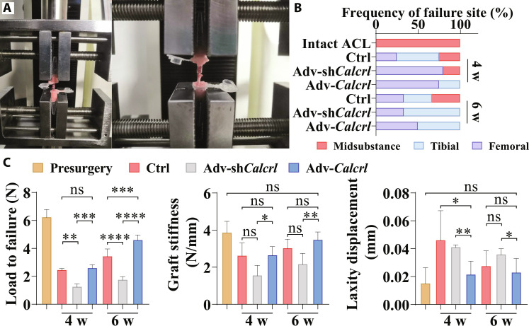
Calcitonin gene-related peptide (CGRP), an osteopromotive neurotransmitter with a short half-life, shows increase while calcitonin receptor-like (CALCRL) level is decreased at the early stage in bone fractures. Therefore, the activation of CALCRL-mediated signaling may be more critical to promote the tendon-bone healing. We found CGRP enhanced osteogenic differentiation of BMSCs through PKA/CREB/JUNB pathway, contributing to improved sonic hedgehog (SHH) expression, which was verified at the tendon-bone interface (TBI) in the mice with Calcrl overexpression. The osteoblast-derived SHH and slit guidance ligand 3 were reported to favor nerve regeneration and type H (CD31hiEMCNhi) vessel formation, respectively. Encouragingly, the activation or inactivation of CALCRL-mediated signaling significantly increased or decreased intensity of type H vessel and nerve fiber at the TBI, respectively. Simultaneously, improved gait characteristics and biomechanical performance were observed in the Calcrl overexpression group. Together, the gene therapy targeting CGRP receptor may be a therapeutic strategy in sports medicine.


Figures

Fig. 1.
Fig. 1.. The effects of CGRP on the osteogenic differentiation capability of mouse BMSCs.
(A) Real-time polymerase chain reaction (PCR) analysis of Sp7, Runx2, osteocalcin (Ocn), osteopontin (Opn), and Calcrl mRNA expression in the bone marrow–derived mesenchymal stem cells (BMSCs) after incubation in OIM with or without CGRP. *P < 0.05, **P < 0.01, and ***P < 0.001; n = 3 independent biological replicates. (B) Western blot (WB) analysis of protein levels of RUNX2, OSX, p-CREB, CREB, CALCRL, and glyceraldehyde-3-phosphate dehydrogenase (GAPDH) in BMSCs with or without treatment of CGRP. (C) Quantification of RUNX2/GAPDH ratio, OSX/GAPDH ratio, and p-CREB/CREB ratio, and CALCRL/GAPDH ratio by WB analysis from (B). ns, not significant, *P < 0.05, **P < 0.01, and ***P < 0.001; n = 3 independent biological replicates. (D) WB analysis of protein levels of OSX, JUNB, SHH, p-CREB, CREB, and GAPDH in BMSCs with or without treatment of CGRP or H-89, a selective inhibitor of cAMP-dependent protein kinase (PKA). d, days. (E) Quantification of OSX/GAPDH ratio, JUNB/GAPDH, p-CREB/CREB ratio, and SHH/GAPDH ratio by WB analysis from (D). *P < 0.05, **P < 0.01, and ***P < 0.001; n = 3 independent biological replicates. The Blank indicates BMSCs without incubation in osteogenic induction medium (OIM).
Fig. 2.
Fig. 2.. Synthesis and characterization of HMPs.
(A) Proton nuclear magnetic resonance (1H NMR) spectra of HB-PEGDA and SH-SA. (B) Schematic diagram of the preparation of HMPs in a poly(dimethylsiloxane)-based microfluidic droplet generator device. (C) Micrograph and diameter distribution of the resulting microdroplet templates. (D) Representative scanning electron microscopy images of the as-obtained HMPs. (E) Viscosity as a function of shear rate for the HMP clusters. (F) Strain-cycle experiment for the HMP clusters, whereby materials are cycled between 0.1 and 1000% stains. (G) The storage (G′) and (G″) modulus were measured under a constant strain of 1% and angular frequency ranging from 0.1 to 100 rad/s at 25°C. (H) Cell viability of BMSCs exposed to gradient concentration of HMPs. n = 4. (I) Live/Dead staining of BMSCs incubated with gradient concentrations of HMPs visualized by a calcein AM/propidium iodide (PI) double-staining assay.
Fig. 3.
Fig. 3.. Transfection efficiency of adenoviral transfer through the HMP delivery system.
(A) Concentration of remaining adenovirus in HMPs or PBS. n = 3 independent experiments. (B) Agarose gel electrophoresis of extracted DNA in the remaining adenovirus in HMPs or PBS. (C) The adenoviral vectors, expressing a green fluorescent protein (GFP), exhibited enhanced gene transfection efficiency in BMSCs for the adenovirus loaded HMPs compared to the adenovirus alone after immersion in PBS for 7 days. (D) Gene and protein expression levels of Calcrl in the BMSCs with or without incubation of adv-shCalcrl or adv-Calcrl after 3 days. ***P < 0.001; n = 3 independent biological replicates. (E) Gene expression levels of Sp7 and Ocn of BMSCs with or without incubation of adv-shCalcrl or adv-Calcrl after osteogenic differentiation in the osteogenic medium with or without addition of CGRP after 7 days. **P < 0.01 and ***P < 0.001; n = 3 independent biological replicates. The Blank indicates BMSCs without incubation in OIM.
Fig. 4.
Fig. 4.. Histological staining examination for the TBI.
(A) Representative hematoxylin and eosin (H&E) and Safranin-O/Fast Green staining images showing tendon-bone healing at 4 and 6 weeks (w) after surgery. The black dashed squares indicated the magnified region at the tendon-bone interzone. Scale bars, 200 μm. (B) Semiquantitative analysis of the tendon-bone healing quality. ns, not significant, *P < 0.05, and **P < 0.01; n = 5.
Fig. 5.
Fig. 5.. CGRP receptor–mediated signaling modulates expression levels of RUNX2 and CALCRL at the TBI in mice.
(A) Representative immunofluorescence staining of RUNX2 and CALCRL at the TBI in mice with various treatments at 4 and 6 weeks after surgery. DAPI, 4′,6-diamidino-2-phenylindole. (B) Quantitative analysis of the relative fluorescence intensities of RUNX2+, CALCRL+, and RUNX2+CALCRL+ area at the TBI in mice with various treatments at 4 and 6 weeks after surgery. *P < 0.05, **P < 0.01, ***P < 0.001, and ****P < 0.0001; n = 3. Dashed lines, tendon graft boundary; B, bone; T, tendon; IF, interface. Scale bars, 200 μm.
Fig. 6.
Fig. 6.. Micro-CT analysis results for peri-tunnel bone in tibia.
(A) Region of interest (ROI) indicated by the yellow cylinder of 0.8 mm in diameter and 0.54 mm in height located around the metaphysis. Scale bar, 1 mm. (B) Representative three-dimensional reconstructed images showing intra-tunnel bone tissue in ROI in different groups at the indicated time points. (C) Quantitative analysis of bone tunnel diameters, relative bone volume (BV/TV), bone mineral density (BMD), trabecular number (Tb. N), trabecular separation (Tb. Sp), and trabecular thickness (Tb. Th) at different groups at the indicated time points. *P < 0.05, **P < 0.01 and ****P < 0.0001; n = 4 to 5.
Fig. 7.
Fig. 7.. CGRP receptor–mediated signaling modulates expression levels of RUNX2, SHH, CGRP, and NF200 at the TBI in mice.
(A) Representative immunofluorescence staining of RUNX2 and SHH at the TBI in mice with various treatments at 1 and 2 weeks after surgery. (B) Representative immunofluorescence staining of CGRP and NF200 at the TBI in mice with various treatments at 1 and 2 weeks after surgery. (C) Quantitative analysis of the relative fluorescence intensities of RUNX2+, SHH+, RUNX2+SHH+, and CGRP+NF200+ area at the TBI in mice with various treatments at 1 and 2 weeks after surgery. *P < 0.05, **P < 0.01, ***P < 0.001, and ****P < 0.0001; n = 3. Dashed lines, tendon graft boundary; B, bone; T, tendon; IF, interface. Scale bars, 200 μm.
Fig. 8.
Fig. 8.. CGRP receptor–mediated signaling modulates expression levels of RUNX2, SLIT3, EMCN, and CD31 at the TBI in mice.
(A) Representative immunofluorescence staining of RUNX2 and SLIT3 at the TBI in mice with various treatments at 1 and 2 weeks after surgery. (B) Representative immunofluorescence staining of EMCN and CD31 at the TBI in mice with various treatments at 1, 2, 4, and 6 weeks after surgery. (C) Quantitative analysis of the relative fluorescence intensities of RUNX2+SLIT3+ and CD31+EMCN+ area at the TBI in mice with various treatments at the indicated time points after surgery. *P < 0.05, **P < 0.01, ***P < 0.001, and ****P < 0.0001; n = 3. Dashed lines, tendon graft boundary; B, bone; T, tendon; IF, interface. Scale bars, 200 μm.
Fig. 9.
Fig. 9.. Gait analysis of mice with various treatment after ACL reconstruction.
(A) Schematic illustration showing measurement of gait parameters. (B to D) Quantification of hind-paw stride length, paw print area, and overlap distance in mice with various treatment at 1, 2, 4, and 6 weeks after surgery. *P < 0.05, **P < 0.01, ***P < 0.001, and ****P < 0.0001; n = 5.
Fig. 10.
Fig. 10.. Mechanical examination of the FTGTC in mice under various treatment.
(A) The fixation of femur–tendon graft–tibia complex (FTGTC) in the customized jigs for dynamic laxity and tensile testing. (B) The failure mode of FTGTC in different groups. (C) Quantification of maximum load to failure, graft stiffness, and laxity displacement of FTGTC in different groups at 4 and 6 weeks after surgery. *P < 0.05, **P < 0.01, ***P < 0.001, and ****P < 0.0001; n = 3 to 5.

References

    1. Sherman S. L., Calcei J., Ray T., Magnussen R. A., Musahl V., Kaeding C. C., Clatworthy M., Bergfeld J. A., Arnold M. P., ACL Study Group presents the global trends in ACL reconstruction: Biennial survey of the ACL Study Group. J. ISAKOS 6, 322–328 (2021). - PubMed
    1. Renstrom P. A., Eight clinical conundrums relating to anterior cruciate ligament (ACL) injury in sport: Recent evidence and a personal reflection. Br. J. Sports Med. 47, 367–372 (2013). - PubMed
    1. Crawford S. N., Waterman B. R., Lubowitz J. H., Long-term failure of anterior cruciate ligament reconstruction. Art Ther. 29, 1566–1571 (2013). - PubMed
    1. Patel S., Caldwell J. M., Doty S. B., Levine W. N., Rodeo S., Soslowsky L. J., Thomopoulos S., Lu H. H., Integrating soft and hard tissues via interface tissue engineering. J. Orthop. Res. 36, 1069–1077 (2018). - PMC - PubMed
    1. Rothrauff B. B., Jorge A., de Sa D., Kay J., Fu F. H., Musahl V., Anatomic ACL reconstruction reduces risk of post-traumatic osteoarthritis: A systematic review with minimum 10-year follow-up. Knee Surg. Sports Traumatol. Arthrosc. 28, 1072–1084 (2020). - PubMed

Publication types

MeSH terms

Substances