. 2024 May:71:103108.
doi: 10.1016/j.redox.2024.103108. Epub 2024 Mar 1.

Human papillomavirus-16 E6 activates the pentose phosphate pathway to promote cervical cancer cell proliferation by inhibiting G6PD lactylation

Affiliations

Human papillomavirus-16 E6 activates the pentose phosphate pathway to promote cervical cancer cell proliferation by inhibiting G6PD lactylation

Qingfei Meng et al. Redox Biol. 2024 May.

Abstract

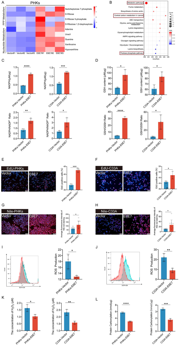
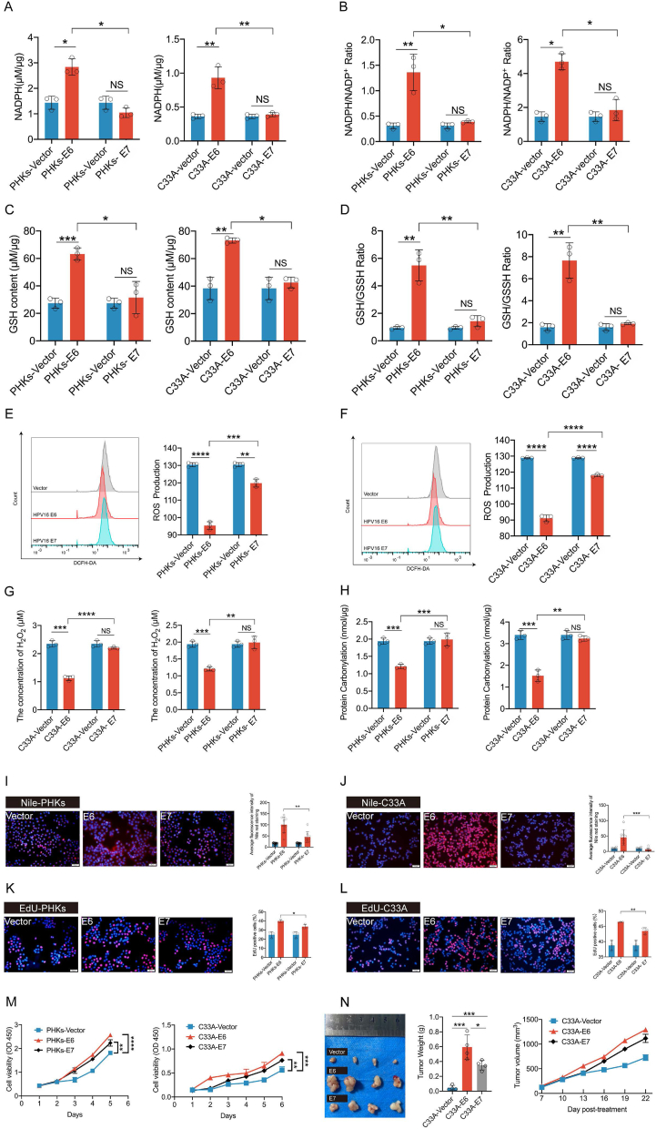
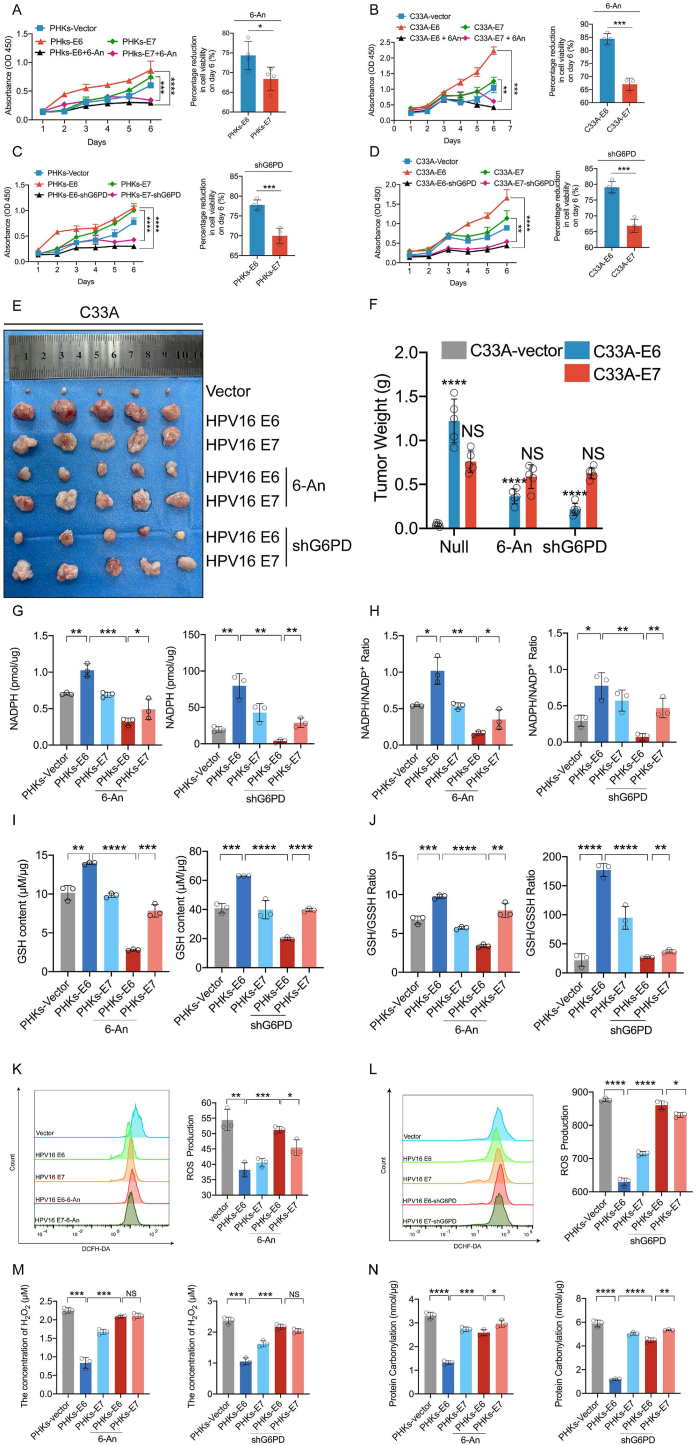
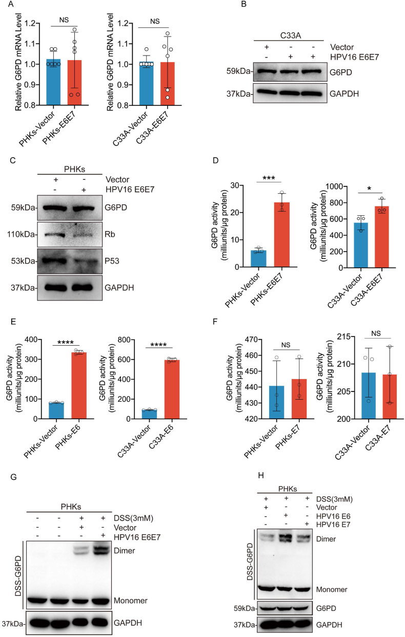
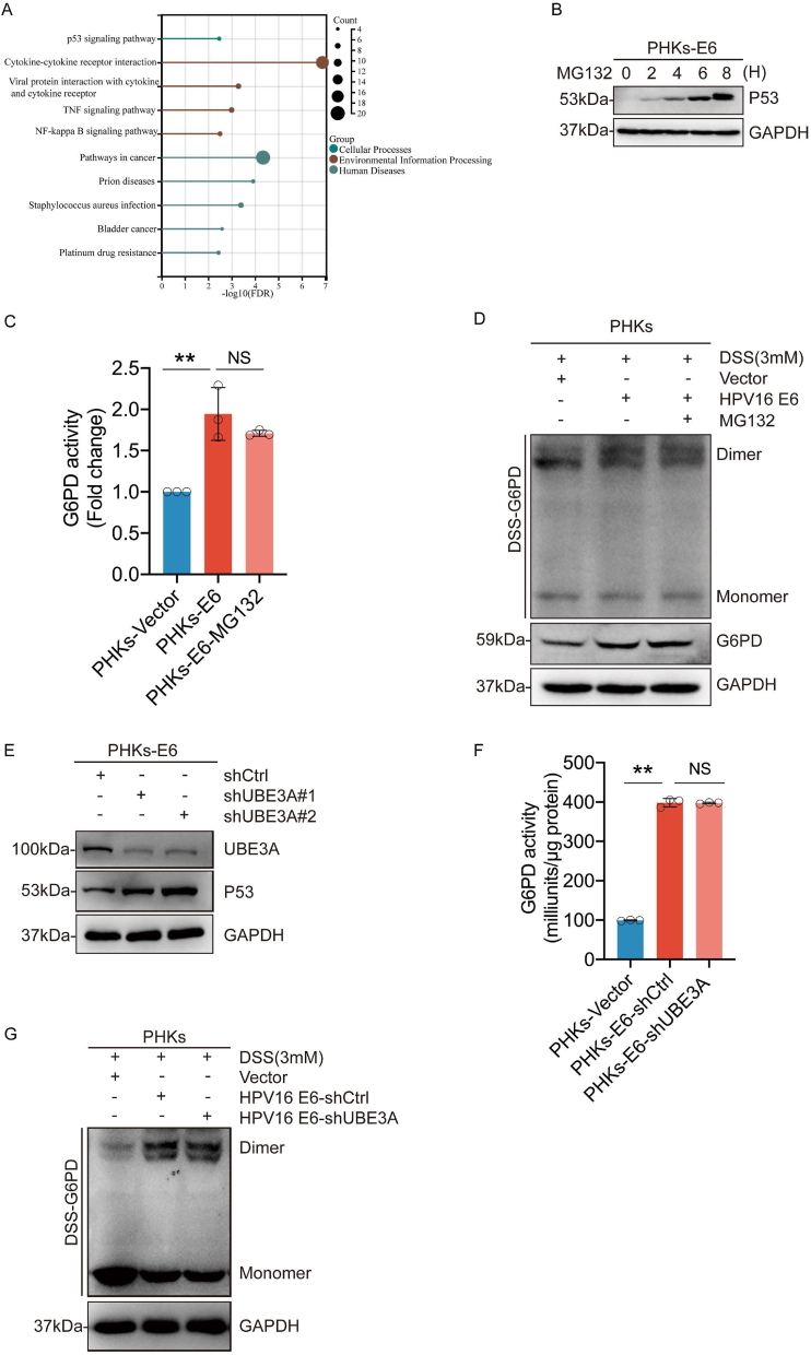
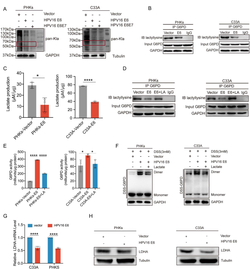
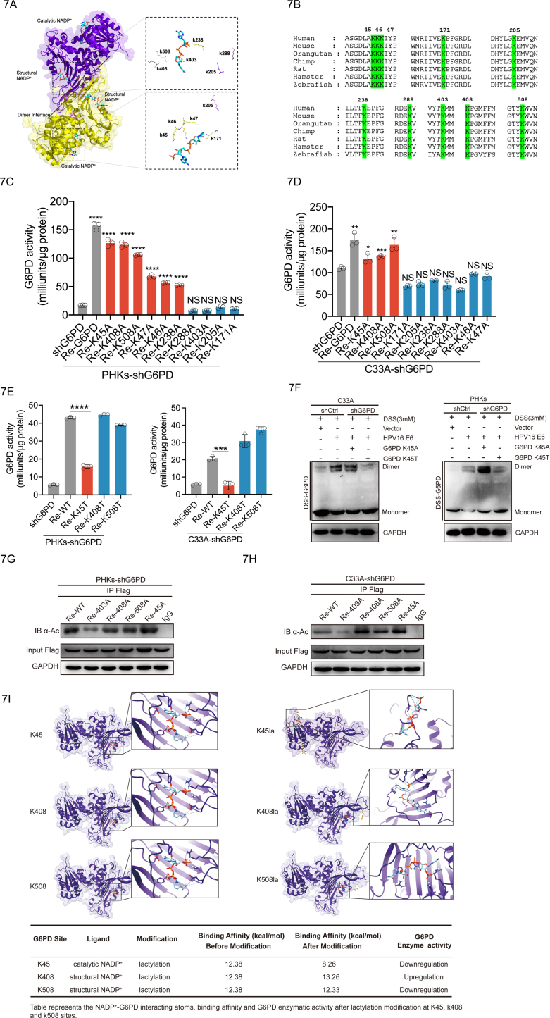
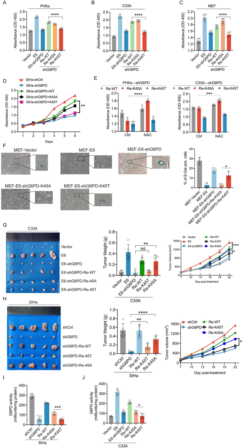
High-risk human papillomaviruses (HPVs) are the causative agents of cervical cancer. Here, we report that HPV16 E6E7 promotes cervical cancer cell proliferation by activating the pentose phosphate pathway (PPP). We found that HPV16 E6 activates the PPP primarily by increasing glucose-6-phosphate dehydrogenase (G6PD) enzyme activity. Mechanistically, HPV16 E6 promoted G6PD dimer formation by inhibiting its lactylation. Importantly, we suggest that G6PD K45 was lactylated during G6PD-mediated antioxidant stress. In primary human keratinocytes and an HPV-negative cervical cancer C33A cells line ectopically expressing HPV16 E6, the transduction of G6PD K45A (unable to be lactylated) increased GSH and NADPH levels and, correspondingly, decreasing ROS levels. Conversely, the re-expression of G6PD K45T (mimicking constitutive lactylation) in HPV16-positive SiHa cells line inhibited cell proliferation. In vivo, the inhibition of G6PD enzyme activity with 6-aminonicotinamide (6-An) or the re-expression of G6PD K45T inhibited tumor proliferation. In conclusion, we have revealed a novel mechanism of HPV oncoprotein-mediated malignant transformation. These findings might provide effective strategies for treating cervical and HPV-associated cancers.

Keywords: High-risk human papillomaviruses; Lactylation; Pentose phosphate pathway; glucose-6-phosphate dehydrogenase.


Conflict of interest statement

Declaration of competing interest The authors declare that they have no known competing financial interests or personal relationships that could have appeared to influence the work reported in this paper.

Figures

Fig. 1
Fig. 1
HPV16 E6E7 enhances the PPP and biosynthesis in PHKs and C33A cells. Stable transduction of PHKs and C33A cells using a lentivirus expression vector or HPV16 E6E7 was performed. (A) Heat map of metabolites for which levels were upregulated in PHKs cells expressing HPV16 E6E7, compared to levels with the vector. (B) Differential metabolites were subjected to KEGG enrichment analysis. (C–D) Intracellular NADPH, NADPH/NADP+, GSH/GSSH, and GSH levels were detected in PHKs and C33A cells exogenously expressing the vector or HPV16 E6E7. (E–F) EdU was used to determine DNA synthesis in PHKs and C33A cells exogenously expressing the vector or HPV16 E6E7. ImageJ was used to analyze the proportion of EdU-positive cells. Representative images (left) and quantification results (right) are shown. (G–H) Detection of lipid synthesis via Nile red staining. Representative images (left) of average fluorescence quantification results (right) are shown. (I–J) DCFH-DA was used to examine ROS in the cytoplasm of C33A and PHKs cells. Representative images detected by performing flow cytometry are shown (left), and the corresponding histograms are shown on the right side of the image. Intracellular H2O2 is shown in (K). PHKs and C33A cells exogenously expressing the vector or HPV16 E6E7. A quantitative spectrophotometric assay involving the derivatization of protein carbonyl groups with 2,4-dinitrophenylhydrazine was used, as described in Section “Materials and Methods.“(L). Each dot represents an independent biological replicate in the plots. Data are presented as mean ± SD. Statistical significance was determined using unpaired two-tailed t-test. *P < 0.05, **P < 0.01, ***P < 0.001, ****P < 0.0001 compared with indicated groups. NS, not significant.
Fig. 2
Fig. 2
HPV16 E6 primarily enhances the PPP and biosynthesis in PHKs and C33A cells. PHKs and C33A cells were stably transduced with a lentiviruses expression vector, HPV16 E6, or HPV16 E7. C33A/PHKs-HPV16 E6, C33A/PHKs -HPV16 E7, and control cells harboring an empty vector were tested for intracellular NADPH (A), NADPH/NADP+ (B), GSH (C) GSH/GSSH (D), ROS (E–F), H2O2 (G) and protein carbonylation (H). (I–J) Detection of lipid synthesis by performing Nile red staining. Representative images (left) of average fluorescence quantification results (right) are shown. (K–L) EdU was used to determine DNA synthesis in PHKs and C33A cells exogenously expressing HPV16 E6, HPV16 E7, or the vector. ImageJ was used to analyze the proportion of EdU-positive cells. Representative images (left) and quantification results (right) are shown. (M) Cell proliferation rates were determined by performing CCK8 assays. (N) Tumor masses in xenograft nude mice injected with C33A-HPV16 E6 (1 × 106) and C33A-HPV16 E7(5 × 106) cells compared to those in mice injected with the control vector cells. Each dot represents an independent biological replicate in the plots. Data are presented as mean ± SD. Statistical significance was determined using unpaired two-tailed t-test. *P < 0.05, **P < 0.01, ***P < 0.001, ****P < 0.0001 compared with indicated groups. NS, not significant.
Fig. 3
Fig. 3
HPV16 E6 promotes cell proliferation mediated by G6PD. PHKs and C33A cells were stably transduced with lentiviruses expressing the vector, HPV16 E6, or HPV16 E7. These cells were then treated with the G6PD inhibitor 6-An (81.06 μM or 26.78 μM) or infected with a lentivirus expressing shG6PD. (A–D) Cell proliferation rates were determined by performing a CCK8 assay (left). The percentage reduction in cell viability on day 6 is shown independently (right). (E) In total, C33A-Vector, C33A-HPV16E6, and C33A-HPV16E7 cells (1 × 106) were inoculated subcutaneously into the right flanks of 4- to 5-week-old female nude mice (n = 5 each). Images are shown of nude mouse xenograft tumors derived from C33A-HPV16 E6 and C33A-HPV16 E7 cells treated with 4 mg/kg/3d 6-An. Tumor sizes were measured every 5 days for 3 weeks. Intracellular NADPH (G), NADPH/NADP+ (H), GSH (I), GSH/GSSH (J) ROS (K–L) H2O2 (M), and protein carbonylation levels (N) were tested. Each dot represents an independent biological replicate in the plots. Data are presented as mean ± SD. *P < 0.05, **P < 0.01, ***P < 0.001, ****P < 0.0001 compared with indicated groups. Statistical significance was determined using unpaired two-tailed t-test. NS, not significant.
Fig. 4
Fig. 4
HPV16 E6 increases G6PD enzyme activity by promoting the formation of G6PD dimers. PHKs and C33A cells were stably transduced with lentiviruses expressing the vector, HPV16 E6, HPV16 E7, or HPV16 E6E7. (A) mRNA levels of G6PD in the transduced cells were determined via qPCR. (B–C) Immunoblots were used to detect G6PD, Rb, and p53 levels in cell lysates. GAPDH served as a loading control. (D–F) G6PD enzyme activity was examined in these cells. (G–I) Cells were harvested and crosslinked using DSS (3 mM), followed by western blotting with an anti-G6PD antibody. Data are presented as mean ± SD. Each dot represents an independent biological replicate in the plots. *P < 0.05, ***P < 0.001, ****P < 0.0001 compared with indicated groups. Statistical significance was determined using unpaired two-tailed t-test. NS, not significant.
Fig. 5
Fig. 5
HPV16 E6 regulates G6PD enzyme activity independent of p53. (A) KEGG enrichment analysis of the differential genes screened based on GEO (GSE58841). (B) PHKs-E6 cells were treated with 5 μM MG132 for the indicated duration. The whole-cell extracts (WCEs) were then collected for immunoblotting to detect p53 levels in the cells. (C) G6PD enzyme activity was assayed after treating PHKs-E6 cells with MG132 (5 μM) for 6 h. (D) PHKs-E6 cells were treated with 5 μM of MG132 for 6 h. Cells were harvested and crosslinked using DSS (3 mM), followed by western blotting with an anti-G6PD antibody. (E) PHKs-E6 cells were stably transduced with lentiviruses expressing shUBE3A. WCEs were analyzed via immunoblotting for UBE3A and p53. (F) PHKs-E6-shUBE3A cells were employed for the detection of G6PD enzyme activity. (G) Crosslinking with DSS followed by immunoblotting to detect dimeric and monomeric G6PD. Each dot represents an independent biological replicate in the plots. Data are presented as mean ± SD. **P < 0.01 compared with indicated groups. Statistical significance was determined using unpaired two-tailed t-test. NS, not significant. HPV, human papilloma virus; G6PD, glucose-6-phosphate dehydrogenase; DSS, disuccinimidyl suberate.
Fig. 6
Fig. 6
HPV16 E6 inhibits G6PD lactylation modifications. (A) Pan-lactyation levels were detected in C33A and PHKs expressing HPV16 E6, HPV16 E7, and the vector via western blotting. (B) Immunoblotting for lactylation in the anti-G6PD immunoprecipitates. The immunoprecipitates were isolated from the C33A and PHKs cells overexpressing the vector and HPV16 E6. (C) Intracellular lactate levels were examined in C33A and PHKs cells overexpressing the vector and HPV16 E6. (D) Immunoblotting for lactylation in the anti-G6PD immunoprecipitates. The immunoprecipitates were isolated from C33A and PHKs cells overexpressing the vector and HPV16 E6 upon NaLa treatment (25 mM) for 24 h. (E) G6PD enzyme activity was assayed in C33A and PHKs cells overexpressing the vector and HPV16 E6 upon NaLa treatment (25 mM) for 24 h. (F) Cells were harvested and crosslinked using DSS (3 mM), followed by western blotting with an anti-G6PD antibody. (G–H) LDHA levels were detected in C33A cells expressing HPV16 E6, HPV16 E7, and the vector. mRNA levels of LDHA in the transduced cells were determined via qPCR. Each dot represents an independent biological replicate in the plots. Data are presented as mean ± SD. *P < 0.05, ****P < 0.0001 compared with indicated groups.
Fig. 7
Fig. 7
The G6PD K45 lactylation modification reduces its enzymatic activity. (A) Schematic diagram of the G6PD structure (PDB: 2BH9). Each G6PD monomer consists of a catalytic NADP+ and a structural NADP+. Dual G6PD monomers are stacked into a dimer. (B) Species conservation analysis of potential l0 lactylation modification sequence sites for G6PD. (C–E) Re-expression of mutation in PHKs and C33A cells with the knockdown G6PD was used to detect G6PD enzyme activity. (F) Crosslinking with DSS, followed by immunoblotting to detect dimeric and monomeric G6PD. (G–H) Acetylation levels were blotted with a pan-anti-acetyllysine antibody (a-Ac). (I) 3D structure of ligand-receptor interactions shown in the left panel. The right panel shows the 2D representation of the interaction with ligands and the receptors in the binding pocket. Each dot represents an independent biological replicate in the plots. Data are presented as mean ± SD. *P < 0.05, **P < 0.01, ***P < 0.001, ****P < 0.0001 compared with indicated groups. Statistical significance was determined using unpaired two-tailed t-test. NS, not significant.
Fig. 8
Fig. 8
Elevated levels of G6PD K45 lactylation inhibit cell proliferation in vivo and in vitro (A–C) PHKs, C33A cells, and MEFs were transfected with the indicated plasmids. Cell proliferation was analyzed via cell viability assays. (D) SiHa (HPV16 positive) cells stably expressing shCtrl or shG6PD were further infected with lentiviruses expressing WT G6PD or its mutation, as indicated. (E) G6PD-knockdown cells or those cells rescued by WT G6PD or the K45T or K45A mutation were treated with NAC (2 mM), and cell proliferation was analyzed 5 days after treatment. (F) β-Galactosidase staining was used to detect the level of senescence in MEF cells. Right panel: analysis of β-galactosidase-positive cells. Left panel: representative images. (G–H) Tumors were weighed after mice were euthanized at the endpoint. (I–J) C33A and SiHa cell xenograft tumors were used to determine G6PD enzyme activity. Each dot represents an independent biological replicate in the plots. Data are presented as mean ± SD. *P < 0.05, **P < 0.01, ***P < 0.001, ****P < 0.0001 compared with indicated groups. Statistical significance was determined using unpaired two-tailed t-test. NS, not significant. G6PD, glucose-6-phosphate dehydrogenase; NAC, N-acetyl-l-cysteine.

Similar articles

Cited by

References

    1. Schiffman M., et al. Carcinogenic human papillomavirus infection. Nat. Rev. Dis. Prim. 2016;2 - PubMed
    1. Liu Y., et al. Nuclear lactate dehydrogenase A senses ROS to produce α-hydroxybutyrate for HPV-induced cervical tumor growth. Nat. Commun. 2018;9(1):4429. - PMC - PubMed
    1. Tommasino M. The human papillomavirus family and its role in carcinogenesis. Semin. Cancer Biol. 2014;26:13–21. - PubMed
    1. Hanahan D., Weinberg R.A. Hallmarks of cancer: the next generation. Cell. 2011;144(5):646–674. - PubMed
    1. Warburg O. On the origin of cancer cells. Science. 1956;123(3191):309–314. - PubMed

MeSH terms

Substances