Review
. 2024 Mar 5;9(3):202-209.
doi: 10.1530/EOR-23-0161.

Treatment of unilateral cervical facet fractures without evidence of dislocation or subluxation: a narrative review and proposed treatment algorithm

Affiliations
Review

Treatment of unilateral cervical facet fractures without evidence of dislocation or subluxation: a narrative review and proposed treatment algorithm

Juan Ignacio Cirillo et al. EFORT Open Rev. .

Abstract

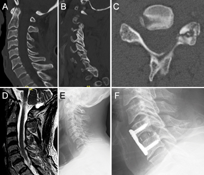
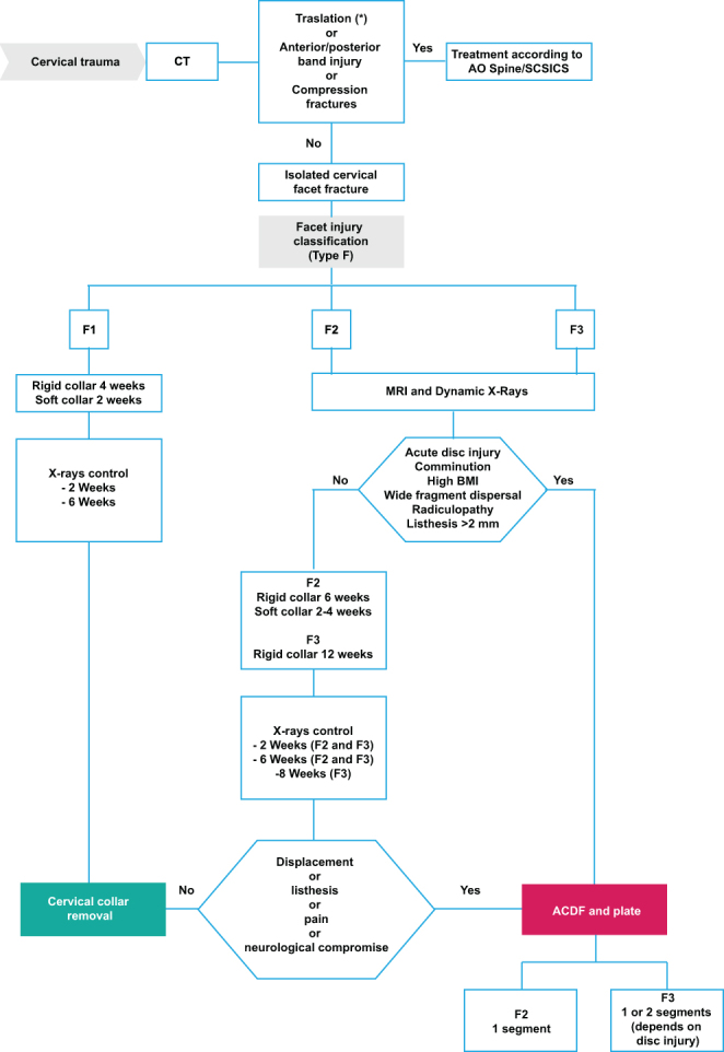
Isolated cervical spine facet fractures are often overlooked. The primary imaging modality for diagnosing these injuries is a computed tomography scan. Treatment of unilateral cervical facet fractures without evidence of dislocation or subluxation remains controversial. The available evidence regarding treatment options for these fractures is of low quality. Risk factors associated with the failure of nonoperative treatment are: comminution of the articular mass or facet joint, acute radiculopathy, high body mass index, listhesis exceeding 2 mm, fragmental diastasis, acute disc injury, and bilateral fractures or fractures that adversely affect 40% of the intact lateral mass height or have an absolute height of 1 cm.

Keywords: Spine; cervical facet fracture; isolated; non-displaced; trauma.


Conflict of interest statement

The authors declare that there is no conflict of interest that could be perceived as prejudicing the impartiality of this review.

Figures

Figure 1
Figure 1
Case example (A–F): 55 years old man with a F2 C6 left facet fracture. (A) Cervical CT sagittal view without evidence of cervical listhesis. (B) Cervical CT sagittal view showing F2 fracture at C6. (C) Cervical CT axial view showing lateral mass fracture with comminution. (D) Evidence of C5–C6 acute disc injury on MRI (T2) sagittal view. (E) X-ray control at 3 weeks with evidence of C5–C6 progressive listhesis. (F) C5–C6 anterior cervical discectomy and fusion.
Figure 2
Figure 2
Measurement of the absolute height of the intact lateral mass and the fracture fragment according to Spector et al. (4) (A) The absolute height of the contralateral intact lateral mass was defined as the maximum cephalocaudal tip-to-tip height measured on sequential CT sagittal images; (B) The absolute height of the fracture fragment was defined as the maximum tip-to-tip cephalocaudal height measured on sequential sagittal CT images. The percentage of the absolute height of the fracture fragment related to the intact contralateral mass can be estimated: (b/a) × 100.
Figure 3
Figure 3
AO Spine cervical facet fracture classification. F1 is a nondisplaced facet fracture (either superior or inferior facets). Fracture fragments are smaller than 1 cm and comprise less than 40% of the lateral mass. F2 is a facet fracture with fragments either larger than 1 cm, comprise more than 40% of the lateral mass, or there are signs of displacement. F3 (floating lateral mass) is a disruption of the pedicle and lamina resulting in disconnection of superior and inferior articular processes at a given level or set of levels. F4 is a traumatic subluxation or perched/dislocated facet (30).
Figure 4
Figure 4
Treatment algorithm. SCSICS, Subaxial Cervical Spine Injury Classification System (30); ACDF, anterior cervical discectomy and fusion; CT, computed tomography; MRI, magnetic resonance imaging; BMI, body mass index. *Including F4 isolated facet fractures.

Similar articles

Cited by

References

    1. Jaumard NV Welch WC & Winkelstein BA. Spinal facet joint biomechanics and mechanotransduction in normal, injury and degenerative conditions. Journal of Biomechanical Engineering 2011133071010. (10.1115/1.4004493) - DOI - PMC - PubMed
    1. Pal GP & Routal RV. A study of weight transmission through the cervical and upper thoracic regions of the vertebral column in man. Journal of Anatomy 1986148245–261. - PMC - PubMed
    1. White AA Johnson RM Panjabi MM & Southwick WO. Biomechanical analysis of clinical stability in the cervical spine. Clinical Orthopaedics and Related Research 197510985–96. (10.1097/00003086-197506000-00011) - DOI - PubMed
    1. Spector LR Kim DH Affonso J Albert TJ Hilibrand AS & Vaccaro AR. Use of computed tomography to predict failure of nonoperative treatment of unilateral facet fractures of the cervical spine. Spine 2006312827–2835. (10.1097/01.brs.0000245864.72372.8f) - DOI - PubMed
    1. Lowery DW, Wald MM, Browne BJ, Tigges S, Hoffman JR, Mower WR. & NEXUS Group. Epidemiology of cervical spine injury victims. Annals of Emergency Medicine 20013812–16. (10.1067/mem.2001.116149) - DOI - PubMed

LinkOut - more resources