. 2024 Jun:510:1-7.
doi: 10.1016/j.ydbio.2024.03.001. Epub 2024 Mar 6.

Anatomy, development and regeneration of zebrafish elasmoid scales

Affiliations

Anatomy, development and regeneration of zebrafish elasmoid scales

Andrew J Aman et al. Dev Biol. 2024 Jun.

Abstract

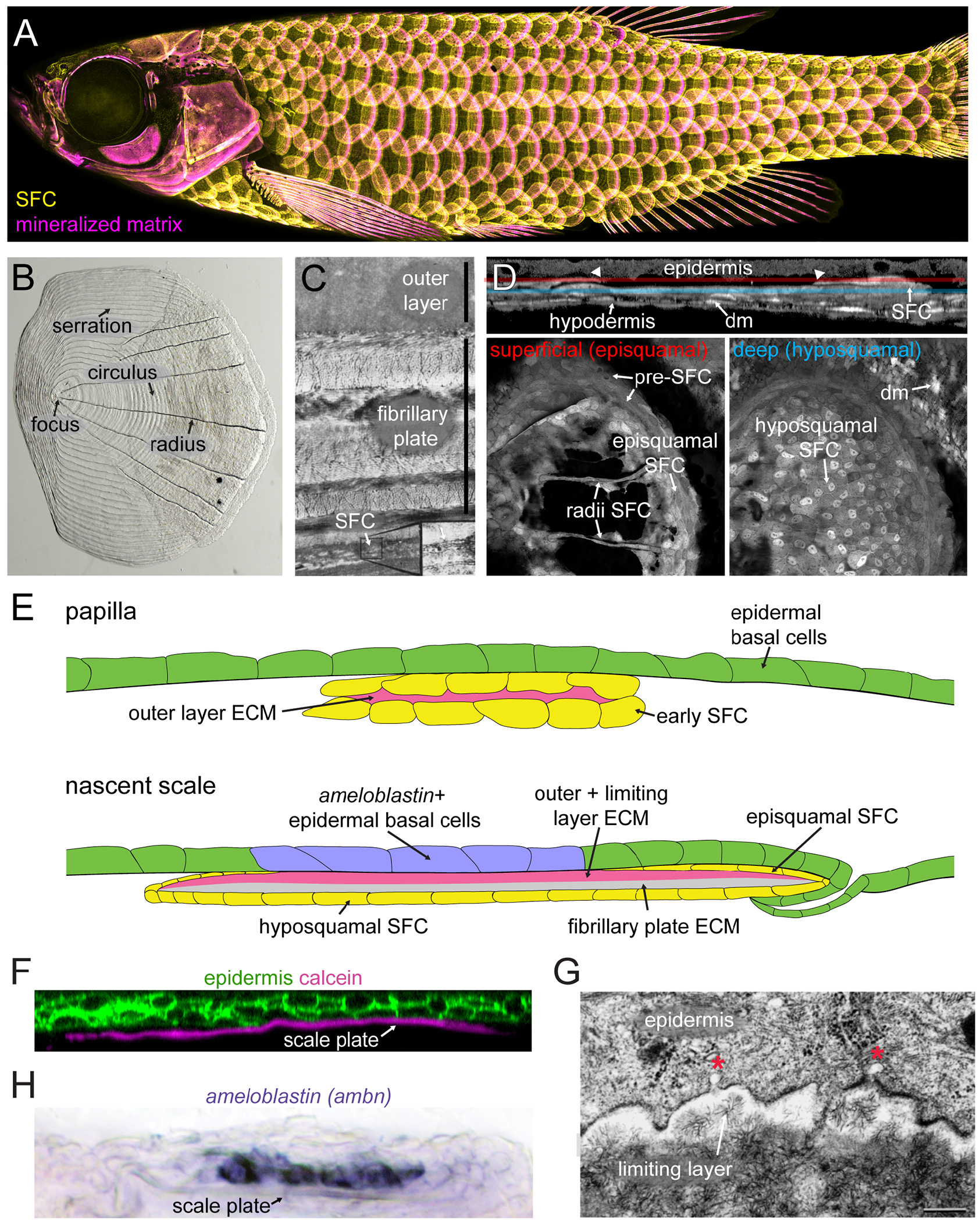
Vertebrate skin appendages - particularly avian feathers and mammalian hairs, glands and teeth - are perennially useful systems for investigating fundamental mechanisms of development. The most common type of skin appendage in teleost fishes is the elasmoid scale, yet this structure has received much less attention than the skin appendages of tetrapods. Elasmoid scales are thin, overlapping plates of partially mineralized extracellular matrices, deposited in the skin in a hexagonal pattern by a specialized population of dermal cells in cooperation with the overlying epidermis. Recent years have seen rapid progress in our understanding of elasmoid scale development and regeneration, driven by the deployment of developmental genetics, live imaging and transcriptomics in larval and adult zebrafish. These findings are reviewed together with histological and ultrastructural approaches to understanding scale development and regeneration.


Figures

Fig. 1.
Fig. 1.
Zebrafish elasmoid scale anatomy and development. (A) Squamation of a live, juvenile fish. Scale-forming cells (SFC) visualized by sp7:EGFP expression (yellow) and mineralized matrix visualized using Alizarin Red S vital dye (magenta). (B) Brightfield image of freshly plucked adult zebrafish scale. Posterior to the left. Circuli and serrations are found circumferentially around the focus, whereas radii extend radially from the focus in the posterior part of the scale. (C) Transmission electron micrograph of a cross section through a mature, decalcified scale stained with uranyl acetate and lead, showing the collagen-poor, hypermineralized outer layer, and the collagenous fibrillary plate above a thin layer of SFCs [reprinted with permission from Waterman (1970)]. (D) Live imaging of a growing scale visualized with ET37:EGFP; sp7:EGFP transgenes (DeLaurier et al., 2010; Parinov et al., 2004). Top panel shows a longitudinal optical section through the scale, posterior to the right. Bottom panels show single optical slices through the superficial, episquamal (red) and deep, hyposquamal (blue) surfaces. dm, non-SFC dermal mesenchyme. (E) Schematic representation of longitudinal sections through an early scale papilla (top) and a nascent scale (bottom) showing early SFCs and later episquamal and hyposquamal SFCs (yellow) in relation to the mineralized outer and limiting layers (magenta) and unmineralized fibrillary plate (tan). (F) Optical section from live fish at similar stage as the nascent scale in (E) showing close proximity of scale plate, vitally dyed with calcein (magenta), and epidermal cells, expressing the krtt1c19e:palm-tdTomato transgene (green)(Lee et al., 2014). (G) Transmission electron micrograph showing radially arranged mineral crystals in the external layer in close proximity to epidermal cell invaginations (red asterisks) potentially marking secretory vesicles that have fuse with the membrane [reprinted with permission from Sire et al. (1997b)]. (H) In-situ hybridization of an ameloblastin (ambn) riboprobe showing expression in epidermal cells that abut the scale plate ECM. Images in (A,D,F,H) reproduced with permission from Aman et al. (2018) and Aman et al. (2023). Original annotations in Fig 1C,G removed using the generative fill tool of Adobe Photoshop.
Fig. 2.
Fig. 2.
Similar signaling interactions regulate zebrafish scale and mouse hair follicle development. (A) Live imaging of zebrafish scales using the sp7:EGFP transgene and fixed analysis of mouse hair follicle patterning using dkk4 in-situ hybridization shows similar responses to Wnt/β-catenin and Eda-Edar-NF-kB signaling pathway manipulation. Wnt/β-catenin signaling repression (Wnt-) by transgenic expression of Dkk1 (hsp70l:dkk1b-EGFP in zebrafish and krt5-rtTA;tetO-Dkk1 in mouse) prevents skin appendage development. Experimental activation of Eda-Edar-NF-κB signaling (Eda+++; hsp70l:edar-2a-nls-mCherry in zebrafish and krt14-Eda-A1 in mouse) leads to mispatterned and dysmorphic skin appendage primordia, with such induction failing in the absence of Wnt/β-catenin signaling (Wnt-Eda+++). Mouse data reproduced with permission from Zhang et al. (2009). (B) Longitudinal cross section through live zebrafish skin and fixed mouse skin shows similar responses to SHH signaling manipulation. In unmanipulated controls, epidermis (labelled with cldnb:EGFP-CAAX in fish and hematoxylin and eosin staining in mouse) invaginates around the calcified ECM of zebrafish scales (alizarin red vital-staining, magenta) and the dermal papillae of mouse hair follicles (dp), respectively. Arrows indicate extent of epidermal invagination. SHH repression blocks this invagination in both animals [mouse data reproduced with permission from St-Jacques et al. (1998)]. Zebrafish data reproduced with permission from Aman et al. (2018). Original annotations in mouse data removed using the generative fill tool in Adobe Photoshop.
Fig. 3.
Fig. 3.
Integrative fish scale research. (A) Vasculature, labeled with fli1a:EGFP; and sensory axons, labelled with p2rx3a>mCherry, are guided by scale radii. Superficial scale capillaries indicated by blue arrows; scale margin capillaries indicated by yellow arrowheads. Reproduced with permission from (Rasmussen et al., 2018), original annotations removed using the generative fill tool in photoshop. (B) Scale patterning in unmanipulated control (euTH) and hypothyroid (hypoTH) fish, visualized using sp7:EGFP. Hypothyroidism lead to more numerous, miniaturized and relatively disorganized squamation pattern, but individual sale morphology is similar (false colored cyan in euTH and magenta in hypoTH). Reproduced with permission from Aman et al. (2021).

Similar articles

Cited by

References

    1. Alibardi L, 2003. Adaptation to the land: The skin of reptiles in comparison to that of amphibians and endotherm amniotes. J Exp Zool B Mol Dev Evol 298, 12–41. - PubMed
    1. Aman AJ, Fulbright AN, Parichy DM, 2018. Wnt/beta-catenin regulates an ancient signaling network during zebrafish scale development. eLife 7, e37001. - PMC - PubMed
    1. Aman AJ, Kim M, Saunders LM, Parichy DM, 2021. Thyroid hormone regulates abrupt skin morphogenesis during zebrafish postembryonic development. Dev Biol 477, 205–218. - PMC - PubMed
    1. Aman AJ, Saunders LM, Carr AA, Srivatasan S, Eberhard C, Carrington B, Watkins-Chow D, Pavan WJ, Trapnell C, Parichy DM, 2023. Transcriptomic profiling of tissue environments critical for post-embryonic patterning and morphogenesis of zebrafish skin. eLife 12, RP86670. - PMC - PubMed
    1. Arola D, Murcia S, Stossel M, Pahuja R, Linley T, Devaraj A, Ramulu M, Ossa EA, Wang J, 2018. The limiting layer of fish scales: Structure and properties. Acta Biomaterialia 67, 319–330. - PubMed

Publication types

LinkOut - more resources