. 2024 Apr;39(2):309-318.
doi: 10.1016/j.virs.2024.03.001. Epub 2024 Mar 6.

Mast cell degranulation-triggered by SARS-CoV-2 induces tracheal-bronchial epithelial inflammation and injury

Affiliations

Mast cell degranulation-triggered by SARS-CoV-2 induces tracheal-bronchial epithelial inflammation and injury

Jian-Bo Cao et al. Virol Sin. 2024 Apr.

Abstract

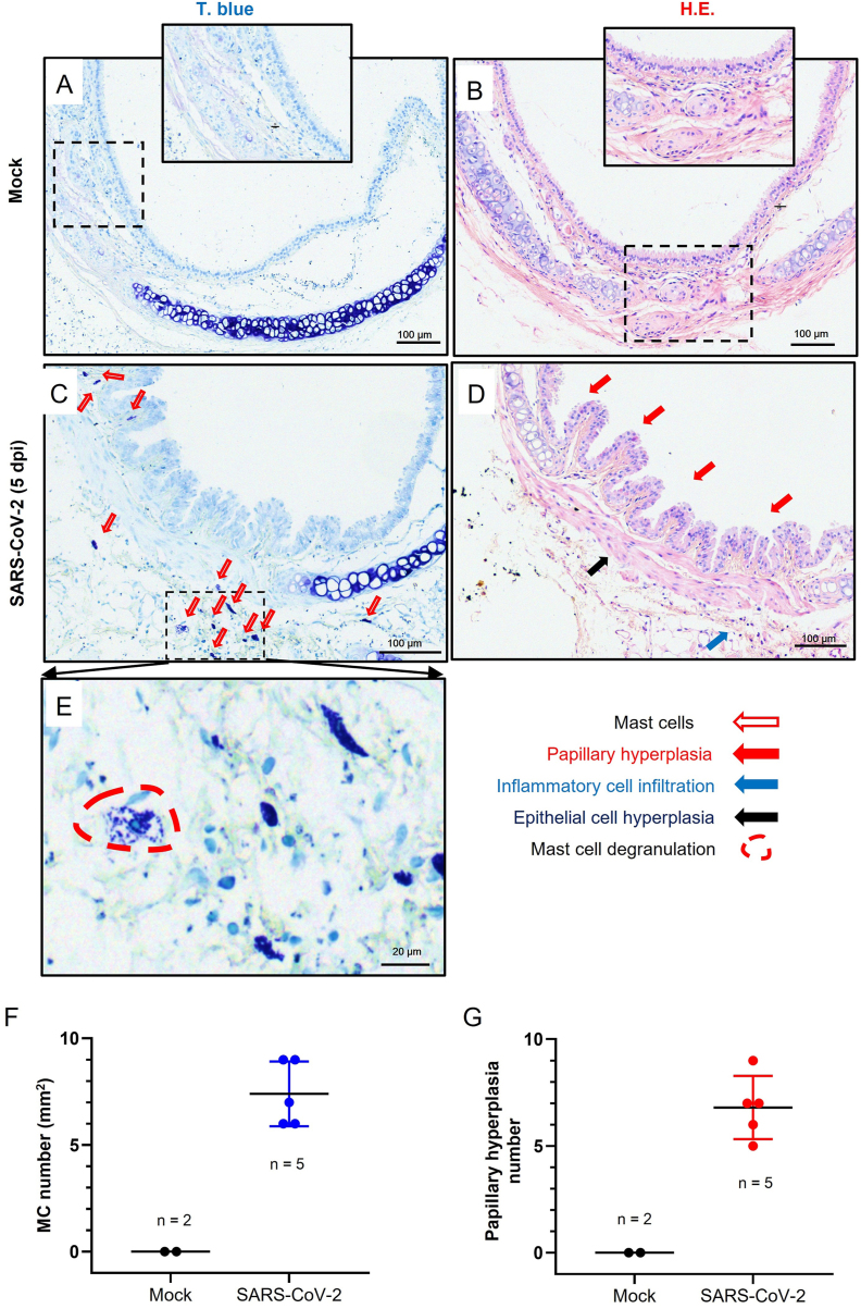
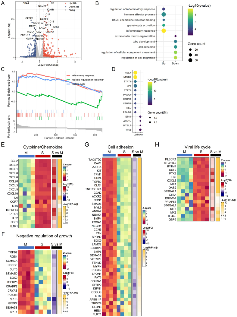
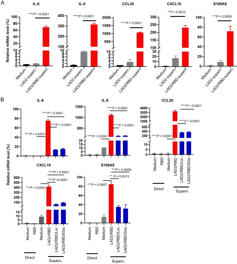
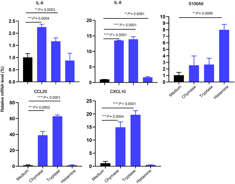
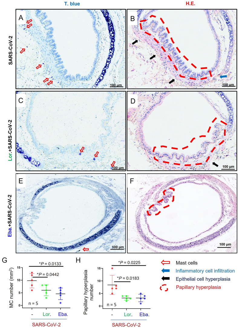
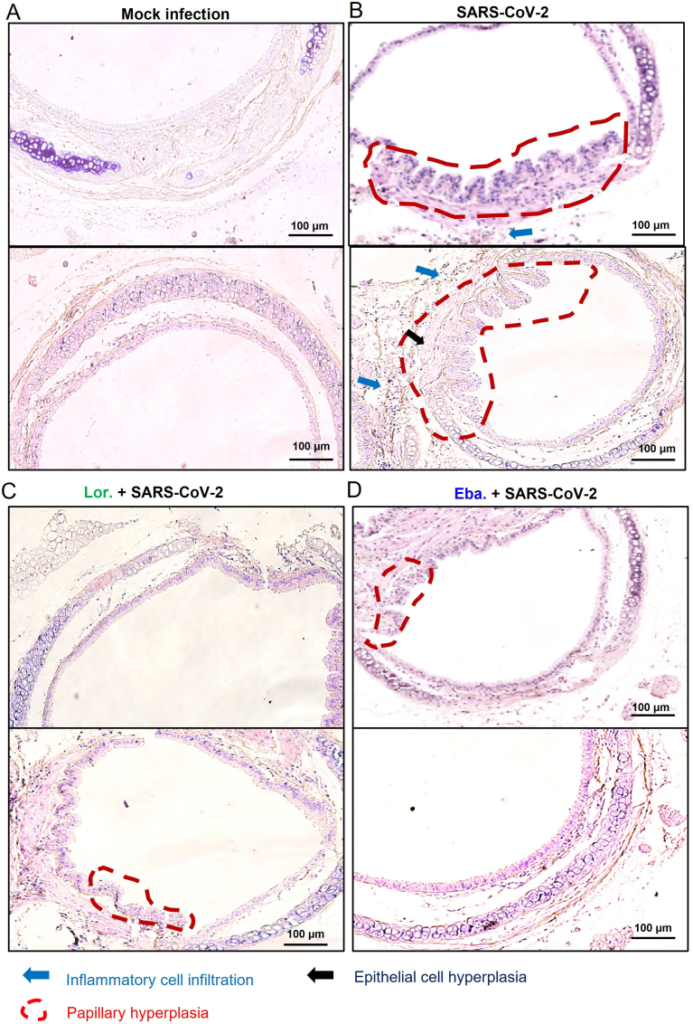
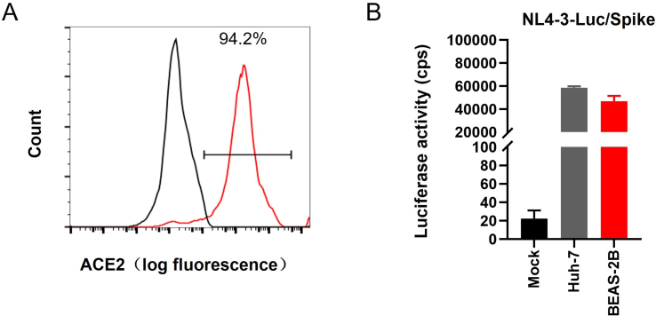
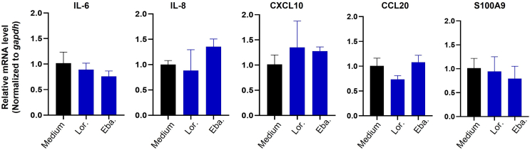
SARS-CoV-2 infection-induced hyper-inflammation is a key pathogenic factor of COVID-19. Our research, along with others', has demonstrated that mast cells (MCs) play a vital role in the initiation of hyper-inflammation caused by SARS-CoV-2. In previous study, we observed that SARS-CoV-2 infection induced the accumulation of MCs in the peri-bronchus and bronchioalveolar-duct junction in humanized mice. Additionally, we found that MC degranulation triggered by the spike protein resulted in inflammation in alveolar epithelial cells and capillary endothelial cells, leading to subsequent lung injury. The trachea and bronchus are the routes for SARS-CoV-2 transmission after virus inhalation, and inflammation in these regions could promote viral spread. MCs are widely distributed throughout the respiratory tract. Thus, in this study, we investigated the role of MCs and their degranulation in the development of inflammation in tracheal-bronchial epithelium. Histological analyses showed the accumulation and degranulation of MCs in the peri-trachea of humanized mice infected with SARS-CoV-2. MC degranulation caused lesions in trachea, and the formation of papillary hyperplasia was observed. Through transcriptome analysis in bronchial epithelial cells, we found that MC degranulation significantly altered multiple cellular signaling, particularly, leading to upregulated immune responses and inflammation. The administration of ebastine or loratadine effectively suppressed the induction of inflammatory factors in bronchial epithelial cells and alleviated tracheal injury in mice. Taken together, our findings confirm the essential role of MC degranulation in SARS-CoV-2-induced hyper-inflammation and the subsequent tissue lesions. Furthermore, our results support the use of ebastine or loratadine to inhibit SARS-CoV-2-triggered degranulation, thereby preventing tissue damage caused by hyper-inflammation.

Keywords: Bronchial epithelial cell; Inflammation; Mast cell (MC); SARS-CoV-2; Tracheal injury.


Conflict of interest statement

Conflict of interest The authors declare no conflict of interest. Prof. Ling Chen and Prof. Jian-Hua Wang are editorial board members for Virologica Sinica and were not involved in the editorial review or the decision to publish this article.

Figures

Fig. 1
Fig. 1
SARS-CoV-2 induces MC degranulation and trachea lesions in hACE2-humanized mice. The mice of C57BL/6N-ACE2em2(hACE2-WPRE, pgk-puro)/CCLA were intratracheally inoculated with SARS-CoV-2 (strain 107) at a dose of 5 ​× ​106 TCID50. At day 5 post-infection, mice were anaesthetized and the trachea tissue were harvested for histological analysis. Toluidine blue (T. blue) staining was used to observe MCs and degranulation (A, C, E). Hematoxylin and Eosin (H.E.) staining was used to observe trachea injury (B, D). The PBS was used as the mock infection (A, B). (FG) The counts of MC and papillary hyperplasia in trachea section were summarized. Scale bar: 100 or 20 ​μm. The number (n) of mouse used in tests was noted.
Fig. 2
Fig. 2
Transcriptome analysis of BEAS-2B cells. BEAS-2B cells were treated with the culture supernatants (the sample “S”) from SARS-CoV-2 spike/RBD protein-triggered LAD2 degranulation cells, or with the LAD2 cell normal culture supernatants (the sample “M”), for 12 ​h. BEAS-2B cells were collected and the transcriptome analysis was performed. The data summarized four independent experimental repeats. A Volcano plot of DEGs comparing “S” versus “M” samples. The symbols of top 10 up-regulated and down-regulated genes are shown. B GO functional enrichment analysis of DEGs. Color bar indicates minus logarithm of q values, and bubble size indicates absolute gene counts enriched in GO terms. C GSEA of distribution of gene sets related to inflammatory response, negative regulation of cell growth, viral life cycle and the enrichment scores based on DEGs. D Transcription-factor enrichment analysis of DEGs. The color bar indicates the minus logarithm of q values, and bubble size indicates the gene enrichment ratio regulated by a transcription factor. (EH) Heatmaps showing relative expression level (left panel), fold-change (middle panel), and adjusted P-values (right panel) for sets of cytokine/chemokine-related genes (E), negative regulation of growth genes (F), cell adhesion (G), viral life cycle (H).
Fig. 3
Fig. 3
Blocking MC degranulation reduces the capacity to induce inflammatory factors in BEAS-2B cells. BEAS-2B cells were treated with either LAD2/RBD co-culture supernatants (LAD2/RBD-supern.), LAD2 cell culture supernatants (LAD2-supern.), or medium for 12 ​h (A); or LAD2 cells were prior-treated with loratadine (Lor., 5 ​μg/mL) or ebastine (Eba., 3 ​μg/mL) for 20 ​h, and then cells were treated with SARS-CoV-2 spike/RBD (5 ​μg/mL) for 2 ​h, and the culture supernatants were harvested to treat BEAS-2B cells for additional 12 ​h (B). The direct treatments of BEAS-2B cells with spike/RBD (5 ​μg/mL) or medium were also performed. The mRNA levels of inflammatory factors were detected with real time qRT-PCR, and normalized to gapdh mRNA. One representative data from 3 independent repeats are shown, data are mean ​± ​standard deviation (SD). ∗∗P ​< ​0.01, ∗∗∗P ​< ​0.001, and ∗∗∗P ​< ​0.0001 are considered significant differences in a Student's unpaired t-test.
Fig. 4
Fig. 4
MC released mediators-induced expression of inflammatory factors. BEAS-2B cells (2 ​× ​105) were stimulated with tryptase, chymase or histamine (5 ​μg/mL for each) for 12 ​h, and the cells were collected to detect the mRNA levels of cytokines and chemokines. One representative data from 3 independent repeats are shown, data are mean ​± ​SD. ∗∗P ​< ​0.01, ∗∗∗P ​< ​0.001, and ∗∗∗∗P ​< ​0.0001 are considered significant differences in a Student's unpaired t-test.
Fig. 5
Fig. 5
The prior-treatment with loratadine and ebastine reduces SARS-CoV-2-mediated tracheal injury in mice. The mice of C57BL/6N-ACE2em2(hACE2-WPRE, pgk-puro)/CCLA were infected intranasally with SARS-CoV-2 (strain 107) at a dose of 5 ​× ​106 TCID50 (AF). The loratadine (Lor., 10 ​mg/kg) (C, D) or ebastine (Eba., 5 ​mg/kg) (E, F) was administered one day before infection, and the treatments were continued daily throughout the infection (five mice for each treatment groups). Mice were euthanized and trachea were harvested for pathological analysis at 5 dpi. Toluidine blue (T. blue) staining (A, C, E) to observe MC degranulation, and Hematoxylin and Eosin (H.E.) staining (B, D, F) to observe the trachea injury. G, H MC and papillary hyperplasia counts in trachea sections were summarized. Scale bar: 100 ​μm. Data were presented as the summary from 5 mice in each group. ∗P ​< ​0.05 and ∗∗P ​< ​0.01 are considered significant differences in a Student's unpaired t-test.
figs1
figs1
figs2
figs2
figs3
figs3

Similar articles

Cited by

References

    1. Abraham S.N., St John A.L. Mast cell-orchestrated immunity to pathogens. Nat. Rev. Immunol. 2010;10:440–452. - PMC - PubMed
    1. Ahn J.H., Kim J., Hong S.P., Choi S.Y., Yang M.J., Ju Y.S., Kim Y.T., Kim H.M., Rahman M.D.T., Chung M.K., Hong S.D., Bae H., Lee C.S., Koh G.Y. Nasal ciliated cells are primary targets for SARS-CoV-2 replication in the early stage of COVID-19. J. Clin. Invest. 2021;131 - PMC - PubMed
    1. Altmann D.M., Whettlock E.M., Liu S., Arachchillage D.J., Boyton R.J. The immunology of long COVID. Nat. Rev. Immunol. 2023;23:618–634. - PubMed
    1. Baker S.A., Kwok S., Berry G.J., Montine T.J. Angiotensin-converting enzyme 2 (ACE2) expression increases with age in patients requiring mechanical ventilation. PLoS One. 2021;16 - PMC - PubMed
    1. Barral M., Sirol M., El Hajjam M., Zhang N., Petit A., Cornelis F.H. Bronchial artery embolization performed in COVID-19 patients: tolerance and outcomes. Cardiovasc. Intervent. Radiol. 2020;43:1949–1951. - PMC - PubMed