. 2024 Mar 8;7(5):e202302229.
doi: 10.26508/lsa.202302229. Print 2024 May.

C5aR plus MEK inhibition durably targets the tumor milieu and reveals tumor cell phagocytosis

Affiliations

C5aR plus MEK inhibition durably targets the tumor milieu and reveals tumor cell phagocytosis

Melissa R Perrino et al. Life Sci Alliance. .

Abstract

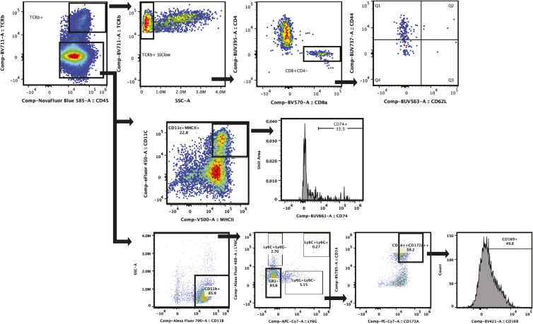
Plexiform neurofibromas (PNFs) are nerve tumors caused by loss of NF1 and dysregulation of RAS-MAPK signaling in Schwann cells. Most PNFs shrink in response to MEK inhibition, but targets with increased and durable effects are needed. We identified the anaphylatoxin C5a as increased in PNFs and expressed largely by PNF m acrophages. We defined pharmacokinetic and immunomodulatory properties of a C5aR1/2 antagonist and tested if peptide antagonists augment the effects of MEK inhibition. MEK inhibition recruited C5AR1 to the macrophage surface; short-term inhibition of C5aR elevated macrophage apoptosis and Schwann cell death, without affecting MEK-induced tumor shrinkage. PNF macrophages lacking C5aR1 increased the engulfment of dying Schwann cells, allowing their visualization. Halting combination therapy resulted in altered T-cell distribution, elevated Iba1+ and CD169+ immunoreactivity, and profoundly altered cytokine expression, but not sustained trumor shrinkage. Thus, C5aRA inhibition independently induces macrophage cell death and causes sustained and durable effects on the PNF microenvironment.


Conflict of interest statement

The authors declare that they have no conflict of interests.

Figures

Figure 1.
Figure 1.. The inflammatory protein C5a is expressed in mouse and human neurofibromas; C5a receptors are expressed mainly on nerve macrophages.
(A) Cytokine expression in mouse PNF lysates collected from PNF-bearing mice treated for 60 d with either vehicle (control) or MEKi (PD0325901). Pixel intensity for each factor was normalized to values vehicle treated controls. C5a expression (red) was unchanged, whereas most other factors were reduced or increased by MEKi treatment. (B) Heatmap of C5, C5AR1, and C5AR2 mRNAs, showing variable expression in normal nerve (N) and neurofibroma (PNF). Z-Score scale shown. (C) Paraffin sections of human normal nerve (top) and human PNF (bottom) stained with hematoxylin and eosin (H&E), anti-S100b (brown), or anti-C5aR1 (brown). Immunohistochemistry shows increased C5aR1 protein expression in PNF. Scale bars = 50 μm. (D) Flow cytometry shows increased numbers of live CD88/C5aR1+ cells in mouse PNF versus DRG/nerve. (E, F, G) Quantification of flow cytometry data. CD88/C5aR1+ macrophages (gated as Live, CD45+; TCRb−;CD11b+; SSClow; CD14++; CD172a++; Ly6C−;Ly6G−;F480+) are increased in pNF (* = P < 0.05 by t test; each data point shows data from a single mouse (n = 3–6/condition). (E) Total macrophages are increased as a percent of live cells in PNF. (F) Similar percentages of macrophages express C5aR1 in DRG/nerve and PNF. (G, H) Percentages of subtypes of immune cells that express CD88/C5aR1+ in DRG/nerve and PNF shown as a bar chart (G) and cell their frequency in PNF (H).
Figure S1.
Figure S1.. Original blots showing cytokine analyses.
(A) Cytokine array blot in mice after 5 d of treatment imaged on film. (B) Cytokine array blot in mice after 60 d of treatment imaged electronically.
Figure S2.
Figure S2.. Flow cytometry panels and representative gating of flow cytometry.
(A) Antibody panels used in flow cytometry; antibodies highlighted in blue were performed with internalization. (B) Representative gating of antibody panel 1.
Figure 2.
Figure 2.. Genetic depletion of C5aR1 does not alter neurofibroma formation, but MEK inhibition (MEKi) increases cell surface CD88/C5aR1 expression on PNF macrophages.
(A) Gross dissections of representative Nf1fl/fl;DhhCre mouse spinal cords with attached DRG/nerves from C5aR1+/− (WT) and C5aR1−/− littermates, which form neurofibromas. (B) Tumor volume (cm3) measured by volumetric MRI in 8-mo-old Nf1fl/fl;DhhCre;C5aR1+/− (WT) and Nf1fl/fl;DhhCre;C5aR1−/− neurofibroma-bearing mice. Each point shows data from a single mouse. (C) Kaplan-Meier curve demonstrates that neither genetic reduction nor ablation of C5aR1 alters survival of Nf1fl/fl;DhhCre mice. (D) Representative flow cytometry showing the absence of C5aR1 in immune cells of C5aR1 null mice versus WT mice. (E) Flow cytometry shows that genetic deletion of C5aR1 does not alter total immune (live; CD45+) cell, macrophages (live; CD45+; TCRb−; CD11b+ SSClow; Ly6c/ly6G−; CD14+++; CD172a++), or dendritic cells (live; CD45+; TCRb−; CD11c+; MHCII+). (F) Nf1fl/fl;DhhCre mice treated with vehicle (VEH) or MEKi for 5 d do not alter percentages of tumor macrophages expressing C5aR1/CD88. Each point shows data from one tumor. (* = P < 0.05 by t test). (G, F) Intensity of C5aR1 on tumor macrophages increases after 5 d of MEKi treatment; histogram at right shows concatenated data from samples in (F), quantifying C5aR1 intensity. (H) Imaging cytometry of BMD-macrophages from tumor-bearing mice treated for 1 h ex vivo with vehicle or MEK inhibitor, showing an increase in cell surface CD88 after MEK inhibition.
Figure S3.
Figure S3.. Representative gating of imaging cytometry and cleaved caspase 3 (CC3, apoptosis).
(A) Representative gating of imaging cytometry. (B) Representative gating of cleaved caspase 3 (CC3, apoptosis) by cell type in neurofibroma tumors.
Figure 3.
Figure 3.. Reducing C5aR1 in combination with short-term MEK inhibition (MEKi) reduces PNF cell proliferation and induces PNF cell apoptosis.
(A) Heatmap shows similar changes in immune proteins by treatment for 5 d with PD0325901 (MEKi), A8Δ71–73 (C5AR1/2 antagonist), or the A8Δ71–73 + PD0325901 combination. C5/C5a is indicated in red and is unchanged. (B) Western blot shows increased apoptotic cell death (cleavage of PARP) in PNF lysates from mice treated with vehicle, MEKi, C5aRA, or the combination collected 2 h a final dose (pooled lysates, n = 3 mice per condition). (C, D, E) Flow cytometry of tumor from C5aR1-null or WT neurofibroma-bearing mice. In (C), increase percentages of cleaved caspase 3+ cells (CC3, apoptosis); percentages of proliferative cells (Ki67+ cells) were unchanged. (D) Histogram of concatenated (pooled) samples (left) and quantification (right) shows an increase in cleaved caspase 3 (CC3) in tumor macrophages lacking C5aR1, with no additional effect after short-term (5 d) MEK inhibition. (E) Histogram of concatenated (pooled) samples (left) and quantification (right) showing an increase in CC3+ in Schwann cells (CD45; S100b+), which was prevented in C5aR1 null mice by 5 d of MEKi treatment. Each point shows data from one mouse (* = P < 0.05, two-way ANOVA). (F, G) Pharmacologic inhibition of C5aR results in cell death. (F) TUNEL staining of PNF sections demonstrates increased cell death in C5aRA and combination groups. (G) Quantification of percentage of TUNEL+/DAPI+ cells; each dot shows data from a single mouse, 3–5 hpf/mouse. **P = <0.01, ***P = <0.001; Tukey’s multiple comparisons ANOVA.
Figure 4.
Figure 4.. 60 d of combination treatment reduces PNF volume and PNF cell proliferation and increases PNF apoptotic cell death.
(A) Pharmacokinetic profile of hC5a A8Δ71–73 in plasma following a single i.p. administration of human C5 A8Δ71–73 (5 mg/kg in WT mice). Time after dosing is shown on the x axis. Each point represents data from one mouse. (B) Change in weight (%) by treatment group Inset shows no significant change in body weight after 60 d of treatment. (C) Waterfall plot showing change in tumor volume among treatment groups between 7–9 mo. of age (treatment period). Each bar shows data from a single mouse. (D) Representative MRI images of tumor volume change after treatment in select animals. (C, E, F) TUNEL staining by immunofluorescence (red) showing apoptotic cell death in treatment groups after 60 d of treatment in sections from animals from (C). (F) Quantification of dying cells (TUNEL staining). **P = <0.01; ***P = <0.001; ****P =<0.0001 by Tukey’s multiple comparison test. (G) Quantification of cell proliferation (percent Ki67+ cells) by immunofluorescence shows decreased proliferation in all treatment groups. ****P = <0.0001 by Tukey’s multiple comparisons test. *P = <0.01 by Tukey’s multiple comparison test.
Figure S4.
Figure S4.. PNF T cells in treatment- and durability-group mice.
(A) Tumor C5a concentration after 60 d of treatment. (B) Quantification and immunohistochemistry representation of Iba1 within treatment groups (* = P < 0.05, one-way ANOVA). A representative image of hematoxylin-stained paraffin sections, anti-Iba1 (brown). Quantification of neurofibroma T cells (gated as Live, CD45+, TCRb+, CD3+), CD4+ T cells (gated as Live, CD45+, TCRb+, CD4+, CD8), CD8 T cells (gated as Live, CD45+, TCRb+, CD8+, CD84) each expressed as percent of Live cells among treatment groups in PNF treatment groups. (C) 60-d drug treatment. (D) 30 d on and 30 d off treatment (* = P < 0.05, one-way ANOVA). (C) Tumor C5a levels at time of PNF excision (2 h after last dose) are low and do not differ significantly among treatment groups. (E) Quantification from immunohistochemistry CD3+ cell per high-powered field in each treatment groups (* = P < 0.05, one-way ANOVA). Hematoxylin-stained paraffin section showing clumps of lymphocytes stained with anti-CD3 (brown) in a PNF from the durability study.
Figure 5.
Figure 5.. Phagocytosis of Schwann cell is increased by pharmacologic C5aR blockade or MEKi.
(A) Schematic at left shows mixing of labeled cells from PNF to enable detection of cell phagocytosis. Middle, representative flow cytometric result indicating quadrant containing violet and green single live cells (e.g., phagocytosis). Right, Quantification at designated times showing that MEKi reduces detection of phagocytosed cells. (B) Photomicrograph shows a representative image of BMDM In vitro, identified using CD172a/SIRPa immunoreactivity (white), engulfing a CFSE-labeled embryonic Nf1−/− Schwann cell (green) (left). At right, quantification (as defined by CSFE signal in macrophages) showing increased Schwann cell phagocytosis by both inhibitors and combination (* = P < 0.05, three technical and two experimental replicates one-way ANOVA). (C) Immunofluorescent co-labeling of cells in tissue sections from mice in Fig 4C, stained with anti-CNPase (Schwann cells) or DhhCre; Nf1fl/fl-lineage traced GFP+ cells (Schwann cells), anti-CD11b (macrophages and some dendritic cells), and cleaved caspase 3 (CC3, apoptosis). Insets (left) highlight an apoptotic Schwann cell being phagocytized by a macrophage after 60 d of treatment. Right, quantification of cell death in a stacked bar graph subclassified by types of cells dying. Each data point was generated from an average 10 hpfs/PNF, in mice n = 7 mice/condition.
Figure 6.
Figure 6.. Mice treated with combination therapy for 1 mo and then observed for 1 mo show rebound tumor volume but not increased cell proliferation.
(A) Treatment regimen scheme; in some mice, treatment was discontinued after 1 mo (durability group). Solid line denotes months 7–8, when mice were given combination treatment. Dashed line denotes month 8–9, when mice were observed without treatment before final MRI and tumor excision at month 9. (B) Tumor change volume over time for mice treated for 2 mo (PD0325901 and A8Δ71–73 + PD0325901) versus durability group where combination treatment was stopped after 1 mo. (C) Quantification of Ki67 immunofluorescence of durability-group tumors compared to tumors with continued treatment (from Fig 4). (D) Heatmap shows cytokine protein levels (normalized to vehicle treatment) after 60 d of single agent PD or A8Δ71–73, A8Δ71–73 + PD0325901 combination treatment or durability treatment shows profound changes in the durability group. (E) H&E staining of sections from durability-group PNF, demonstrating chaotic architecture with clumps of immune-cell infiltration. Scale bars = 50 μm. Inset, H&E of vehicle treated tumor. (F) Quantification and immunohistochemistry show immunoregulatory macrophages (CD169+) within treatment groups. *P = <0.05; **P = <0.01; ***P = <0.001; ****P = <0.0001 by Tukey’s multiple comparisons test. (G) Quantification from flow cytometry of MHCII+ myeloid cells (live, TCRb−, CD3, CD11b+, MHCII+) and dendritic cells (live, TCRb−, CD3, CD11c+, MHCII+) (*P = <0.05, one-way ANOVA) verifies an increase in MHCII high myeloid cells and an increase in CD11c+ DC in the durability setting.
Figure 7.
Figure 7.. Mice treated with single-agent therapy for 1 mo and then observed for 4 mo show persistent changes in the immune environment.
(A) Treatment regimen scheme and waterfall plots showing percent change in tumor volume in mice on treatment for month (imaging at 5 mo; left), these same mice were re-imaged 4 mo later, showing tumor growth in all groups (right). (B) Quantification of flow cytometetric data, showing reduction in total immune cells (CD45+) in tumors from mice who previously had received A8Δ71–73. (C) Quantification of flow cytometetric data showing changes in immune-cell populations and phenotypes after 4 mo off treatment, selective for mice treated with A8Δ71–73; reductions in macrophages were also present in mice treated with the combination.
Figure S5.
Figure S5.. Representative gating of CD8 T-effector cells.
Low-side scatter TCRβ+ cells include CD8+ and CD4+ T cells; of CD8+ cells, CD44hi; CD62L− cells are effector T cells.

References

    1. Baumann D, Hägele T, Mochayedi J, Drebant J, Vent C, Blobner S, Noll JH, Nickel I, Schumacher C, Boos SL, et al. (2020) Proimmunogenic impact of MEK inhibition synergizes with agonist anti-CD40 immunostimulatory antibodies in tumor therapy. Nat Commun 11: 2176. 10.1038/s41467-020-15979-2 - DOI - PMC - PubMed
    1. Breivik T, Gundersen Y, Gjermo P, Taylor SM, Woodruff TM, Opstad PK (2011) Oral treatment with complement factor C5a receptor (CD88) antagonists inhibits experimental periodontitis in rats. J Periodontal Res 46: 643–647. 10.1111/j.1600-0765.2011.01383.x - DOI - PubMed
    1. Cain SA, Monk PN (2002) The orphan receptor C5L2 has high affinity binding sites for complement fragments C5a and C5a des-Arg(74). J Biol Chem 277: 7165–7169. 10.1074/jbc.C100714200 - DOI - PubMed
    1. Canavese F, Krajbich JI (2011) Resection of plexiform neurofibromas in children with neurofibromatosis type 1. J Pediatr Orthop 31: 303–311. 10.1097/BPO.0b013e31820cad77 - DOI - PubMed
    1. Choi K, Komurov K, Fletcher JS, Jousma E, Cancelas JA, Wu J, Ratner N (2017) An inflammatory gene signature distinguishes neurofibroma Schwann cells and macrophages from cells in the normal peripheral nervous system. Sci Rep 7: 43315. 10.1038/srep43315 - DOI - PMC - PubMed

Substances