

Exosomes derived from human umbilical cord mesenchymal stem cells decrease neuroinflammation and facilitate the restoration of nerve function in rats suffering from intracerebral hemorrhage
- PMID: 38459276
- PMCID: PMC11695394
- DOI: 10.1007/s11010-024-04954-w
Exosomes derived from human umbilical cord mesenchymal stem cells decrease neuroinflammation and facilitate the restoration of nerve function in rats suffering from intracerebral hemorrhage
Abstract
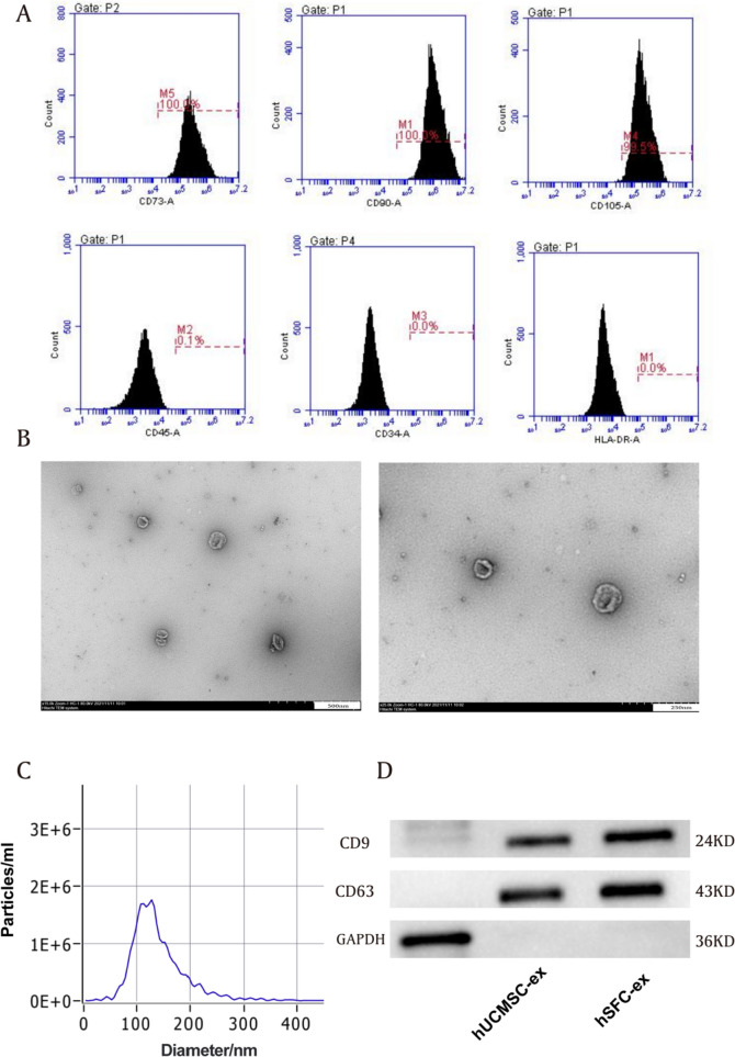
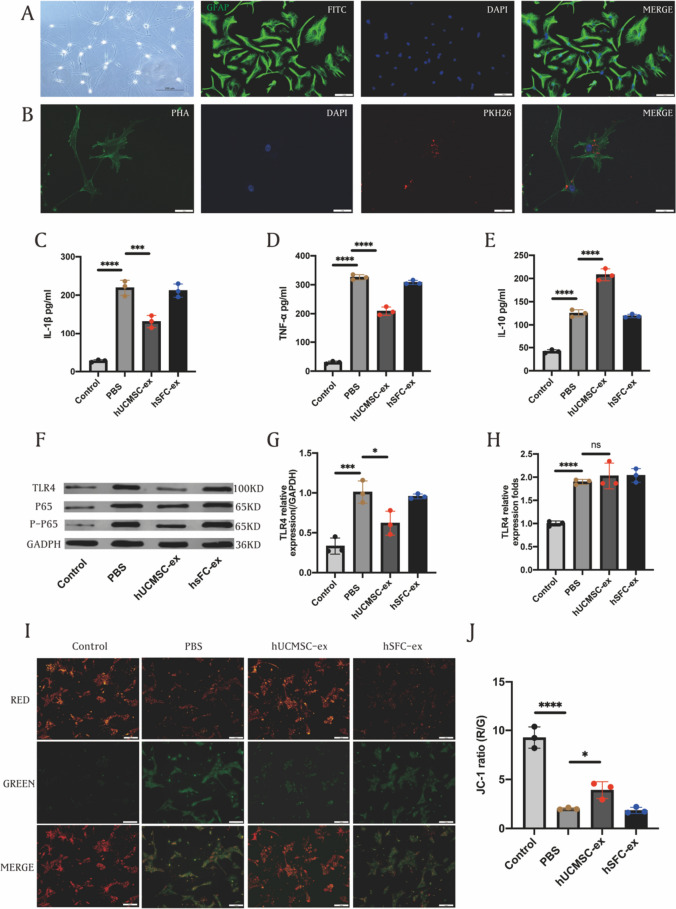
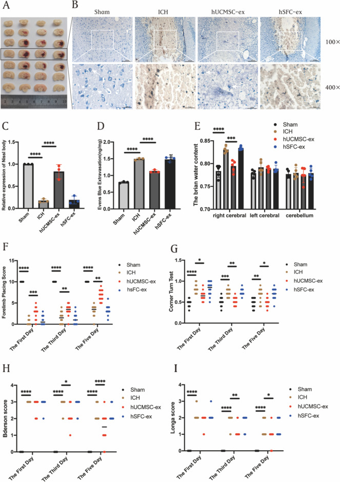
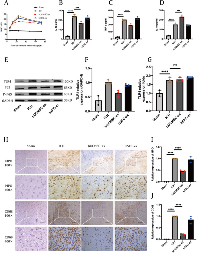
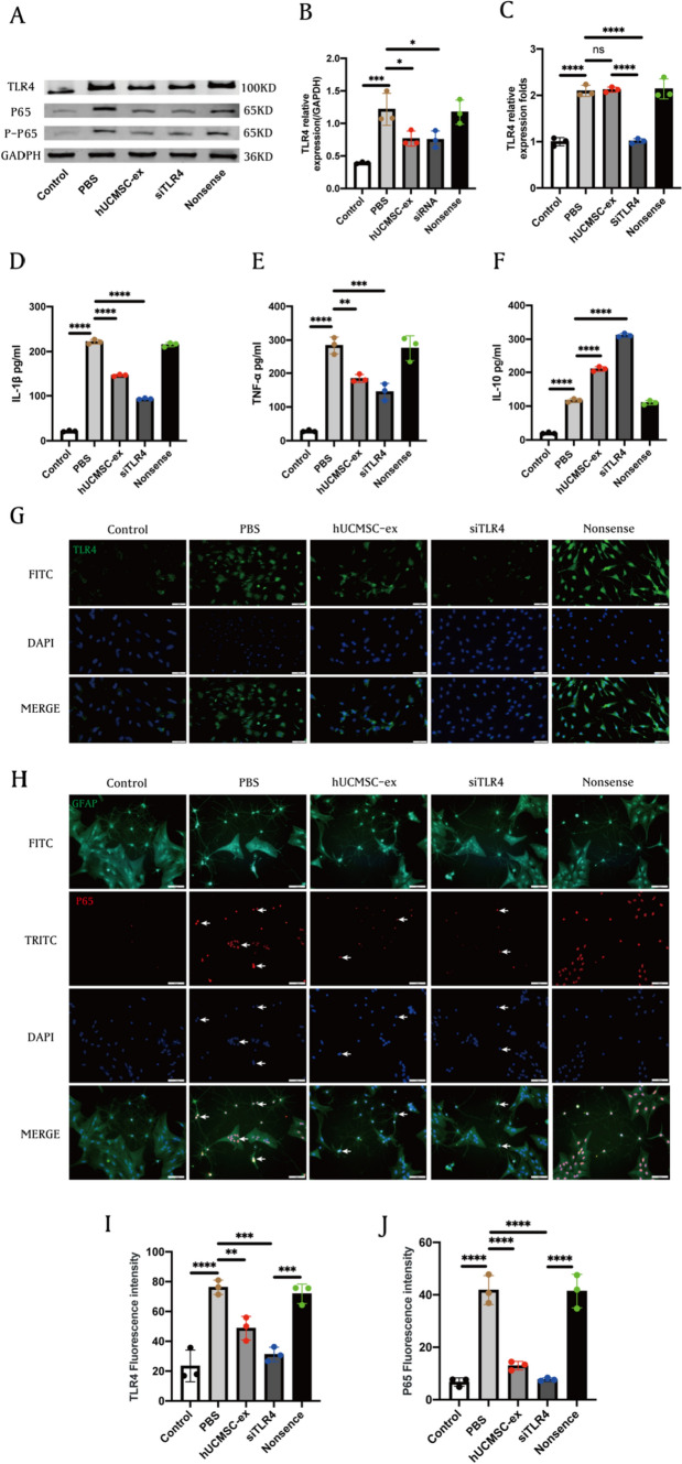
Exosomes derived from human umbilical cord mesenchymal stem cells (hUCMSC-ex) have become a hopeful substitute for whole-cell therapy due to their minimal immunogenicity and tumorigenicity. The present study aimed to investigate the hypothesis that hUCMSC-ex can alleviate excessive inflammation resulting from intracerebral hemorrhage (ICH) and facilitate the rehabilitation of the nervous system in rats. In vivo, hemorrhagic stroke was induced by injecting collagenase IV into the striatum of rats using stereotactic techniques. hUCMSC-ex were injected via the tail vein at 6 h after ICH model establishment at a dosage of 200 µg. In vitro, astrocytes were pretreated with hUCMSC-ex and then stimulated with hemin (20 μmol/mL) to establish an ICH cell model. The expression of TLR4/NF-κB signaling pathway proteins and inflammatory factors, including TNF-α, IL-1β, and IL-10, was assessed both in vivo and in vitro to investigate the impact of hUCMSC-ex on inflammation. The neurological function of the ICH rats was evaluated using the corner turn test, forelimb placement test, Longa score, and Bederson score on the 1st, 3rd, and 5th day. Additionally, RT-PCR was employed to examine the mRNA expression of TLR4 following hUCMSC-ex treatment. The findings demonstrated that hUCMSC-ex downregulated the protein expression of TLR4, NF-κB/P65, and p-P65, reduced the levels of pro-inflammatory cytokines TNF-α and IL-1β, and increased the expression of the anti-inflammatory cytokine IL-10. Ultimately, the administration of hUCMSC-ex improved the behavioral performance of the ICH rats. However, the results of PT-PCR indicated that hUCMSC-ex did not affect the expression of TLR4 mRNA induced by ICH, suggesting that hUCMSCs-ex may inhibit TLR4 translation rather than transcription, thereby suppressing the TLR4/NF-κB signaling pathway. We can conclude that hUCMSC-ex mitigates hyperinflammation following ICH by inhibiting the TLR4/NF-κB signaling pathway. This study provides preclinical evidence for the potential future application of hUCMSC-ex in the treatment of cerebral injury.
Keywords: Exosomes; Human umbilical cord mesenchymal stem cells; Inflammation; Intracerebral hemorrhage; TLR4/NF-κB.
© 2024. The Author(s).
Conflict of interest statement
Declarations. Conflicts of interest: The authors declare that they have no conflict of interest. Ethical approval: The studies involving human participants were reviewed and approved by the Ethics Committee of the second Hospital of Hebei Medical University. The patients/participants provided their written informed consent to participate in this study. The animal study was reviewed and approved by the Ethics Committee of The Second Hospital of Hebei Medical University.
Figures
Similar articles
-
Human umbilical cord mesenchymal stem cell-derived exosomes inhibit inflammation and fibrotic scar formation after intracerebral hemorrhage.Mol Cell Biochem. 2025 Aug;480(8):4829-4847. doi: 10.1007/s11010-025-05259-2. Epub 2025 Apr 25. Mol Cell Biochem. 2025. PMID: 40279088 Free PMC article.
-
Human umbilical cord mesenchymal stem cell conditioned medium attenuates renal fibrosis by reducing inflammation and epithelial-to-mesenchymal transition via the TLR4/NF-κB signaling pathway in vivo and in vitro.Stem Cell Res Ther. 2018 Jan 12;9(1):7. doi: 10.1186/s13287-017-0760-6. Stem Cell Res Ther. 2018. PMID: 29329595 Free PMC article.
-
Effects of 3D-printed exosome-functionalized brain acellular matrix hydrogel on neuroinflammation in rats following cerebral hemorrhage.Stem Cell Res Ther. 2025 Apr 20;16(1):196. doi: 10.1186/s13287-025-04332-3. Stem Cell Res Ther. 2025. PMID: 40254565 Free PMC article.
-
Preclinical and clinical research on inflammation after intracerebral hemorrhage.Prog Neurobiol. 2010 Dec;92(4):463-77. doi: 10.1016/j.pneurobio.2010.08.001. Epub 2010 Aug 14. Prog Neurobiol. 2010. PMID: 20713126 Free PMC article. Review.
-
[Research progress of mesenchymal stem cell exosomes in the treatment of intraventricular hemorrhage in premature infants].Zhonghua Er Ke Za Zhi. 2025 Jun 2;63(6):691-694. doi: 10.3760/cma.j.cn112140-20250211-00107. Zhonghua Er Ke Za Zhi. 2025. PMID: 40393770 Review. Chinese.
Cited by
-
Human umbilical cord mesenchymal stem cell-derived exosomes inhibit inflammation and fibrotic scar formation after intracerebral hemorrhage.Mol Cell Biochem. 2025 Aug;480(8):4829-4847. doi: 10.1007/s11010-025-05259-2. Epub 2025 Apr 25. Mol Cell Biochem. 2025. PMID: 40279088 Free PMC article.
-
The role of potential oxidative biomarkers in the prognosis of intracerebral hemorrhage and the exploration antioxidants as possible preventive and treatment options.Front Mol Biosci. 2025 Feb 4;12:1541230. doi: 10.3389/fmolb.2025.1541230. eCollection 2025. Front Mol Biosci. 2025. PMID: 39967652 Free PMC article. Review.
-
Therapeutic potential of EVs loaded with CB2 receptor agonist in spinal cord injury via the Nrf2/HO-1 pathway.Redox Rep. 2024 Dec;29(1):2420572. doi: 10.1080/13510002.2024.2420572. Epub 2024 Oct 28. Redox Rep. 2024. PMID: 39466990 Free PMC article.
-
3D biological scaffold delivers Bergenin to reduce neuroinflammation in rats with cerebral hemorrhage.J Transl Med. 2024 Oct 17;22(1):946. doi: 10.1186/s12967-024-05735-1. J Transl Med. 2024. PMID: 39420402 Free PMC article.
-
Therapeutic Efficacy and Promise of Human Umbilical Cord Mesenchymal Stem Cell-Derived Extracellular Vesicles in Aging and Age-Related Disorders.Int J Mol Sci. 2024 Dec 30;26(1):225. doi: 10.3390/ijms26010225. Int J Mol Sci. 2024. PMID: 39796081 Free PMC article. Review.
References
-
- Chen G, Leak RK, Sun Q, Zhang JH, Chen J (2018) Neurobiology of stroke: research progress and perspectives. Prog Neurobiol 163–164:1–4 - PubMed
-
- Babu R, Bagley JH, Di C, Friedman AH, Adamson C (2012) Thrombin and hemin as central factors in the mechanisms of intracerebral hemorrhage-induced secondary brain injury and as potential targets for intervention. Neurosurg Focus 32(4):E8 - PubMed
MeSH terms
Substances
Grants and funding
- H2021206003/the Provincial Natural Science Foundation of Hebei
- 81870984/National Outstanding Youth Science Fund Project of National Natural Science Foundation of China
- 2021JH026/the Innovation Fund for Industry-University Research for Chinese Universities
- 236Z7752G/the central government guides local funds for science and technology development
LinkOut - more resources
Full Text Sources
