. 2024 Mar 8;29(1):34.
doi: 10.1186/s11658-024-00548-y.

A multiplex RPA-CRISPR/Cas12a-based POCT technique and its application in human papillomavirus (HPV) typing assay

Affiliations

A multiplex RPA-CRISPR/Cas12a-based POCT technique and its application in human papillomavirus (HPV) typing assay

Yan Liu et al. Cell Mol Biol Lett. .

Erratum in

Abstract

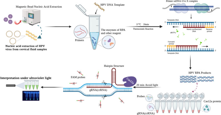
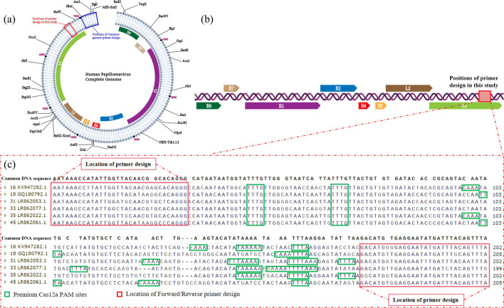
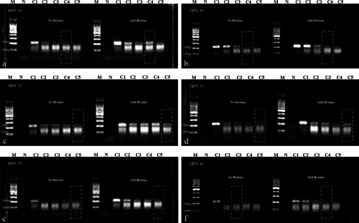
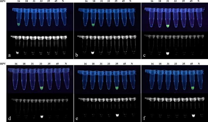
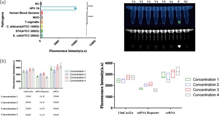
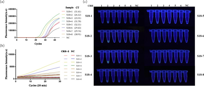
Persistent infection with high-risk human papillomavirus (HR-HPV) is the primary and initiating factor for cervical cancer. With over 200 identified HPV types, including 14 high-risk types that integrate into the host cervical epithelial cell DNA, early determination of HPV infection type is crucial for effective risk stratification and management. Presently, on-site immediate testing during the HPV screening stage, known as Point of Care Testing (POCT), remains immature, severely limiting the scope and scenarios of HPV screening. This study, guided by the genomic sequence patterns of HPV, established a multiplex recombinase polymerase amplification (RPA) technology based on the concept of "universal primers." This approach achieved the multiple amplification of RPA, coupled with the CRISPR/Cas12a system serving as a medium for signal amplification and conversion. The study successfully constructed a POCT combined detection system, denoted as H-MRC12a (HPV-Multiple RPA-CRISPR/Cas12a), and applied it to high-risk HPV typing detection. The system accomplished the typing detection of six high-risk HPV types (16, 18, 31, 33, 35, and 45) can be completed within 40 min, and the entire process, from sample loading to result interpretation, can be accomplished within 45 min, with a detection depth reaching 1 copy/μL for each high-risk type. Validation of the H-MRC12a detection system's reproducibility and specificity was further conducted through QPCR on 34 clinical samples. Additionally, this study explored and optimized the multiplex RPA amplification system and CRISPR system at the molecular mechanism level. Furthermore, the primer design strategy developed in this study offers the potential to enhance the throughput of H-MRC12a detection while ensuring sensitivity, providing a novel research avenue for high-throughput detection in Point-of-Care molecular pathogen studies.

Keywords: CRISPR/Cas12a; HR-HPV; Multiplex RPA; POCT.


Conflict of interest statement

The authors declare that they have no competing interests.

Figures

Scheme 1
Scheme 1
Schematic view of the nucleic acid extraction and HPV-Multiple RPA-CRISPR/Cas12a(H-MRC12a) typing assay
Fig. 1
Fig. 1
Results of the first round of primer and amplicon region screening. A1, B1 Primer screening results for plasmids of HPV type 16 at concentrations of 107 copies/μL and 105 copies/μL, respectively. A2, B2 Primer screening results for plasmids of HPV type 18 at concentrations of 107 copies/μL and 105 copies/μL, respectively. P1–P6: Primer combinations HF3/HR4, HF3/HR5, HF4/HR4, HF4/HR5, HF5/HR4, HF5/HR5. N Negative control. As the exact lengths of the amplicon bands are not yet determined, a separate positive control for this section has not been individually included. The positive results from P1 to P6 can serve as inherent positive controls for this experiment. Gel electrophoresis results indicate that among all designed primer combinations, only the HF5/HR4 combination exhibits higher sensitivity for the amplification of HPV types 16 and 18
Fig. 2
Fig. 2
Design positions of multiple RPA primers and crRNA for HPV in this study. a Circular representation of the HPV whole genome, with the red box indicating the primer design positions in this study. b Schematic representation of the unfolded HPV whole genome. c Specific sequences of the 6 HR-HPV amplicon regions and primers along with the PAM sites in this study. The green annotations indicate the positions of PAM sites, while the red annotations indicate the positions of forward/reverse primer designs
Fig. 3
Fig. 3
Results of the second round of single RPA primer screening for various HPV subtypes. a, b, d Amplification of plasmids of HPV types 16, 18, and 33 at different concentrations using the HF5-1-1/HR4 primer combination. c Amplification of plasmids of HPV type 31 at different concentrations using the HF5-1-3/HR4 primer combination. e Amplification of plasmids of HPV type 35 at different concentrations using the HF5-3-2/HR4 primer combination. f Amplification of plasmids of HPV type 45 at different concentrations using the HF5-2-2/HR4 primer combination. The forward primers in the above combinations share the same reverse primer, HR4. g Amplification of plasmids of HPV type 33 at different concentrations using the HF5-3-2/HR4-33Q2 primer combination. h Amplification of plasmids of HPV type 18 at different concentrations using the HF5-18Y/HR4-18Q2 primer combination. C1–C5: Plasmid concentrations of 104 copies/μL, 103 copies/μL, 102 copies/μL, 10 copies/μL, and 1 copy/μL respectively. N Negative control. The RPA amplification sensitivity for HPV types 18, 31, 33, and 35 is achieved at 1 copy/μL, while for HPV types 16 and 45, it is achieved at 10 copies/μL
Fig. 4
Fig. 4
Experimental results exploring the efficiency of primer depletion for Cas12a protein cleavage and validation of structure-specificity in the hairpin structure. a Fluorescence curves and endpoint bar charts comparing three parallel CRISPR reactions of RPA products with and without residual primers. Fluorescence was observed immediately under UV light after a 15-min reaction. Purified: RPA products with residual primers removed; Unpurified: RPA products without removing residual primers. b Fluorescence curves and endpoint fluorescence values of CRISPR reactions using crRNA with different numbers of mismatched bases compared to the original sequence when connected to a specific hairpin structure. The asterisk indicates significant differences as determined by two-tailed Student’s t-test (****P < 0.0001). ns denotes no statistical significance. NC/NTC negative control reaction
Fig. 5
Fig. 5
Gel electrophoresis results before and after the addition of betaine in the multiple RPA system. af Gel electrophoresis results of the multiple RPA system amplifying plasmids of HPV 16, 18, 31, 33, 35, and 45 at different concentrations before and after the addition of betaine. C1–C5: Plasmid concentrations of 104 copies/μL, 103 copies/μL, 102 copies/μL, 10 copies/μL, and 1 copy/μL respectively. N negative control. Sensitivity for HPV types 16 and 18 reaches 10 copies/μL both before and after the addition of betaine, with brighter bands observed after addition. Sensitivity for HPV types 31, 33, and 35 reaches 1 copy/μL both before and after the addition of betaine, with brighter bands observed after addition. Sensitivity for HPV type 45 surpasses 102 copies/μL after the addition of betaine, reaching 10 copies/μL
Fig. 6
Fig. 6
Cross-validation results for the RNA specificity in the crRNA pool of the constructed CRISPR system. af Specificity cross-validation results for CRH1 to CRH6, respectively, against various HPV types. DNA templates for HPV 16–45 were added at concentrations of 104 copies/μL each for the cross-validation. N negative control. The experimental results demonstrate that each crRNA shows no cross-reaction with HPV types other than its corresponding type
Fig. 7
Fig. 7
Specificity validation of the dual detection system H-MRC12a and optimization results for the components of the CRISPR system. a Specificity validation results for the H-MRC12a detection system against common pathogens in the female reproductive tract. After extracting nucleic acids from each pathogen, they were added to the reaction system, and fluorescence was observed immediately under UV light after a 35-min reaction. The results demonstrate no cross-reaction with the following five pathogens and the human blood genome: V1: E. coli (ATCC 25922), V2: S. aureus (ATCC 25923), V3: C. albicans (ATCC 14053), V4: T. vaginalis, V5: N. gonorrhoeae (NGO), V6: Human Blood Genome, NC negative control reaction, P positive control using HPV 18 as the DNA template. The asterisk indicates significant differences compared to P, as determined by two-tailed Student’s t-test (****P < 0.0001). b Comparison of fluorescence intensity for different concentrations of Lba Cas12a protein, ssDNA reporter, and crRNA in the CRISPR system. The optimal component concentrations were determined as follows: Lba Cas12a 250 nM, ssDNA reporter 4 μM, and crRNA 500 nM
Fig. 8
Fig. 8
Sensitivity validation of the dual detection system H-MRC12a with a reaction time of 40 min (Multiple RPA 20 min + CRISPR 20 min). af Sensitivity validation results for different concentrations of plasmids for HPV types 16, 18, 31, 33, 35, and 45, respectively. The reaction results were observed immediately under UV light after completion. The asterisk indicates significant differences compared to the negative control, as determined by two-tailed Student’s t-test (*P < 0.1, ***P < 0.001, ****P < 0.0001). ns denotes no statistical significance. NC negative control reaction
Fig. 9
Fig. 9
Comparison of experimental results between the H-MRC12a combined detection system and a commercially available QPCR genotyping kit for detecting 8 clinical HPV 16-positive samples. Clinical samples were heat-inactivated and subjected to nucleic acid extraction using the nanomagnetic bead method. The extracted nucleic acids were then incubated in the H-MRC12a combined detection system for 40 min (Multiple RPA incubation for 20 min, followed by CRISPR incubation for an additional 20 min) and immediately observed under 300 nm UV light. a Fluorescence curves and CT values obtained from the commercially available QPCR genotyping kit for the 8 clinical samples. b Real-time fluorescence signals (recorded using the Q160 LongGene portable QPCR instrument) of the H-MRC12a dual detection system for the clinical samples and negative control after 20 min of incubation in the CRISPR step. c Photographs taken under UV light of the 8 clinical samples incubated for 40 min in the H-MRC12a detection system. S16-1~S16-8: Identification numbers of the 8 HPV 16-positive clinical samples; CRH1~6: Tubes containing crRNA specific for HPV 16, 18, 31, 33, 35, and 45, respectively, for genotyping detection; NC: Negative control; PC: Positive control
Fig. 10
Fig. 10
Comparison of experimental results between the H-MRC12a combined detection system and a commercially available QPCR genotyping kit for detecting 8 clinical HPV 18-positive samples. Clinical samples were heat-inactivated and subjected to nucleic acid extraction using the nanomagnetic bead method. The extracted nucleic acids were then incubated in the H-MRC12a combined detection system for 40 min (Multiple RPA incubation for 20 min, followed by CRISPR incubation for an additional 20 min) and immediately observed under 300 nm UV light. a Fluorescence curves and CT values obtained from the commercially available QPCR genotyping kit for the 8 clinical samples. b Real-time fluorescence signals (recorded using the Q160 LongGene portable QPCR instrument) of the H-MRC12a dual detection system for the clinical samples and negative control after 20 min of incubation in the CRISPR step. c Photographs taken under UV light of the 8 clinical samples incubated for 40 min in the H-MRC12a detection system. S18-1~S18-8: Identification numbers of the 8 HPV 18-positive clinical samples; CRH1~6: Tubes containing crRNA specific for HPV 16, 18, 31, 33, 35, and 45, respectively, for genotyping detection; NC: Negative control
Fig. 11
Fig. 11
Comparison of experimental results between the H-MRC12a combined detection system and a commercially available QPCR genotyping kit for detecting clinical HPV 31, 33, 35, and 45-positive samples. Clinical samples were heat-inactivated and subjected to nucleic acid extraction using the nanomagnetic bead method. The extracted nucleic acids were then incubated in the H-MRC12a combined detection system for 40 min (Multiple RPA incubation for 20 min, followed by CRISPR incubation for an additional 20 min) and immediately observed under 300 nm UV light. a Fluorescence curves and CT values obtained from the commercially available QPCR genotyping kit for the clinical samples. b Real-time fluorescence signals (recorded using the Q160 LongGene portable QPCR instrument) of the H-MRC12a dual detection system for the clinical samples and negative control after 20 min of incubation in the CRISPR step. c Photographs taken under UV light of the clinical samples of each type incubated for 40 min in the H-MRC12a detection system. S31-1~S31-3: Identification numbers of 3 HPV 31-positive clinical samples; S33-1/S35-1: Identification numbers of HPV 33-positive and HPV 35-positive clinical samples; S31/45: Identification numbers of HPV 31/45 double-positive clinical samples; CRH1~6: Tubes containing crRNA specific for HPV 16, 18, 31, 33, 35, and 45, respectively, for genotyping detection; NC: Negative control
Fig. 12
Fig. 12
Fluorescence curves of clinical samples with different HPV subtypes detected using a commercially available QPCR genotyping kit. a Fluorescence curve of a clinical sample double-positive for HPV 39/53. b Fluorescence curve of a clinical sample double-positive for HPV 51/52. c Fluorescence curve of a clinical sample double-positive for HPV 58/6. d Fluorescence curves of three clinical samples positive for HPV 68, 81, and 82, respectively

References

    1. Andersson S, Safari H, Mints M, Lewensohn-Fuchs I, Gyllensten U, Johansson B. Type distribution, viral load and integration status of high-risk human papillomaviruses in pre-stages of cervical cancer (CIN) Br J Cancer. 2005;92(12):2195–2200. doi: 10.1038/sj.bjc.6602648. - DOI - PMC - PubMed
    1. World Health Organization . Global strategy to accelerate the elimination of cervical cancer as a public health problem. Geneva: World Health Organization; 2020.
    1. World Health Organization . WHO guideline for screening and treatment of cervical pre-cancer lesions for cervical cancer prevention. Geneva: World Health Organization; 2021. - PubMed
    1. Hawkes D, Keung MHT, Huang YP, McDermott TL, Romano J, Saville M, et al. Self-collection for cervical screening programs: from research to reality. Cancers. 2020;12(4):1053. doi: 10.3390/cancers12041053. - DOI - PMC - PubMed
    1. Li GR, Lamsisi M, Chenafi S, Moniod L, Benlghazi A, Ennaji MM, et al. Urine-based detection of HPV for cervical cancer screening: time for standardized tests. J Med Virol. 2023;95(4):e28737. doi: 10.1002/jmv.28737. - DOI - PubMed