. 2024 Mar 8;13(1):31.
doi: 10.1186/s13756-024-01386-5.

Uncovering the spread of drug-resistant bacteria through next-generation sequencing based surveillance: transmission of extended-spectrum β-lactamase-producing Enterobacterales by a contaminated duodenoscope

Affiliations

Uncovering the spread of drug-resistant bacteria through next-generation sequencing based surveillance: transmission of extended-spectrum β-lactamase-producing Enterobacterales by a contaminated duodenoscope

Cansu Cimen et al. Antimicrob Resist Infect Control. .

Abstract

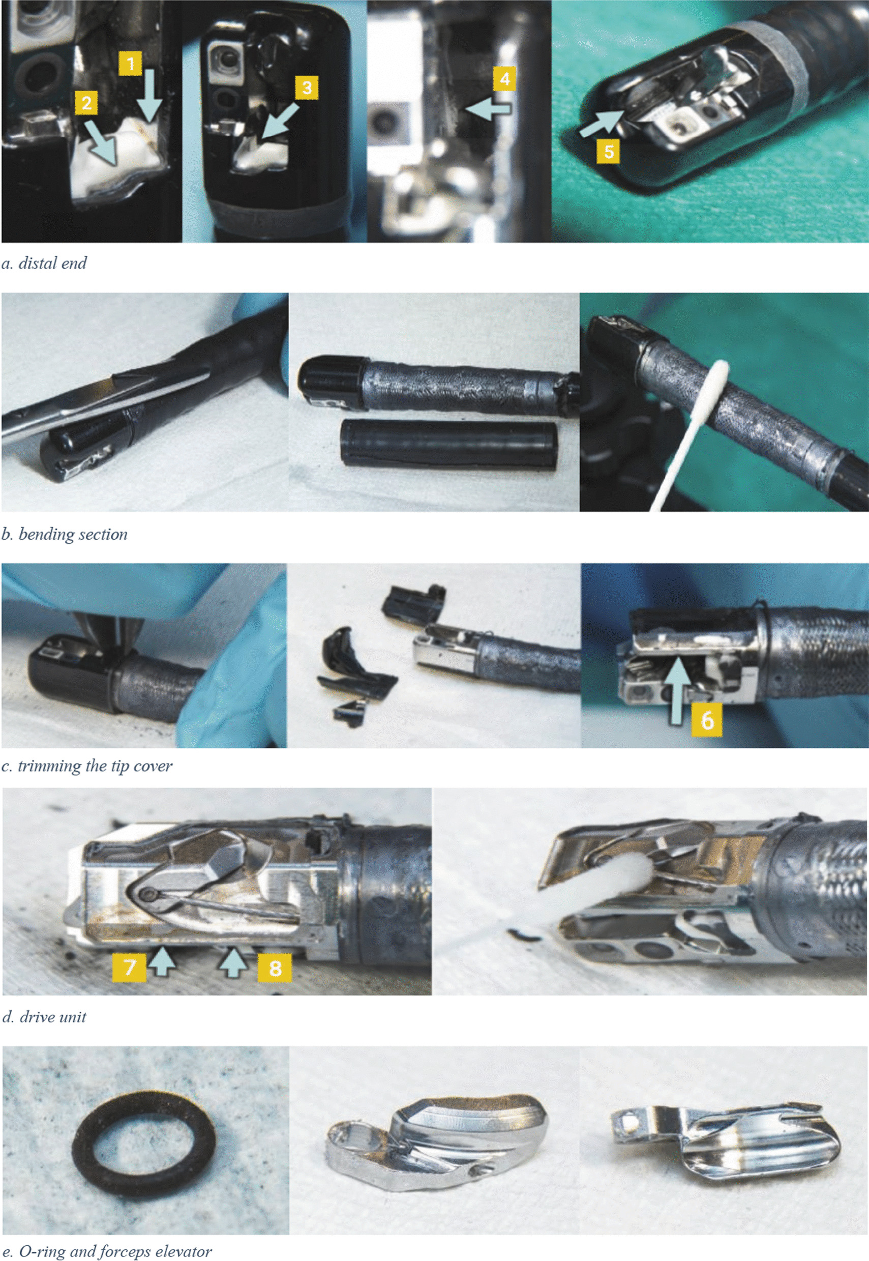
Contamination of duodenoscopes is a significant concern due to the transmission of multidrug-resistant organisms (MDROs) among patients who undergo endoscopic retrograde cholangiopancreatography (ERCP), resulting in outbreaks worldwide. In July 2020, it was determined that three different patients, all had undergone ERCP with the same duodenoscope, were infected. Two patients were infected with blaCTX-M-15 encoding Citrobacter freundii, one experiencing a bloodstream infection and the other a urinary tract infection, while another patient had a bloodstream infection caused by blaSHV-12 encoding Klebsiella pneumoniae. Molecular characterization of isolates was available as every ESBL-producing isolate undergoes Next-Generation Sequencing (NGS) for comprehensive genomic analysis in our center. After withdrawing the suspected duodenoscope, we initiated comprehensive epidemiological research, encompassing case investigations, along with a thorough duodenoscope investigation. Screening of patients who had undergone ERCP with the implicated duodenoscope, as well as a selection of hospitalized patients who had ERCP with a different duodenoscope during the outbreak period, led to the discovery of three additional cases of colonization in addition to the three infections initially detected. No microorganisms were detected in eight routine culture samples retrieved from the suspected duodenoscope. Only after destructive dismantling of the duodenoscope, the forceps elevator was found to be positive for blaSHV-12 encoding K. pneumoniae which was identical to the isolates detected in three patients. This study highlights the importance of using NGS to monitor the transmission of MDROs and demonstrates that standard cultures may fail to detect contaminated medical equipment such as duodenoscopes.

Keywords: Citrobacter freundii; Klebsiella pneumoniae; CTXM-15; Contamination; Duodenoscope; Endoscopic retrograde cholangiopancreatography (ERCP); Extended-spectrum β-lactamase (ESBL); Multi locus sequence typing (MLST); Next-generation sequencing (NGS); Nosocomial transmission; SHV-12.


Conflict of interest statement

The authors declare no competing interests.

Figures

Fig. 1
Fig. 1
Timeline of ERCP procedure and infection and colonization of patients. (*This patient was known to be colonized with blaSHV-12 K. pneumoniae since October 2019; ERCP, endoscopic retrograde cholangiopancreatography; ESBL, Extended-spectrum β-lactamase; P1, patient 1; P2, patient 2; P3, patient 3; P4, patient 4; P5, patient 5; P6, patient 6). Created with Biorender
Fig. 2
Fig. 2
Dismantling and sampling the ERCP duodenoscope. a, red-brown residue in groove (1) on the forceps elevator drive unit side and whitish-grey residue in the border above the white block around the instrument channel exit (2); white–gray residue (3) in groove on camera side; white deposit (4) on the bottom under the forceps elevator; white crystals (5) next to and below the forceps elevator; b, cutting and removing the cardan rubber, sampling the exposed surface; c, trimming the tip cover and the possible break (6) in the cementing around the arm cover.; d, brownish-red deposit and/or discoloration on the frame of the drive unit, with an apparent intrusion trace (7), corresponding to the previously observed possible interruption of cementing (8); f, sampling the instrument channel port; removed instrument channel port viewed from the entrance and port from the side; g, left half the piece of instrument channel with the brownish-yellow area; the ribbed scratch mark shows a kink where there is also a kink or dent in the tubing; Yellow discoloration of the tubing material with grey cloudy discoloration on the inner wall
Fig. 2
Fig. 2
Dismantling and sampling the ERCP duodenoscope. a, red-brown residue in groove (1) on the forceps elevator drive unit side and whitish-grey residue in the border above the white block around the instrument channel exit (2); white–gray residue (3) in groove on camera side; white deposit (4) on the bottom under the forceps elevator; white crystals (5) next to and below the forceps elevator; b, cutting and removing the cardan rubber, sampling the exposed surface; c, trimming the tip cover and the possible break (6) in the cementing around the arm cover.; d, brownish-red deposit and/or discoloration on the frame of the drive unit, with an apparent intrusion trace (7), corresponding to the previously observed possible interruption of cementing (8); f, sampling the instrument channel port; removed instrument channel port viewed from the entrance and port from the side; g, left half the piece of instrument channel with the brownish-yellow area; the ribbed scratch mark shows a kink where there is also a kink or dent in the tubing; Yellow discoloration of the tubing material with grey cloudy discoloration on the inner wall
Fig. 3
Fig. 3
Overview of ESBL-producing C. freundii and K. pneumoniae isolates with ST assigned by cgMLST analysis performed at UMCG during 2018–2020, highlighting different isolate categories and outbreak strains. Strains were selected corresponding to the same lineages. Blue, isolates obtained from patients who underwent endoscopy before the first positive culture; purple, isolates obtained from patients who underwent ERCP before the first positive culture; pink, isolates obtained from the duodenoscope; red dots around the purple-colored isolates, outbreak strains of C. freundii (ST540) and K. pneumoniae (ST17). a, ST22 C. freundii were obtained from the same patient at different times; b, ST628 K. pneumoniae strains were identified in two patients who shared a room, and both underwent ERCP procedures with different duodenoscopes in October 2019
Fig. 4
Fig. 4
Culture results of the ERCP duodenoscope

Similar articles

Cited by

References

    1. Kovaleva J, Peters FT, van der Mei HC, Degener JE. Transmission of infection by flexible gastrointestinal endoscopy and bronchoscopy. Clin Microbiol Rev. 2013;26(2):231–254. doi: 10.1128/cmr.00085-12. - DOI - PMC - PubMed
    1. Rutala WA, Kanamori H, Sickbert-Bennett EE, Weber DJ. What’s new in reprocessing endoscopes: are we going to ensure "the needs of the patient come first" by shifting from disinfection to sterilization? Am J Infect Control. 2019 doi: 10.1016/j.ajic.2019.01.017. - DOI - PubMed
    1. Wang P, Xu T, Ngamruengphong S, Makary MA, Kalloo A, Hutfless S. Rates of infection after colonoscopy and osophagogastroduodenoscopy in ambulatory surgery centres in the USA. Gut. 2018;67(9):1626–1636. doi: 10.1136/gutjnl-2017-315308. - DOI - PubMed
    1. Fraser TG, Reiner S, Malczynski M, Yarnold PR, Warren J, Noskin GA. Multidrug-resistant Pseudomonas aeruginosa cholangitis after endoscopic retrograde cholangiopancreatography: failure of routine endoscope cultures to prevent an outbreak. Infect Control Hosp Epidemiol. 2004;25(10):856–859. doi: 10.1086/502309. - DOI - PubMed
    1. Kovaleva J, Meessen NE, Peters FT, Been MH, Arends JP, Borgers RP, et al. Is bacteriologic surveillance in endoscope reprocessing stringent enough? Endoscopy. 2009;41(10):913–916. doi: 10.1055/s-0029-1215086. - DOI - PubMed

Substances