. 2024 Mar 9;43(1):75.
doi: 10.1186/s13046-024-02998-w.

TRF2 as novel marker of tumor response to taxane-based therapy: from mechanistic insight to clinical implication

Affiliations

TRF2 as novel marker of tumor response to taxane-based therapy: from mechanistic insight to clinical implication

Sara Iachettini et al. J Exp Clin Cancer Res. .

Abstract

Background: Breast Cancer (BC) can be classified, due to its heterogeneity, into multiple subtypes that differ for prognosis and clinical management. Notably, triple negative breast cancer (TNBC) - the most aggressive BC form - is refractory to endocrine and most of the target therapies. In this view, taxane-based therapy still represents the elective strategy for the treatment of this tumor. However, due variability in patients' response, management of TNBC still represents an unmet medical need. Telomeric Binding Factor 2 (TRF2), a key regulator of telomere integrity that is over-expressed in several tumors, including TNBC, has been recently found to plays a role in regulating autophagy, a degradative process that is involved in drug detoxification. Based on these considerations, we pointed, here, at investigating if TRF2, regulating autophagy, can affect tumor sensitivity to therapy.

Methods: Human TNBC cell lines, over-expressing or not TRF2, were subjected to treatment with different taxanes and drug efficacy was tested in terms of autophagic response and cell proliferation. Autophagy was evaluated first biochemically, by measuring the levels of LC3, and then by immunofluorescence analysis of LC3-puncta positive cells. Concerning the proliferation, cells were subjected to colony formation assays associated with western blot and FACS analyses. The obtained results were then confirmed also in mouse models. Finally, the clinical relevance of our findings was established by retrospective analysis on a cohort of TNBC patients subjected to taxane-based neoadjuvant chemotherapy.

Results: This study demonstrated that TRF2, inhibiting autophagy, is able to increase the sensitivity of TNBC cells to taxanes. The data, first obtained in in vitro models, were then recapitulated in preclinical mouse models and in a cohort of TNBC patients, definitively demonstrating that TRF2 over-expression enhances the efficacy of taxane-based neoadjuvant therapy in reducing tumor growth and its recurrence upon surgical intervention.

Conclusions: Based on our finding it is possible to conclude that TRF2, already known for its role in promoting tumor formation and progression, might represents an Achilles' heel for cancer. In this view, TRF2 might be exploited as a putative biomarker to predict the response of TNBC patients to taxane-based neoadjuvant chemotherapy.

Keywords: Autophagy; Drug sensitivity; Predictive marker; TNBC; TRF2; Taxanes.


Conflict of interest statement

The authors declare that they have no competing interests.

Figures

Fig. 1
Fig. 1
TRF2 impairs autophagy induced by chemo-therapeutic agents in TNBC. a Human triple negative breast cancer (TNBC) cell lines MDA-MB-231, over-expressing (pTRF2) or not (pBabe) TRF2 were transfected with EGFP-LC3 for 24 hours and treated with Paclitaxel, Carboplatin and Epirubicin for 24 hours at the indicated doses. The autophagic process was evaluated by the quantitative analysis of punctate vesicular structures by fluorescence experiments. Histogram represented the percentage of LC3 puncta positive cells on total GFP positive cells. b Representative images of the experiment described in a were acquired by deconvolution microscopy (63X magnification). Blue: the nuclei stained with DAPI. Green: EGFP-LC3B. c Western blot analysis of LC3 I-II protein levels in MDA-MB-231 pBabe or pTRF2 treated with the indicated drugs in combination or not with 30 µM of the autophagy inhibitor Chloroquine (CQ) for 3 hours. TRF2 protein levels were monitored to check cell lines and b-actin as loading internal control. The histograms represent the mean values ± S.D. of three independent experiments; *p<0.05, **p<0.01, ***p <0.001.
Fig. 2
Fig. 2
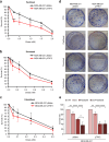
TRF2 enhances TNBC cell sensitivity to taxanes. a-c Cell survival evaluation by clonogenic assay in human triple negative breast cancer (TNBC) cell lines, MDA-MB-231, over-expressing (pTRF2) or not (pBabe) TRF2 and treated with three taxanes (Paclitaxel a, Docetaxel b and Cabazitaxel c) at the indicated doses for 24 hours. d Representative images of the clonogenic assays described in a-c. e Clonogenic assay performed in TNBC cell lines MDA-MB-231 pBabe or pTRF2 pretreated with Chloroquine (CQ) 10 mM for 24 hours before the administration of Paclitaxel 1 nM for 24 hours. The graphs represent the mean values ± S.D. of at least three independent experiments. *p<0.05, **p<0.01, ***p <0.001, ****p<0.0001.
Fig. 3
Fig. 3
Paclitaxel efficacy in xenograft TNBC tumors is enhanced by TRF2 over-expression. a CB17-SCID female immunodeficient mice were injected intramuscularly with 3x10⁶ MDA-MB-231 pBabe and pTRF2 cells. When tumors reached 250 mm3 mice were randomised and treated with Paclitaxel iv at 20 mg/Kg once a week for two weeks. Tumor volumes were measured in two dimensions using a caliper and calculated by the formula a× b2/2, where “a” and “b” are the long and short sizes of the tumor, respectively. Each experimental group included five mice. Error bars represent S.D. and p-values were calculated at day 30 using an unpaired two-tailed t-test; ***p<0.001, ****p<0.00001. b Kaplan-Meier survival curves for mice treated as in a. Statistical significance was assessed by Log-rank test (****p<0.0001). c Histological and Immunohistochemical analysis of primary tumor established from MDA-MB-231 pBabe and pTRF2 treated with Paclitaxel. Representative images of immunostained sections. Scale bar: 200 µm. d Quantification of TRF2 and LC-3B (expressed as ImmunoReactive Score, IRS) and SQSTM1/p62 expression (indicated as percentage of SQSTM1/p62 positive cells). Thirty fields for condition were analyzed. *p<0.05, **p<0.01, ***p <0.001, ****p<0.0001.
Fig. 4
Fig. 4
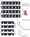
TRF2 over-expression impairs tumor growth and dissemination in advanced orthotopic TNBC models. NSG female immunodeficient mice were orthotopically injected in the mouse mammary gland with 1x10⁶ MDA-MB-231-LUC pBabe and pTRF2 cells. When tumors were palpable (day 10 after cells injection), mice were randomized and treated intravenously with vehicle, or Paclitaxel iv at 20 μg/mouse once a week for two weeks. At the end of the treatment (15 days post treatment) mice were analyzed by the IVIS imaging system 200 series and bioluminescence signals were determined by the number of photons analyzed using the Living image software version 4.3 (PerkinElmer). a Representative pictures of six mice. b Quantitative analyses of bioluminescence signals were shown. Luminescent signals, relative to photons at the beginning of treatment, are expressed as mean of total flux of photons/sec/cm2/steradian (p/s/cm2/sr). Each experimental group included n=8 mice. Error bars represent SEM. P values were calculated using an unpaired two-tailed t-test, ***p<0.001. c,d Fifteen days post treatment, primary tumors were surgically resected and mice treated with Paclitaxel were monitored for lung metastasis appearance by IVIS imaging system 200 series. Representative pictures of six mice, at day 21 post tumor resection c and quantitative analysis of lung metastasis photons d were shown. P values were calculated using an unpaired two-tailed t-test, *p<0.05. e Kaplan-Meier disease free survival curve of mice as in C, with each experimental group included n=8 mice. Statistical significance was assessed by Log-rank test (**p=0.0046)
Fig. 5
Fig. 5
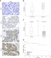
High TRF2 levels in TNBC patients correlate with a better response to neoadjuvant taxane-based therapy. a Representative IHC images of TRF2 score in tumor specimens retrieved from TNBC patients before neoadjuvant therapy. Scale bar: 30 µm. b TRF2-IRS distribution by patients’ response to taxane-based neoadjuvant therapy. Responders showed TRF2 IRS values significantly higher than no responders (Mann-Whitney test, p=0.010). c Analysis of TRF2 IRS value pre- and post- neoadjuvant taxane-based therapy. Post treatment TRF2-IRS values were significantly lower than pre-treatment (Wilcoxon test, p=0.005). d Kaplan-Meier EFS curves of TNBC patients subjected to neoadjuvant taxane-based therapy. Patients with TRF2 IRSHigh had a longer time without disease recurrence than those with TRF2Low (TRF2 IRSHigh median time not estimable vs TRF2Low median time 37 months 95%CI [4.7- 49.3], Log-rank test, p=0.005).

References

    1. Sung H, Ferlay J, Siegel RL, Laversanne M, Soerjomataram I, Jemal A, et al. Global Cancer Statistics 2020: GLOBOCAN Estimates of Incidence and Mortality Worldwide for 36 Cancers in 185 Countries. CA Cancer J Clin. 2021;71:209–49. doi: 10.3322/caac.21660. - DOI - PubMed
    1. Yin L, Duan J-J, Bian X-W, Yu S-C. Triple-negative breast cancer molecular subtyping and treatment progress. Breast Cancer Res. 2020;22:61. doi: 10.1186/s13058-020-01296-5. - DOI - PMC - PubMed
    1. Son J, Lee SE, Kim E-K, Kim S. Prediction of breast cancer molecular subtypes using radiomics signatures of synthetic mammography from digital breast tomosynthesis. Sci Rep. 2020;10:21566. doi: 10.1038/s41598-020-78681-9. - DOI - PMC - PubMed
    1. Bergin ART. Loi S. Triple-negative breast cancer: recent treatment advances. F1000Res. 2019;8:1342. doi: 10.12688/f1000research.18888.1. - DOI - PMC - PubMed
    1. Kwapisz D. Pembrolizumab and atezolizumab in triple-negative breast cancer. Cancer Immunol Immunother. 2021;70:607–17. doi: 10.1007/s00262-020-02736-z. - DOI - PMC - PubMed

MeSH terms