Structural brain abnormalities in Pallister-Killian syndrome: a neuroimaging study of 31 children
- PMID: 38459574
- PMCID: PMC10921669
- DOI: 10.1186/s13023-024-03065-5
Structural brain abnormalities in Pallister-Killian syndrome: a neuroimaging study of 31 children
Abstract
Background: Pallister-Killian syndrome (PKS) is a rare genetic disorder caused by mosaic tetrasomy of 12p with wide neurological involvement. Intellectual disability, developmental delay, behavioral problems, epilepsy, sleep disturbances, and brain malformations have been described in most individuals, with a broad phenotypic spectrum. This observational study, conducted through brain MRI scan analysis on a cohort of patients with genetically confirmed PKS, aims to systematically investigate the neuroradiological features of this syndrome and identify the possible existence of a typical pattern. Moreover, a literature review differentiating the different types of neuroimaging data was conducted for comparison with our population.
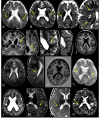
Results: Thirty-one individuals were enrolled (17 females/14 males; age range 0.1-17.5 years old at first MRI). An experienced pediatric neuroradiologist reviewed brain MRIs, blindly to clinical data. Brain abnormalities were observed in all but one individual (compared to the 34% frequency found in the literature review). Corpus callosum abnormalities were found in 20/30 (67%) patients: 6 had callosal hypoplasia; 8 had global hypoplasia with hypoplastic splenium; 4 had only hypoplastic splenium; and 2 had a thin corpus callosum. Cerebral hypoplasia/atrophy was found in 23/31 (74%) and ventriculomegaly in 20/31 (65%). Other frequent features were the enlargement of the cisterna magna in 15/30 (50%) and polymicrogyria in 14/29 (48%). Conversely, the frequency of the latter was found to be 4% from the literature review. Notably, in our population, polymicrogyria was in the perisylvian area in all 14 cases, and it was bilateral in 10/14.
Conclusions: Brain abnormalities are very common in PKS and occur much more frequently than previously reported. Bilateral perisylvian polymicrogyria was a main aspect of our population. Our findings provide an additional tool for early diagnosis.Further studies to investigate the possible correlations with both genotype and phenotype may help to define the etiopathogenesis of the neurologic phenotype of this syndrome.
© 2024. The Author(s).
Conflict of interest statement
The authors declare no competing interests.
Figures


References
-
- Wilkens A, Liu H, Park K, Campbell LB, Jackson M, Kostanecka A et al. Novel clinical manifestations in Pallister-Killian syndrome: Comprehensive evaluation of 59 affected individuals and review of previously reported cases. Am J Med Genet A [Internet]. 2012;158 A:3002–17. 10.1002/ajmg.a.35722. - PubMed
-
- Izumi K, Krantz ID. Pallister-Killian syndrome., Am J, Med Genet C. Semin Med Genet [Internet]. 2014 [cited 2021 Jun 30];166:406–13. Available from: https://onlinelibrary.wiley.com/doi/full/10.1002/ajmg.c.31423. - DOI - PubMed
-
- Izumi K, Conlin LK, Berrodin D, Fincher C, Wilkens A, Haldeman-Englert C, et al. Duplication 12p and Pallister-Killian syndrome: a case report and review of the literature toward defining a Pallister-Killian syndrome minimal critical region. Am J Med Genet A. 2012;158 A:3033–45. doi: 10.1002/ajmg.a.35500. - DOI - PubMed
Publication types
MeSH terms
Supplementary concepts
Grants and funding
LinkOut - more resources
Full Text Sources
Medical
Miscellaneous

