

Primary extraskeletal intradural Ewing sarcoma with acute hemorrhage: a case report and review of the literature
- PMID: 38459600
- PMCID: PMC10924417
- DOI: 10.1186/s13256-024-04384-8
Primary extraskeletal intradural Ewing sarcoma with acute hemorrhage: a case report and review of the literature
Abstract
Background: Spinal cord tumors present a challenge in diagnosis and treatment due to their varied histopathological characteristics. While Ewing sarcoma is a rare malignant tumor typically originating from skeletal bone, cases of primary intradural extraskeletal Ewing sarcoma are exceptionally rare. The similarity of its presentation to other spinal tumors further complicates its identification and management.
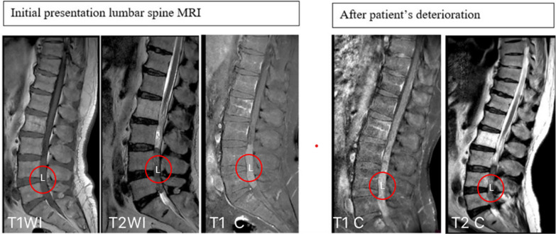
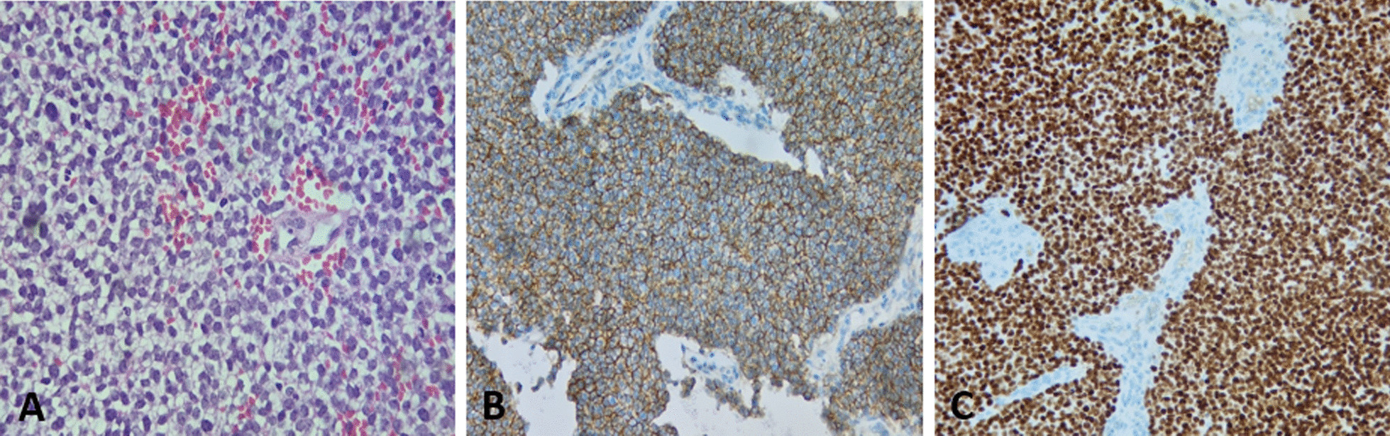
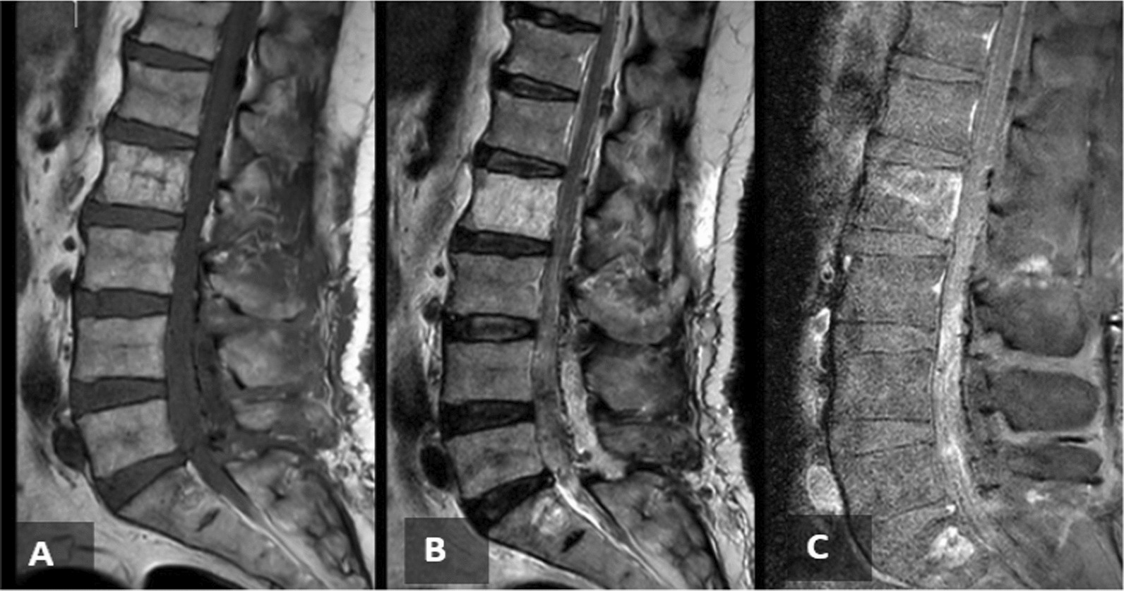
Case presentation: We report a case of a 58-year-old Palestinian male with intradural extraskeletal lumbar Ewing sarcoma. The patient initially presented with lower back pain and bilateral S1 radiculopathy, with more severe symptoms on the left side. Magnetic resonance imaging revealed a 7 cm oval-shaped mass with homogeneous contrast enhancement, obstructing the spinal canal from L3/L4 to L5/S1 levels. Initially, a myxopapillary ependymoma was suspected, but the patient's sensory and motor functions suddenly deteriorated during hospitalization. Repeat magnetic resonance imaging indicated heterogeneous contrast enhancement, indicating acute intratumoral hemorrhage. Consequently, the patient underwent emergent L3-L5 laminotomy, with successful gross total resection of the tumor. Histopathological and immunohistochemical analyses confirmed the diagnosis of intradural extraskeletal Ewing sarcoma. Adjuvant therapy was administered to minimize the risk of local recurrence or distant metastasis. A systematic review of relevant literature, along with retrospective analysis of medical records, operative reports, radiological studies, and histopathological findings of similar cases, was also conducted.
Conclusions: Intradural extraskeletal Ewing sarcoma is an infrequently encountered condition in adult patients, emphasizing the importance of considering it in the differential diagnosis of spinal tumors. Surgeons must possess a comprehensive understanding of this rare entity to ensure accurate staging and optimal management, particularly in the early stages when prompt intervention may improve prognosis.
Keywords: Chemotherapy; Ewing sarcoma; Extra-skeletal; Intradural; Intratumoral hemorrhage; Radiotherapy; Tumor.
© 2024. The Author(s).
Conflict of interest statement
The authors declare that they have no competing interests.
Figures
Similar articles
-
Primary spinal multifocal intradural-extramedullary Ewing sarcoma in children: presentation of a case and review of the literature.Turk J Pediatr. 2021;63(6):1084-1090. doi: 10.24953/turkjped.2021.06.018. Turk J Pediatr. 2021. PMID: 35023660 Review.
-
Primary spinal intradural extraskeletal Ewing sarcoma mimicking a giant nerve sheath tumor: case report and review of the literature.Int J Clin Exp Pathol. 2014 Dec 1;7(12):9081-5. eCollection 2014. Int J Clin Exp Pathol. 2014. PMID: 25674292 Free PMC article. Review.
-
Intradural extramedullary Ewing's sarcoma: A case report and review of the literature.Neurol Neurochir Pol. 2017 Jan-Feb;51(1):106-110. doi: 10.1016/j.pjnns.2016.11.006. Epub 2016 Nov 30. Neurol Neurochir Pol. 2017. PMID: 27939398 Review.
-
Histologic Features and Prognosis of Spinal Intradural Extramedullary Ewing Sarcoma: Case Report, Literature Review, and Analysis of Prognosis.World Neurosurg. 2018 Jul;115:448-452.e2. doi: 10.1016/j.wneu.2018.04.015. Epub 2018 Apr 11. World Neurosurg. 2018. PMID: 29654955 Review.
-
Primary intradural extraosseous Ewing sarcoma of the spine: case report and literature review.Neurosurgery. 2011 Oct;69(4):E995-9. doi: 10.1227/NEU.0b013e318223b7c7. Neurosurgery. 2011. PMID: 21572359 Review.
References
-
- Ewing J. Diffuse endothelioma of bone. Proc N Y Pathol Soc. 1921;21:17–24.
Publication types
MeSH terms
LinkOut - more resources
Full Text Sources
