. 2024 Jun;23(6):e14133.
doi: 10.1111/acel.14133. Epub 2024 Mar 8.

Chronic TNF exposure induces glucocorticoid-like immunosuppression in the alveolar macrophages of aged mice that enhances their susceptibility to pneumonia

Affiliations

Chronic TNF exposure induces glucocorticoid-like immunosuppression in the alveolar macrophages of aged mice that enhances their susceptibility to pneumonia

Katherine L Kruckow et al. Aging Cell. 2024 Jun.

Abstract

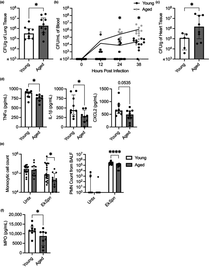
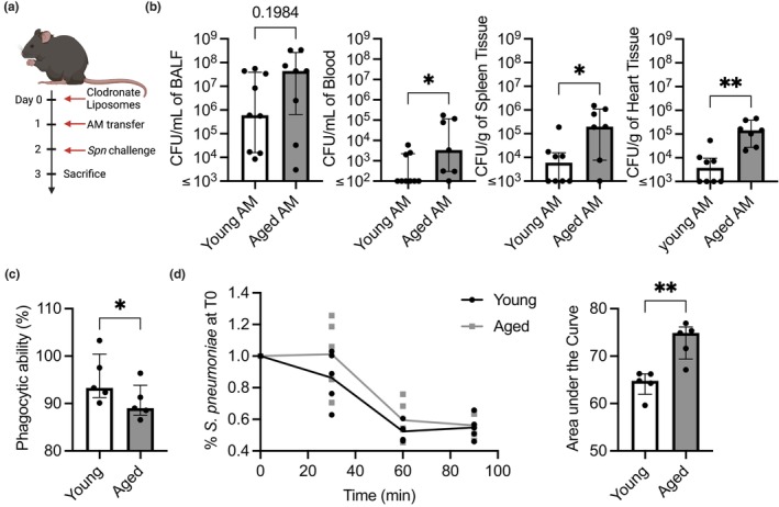
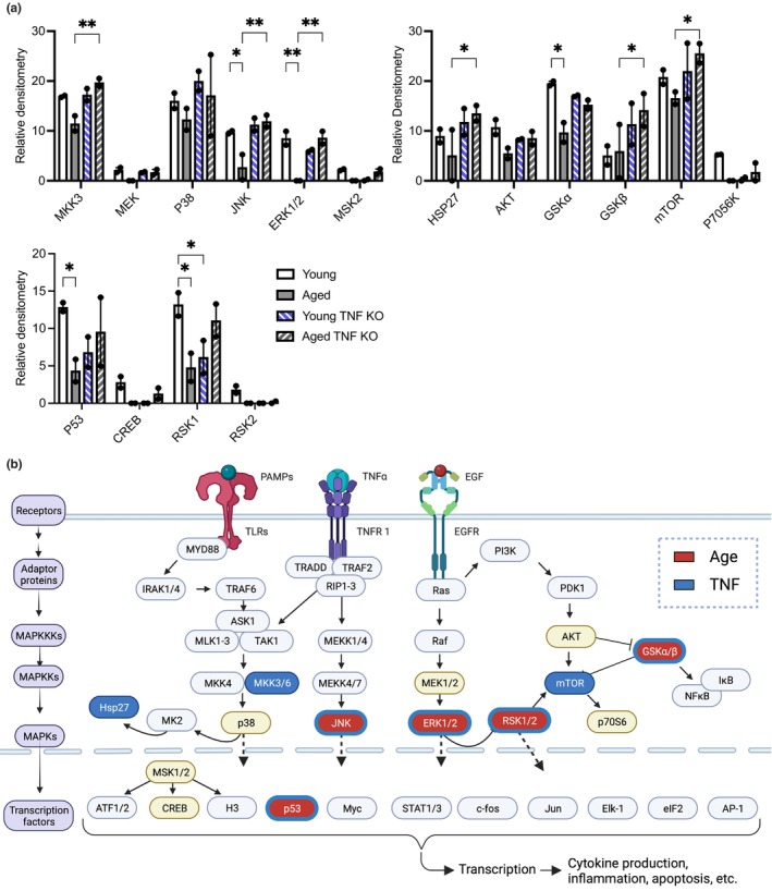
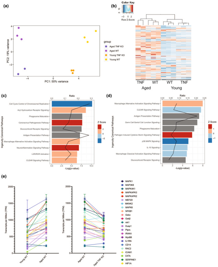
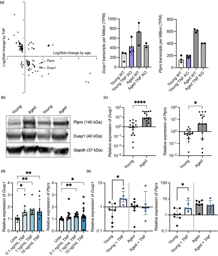
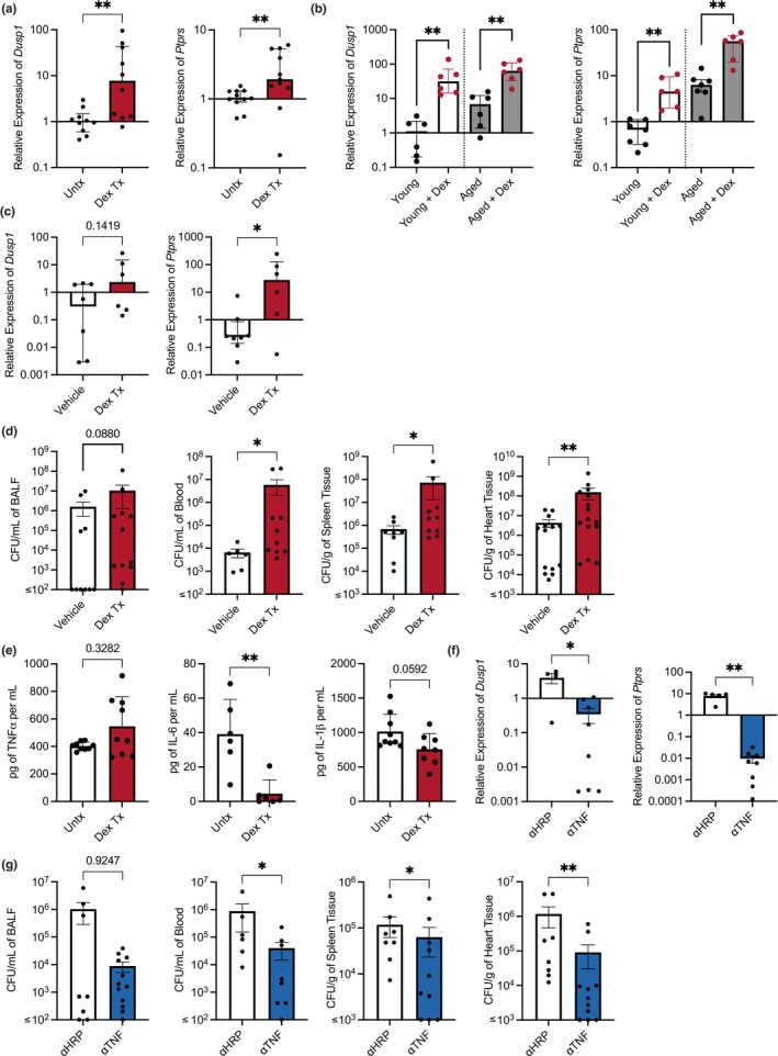
Chronic low-grade inflammation, particularly elevated tumor necrosis factor (TNF) levels, occurs due to advanced age and is associated with greater susceptibility to infection. One reason for this is age-dependent macrophage dysfunction (ADMD). Herein, we use the adoptive transfer of alveolar macrophages (AM) from aged mice into the airway of young mice to show that inherent age-related defects in AM were sufficient to increase the susceptibility to Streptococcus pneumoniae, a Gram-positive bacterium and the leading cause of community-acquired pneumonia. MAPK phosphorylation arrays using AM lysates from young and aged wild-type (WT) and TNF knockout (KO) mice revealed multilevel TNF-mediated suppression of kinase activity in aged mice. RNAseq analyses of AM validated the suppression of MAPK signaling as a consequence of TNF during aging. Two regulatory phosphatases that suppress MAPK signaling, Dusp1 and Ptprs, were confirmed to be upregulated with age and as a result of TNF exposure both ex vivo and in vitro. Dusp1 is known to be responsible for glucocorticoid-mediated immune suppression, and dexamethasone treatment increased Dusp1 and Ptprs expression in cells and recapitulated the ADMD phenotype. In young mice, treatment with dexamethasone increased the levels of Dusp1 and Ptprs and their susceptibility to infection. TNF-neutralizing antibody reduced Dusp1 and Ptprs levels in AM from aged mice and reduced pneumonia severity following bacterial challenge. We conclude that chronic exposure to TNF increases the expression of the glucocorticoid-associated MAPK signaling suppressors, Dusp1 and Ptprs, which inhibits AM activation and increases susceptibility to bacterial pneumonia in older adults.

Keywords: Streptococcus pneumoniae; Dusp1; MAPK; Ptprs; alveolar macrophages; glucocorticoids; inflamm‐aging; pneumonia; tumor necrosis factor.


Conflict of interest statement

We have no conflicts of interest to declare.

Figures

FIGURE 1
FIGURE 1
Aged mice are more susceptible to pneumococcal disease and have defects in their ability to respond to infectious stimuli. Young (3–6 months) and aged (18–24 months) C57BL/6 mice were infected intratracheally with 105 CFU of Spn. Mice were sacrificed at 24 h postinfection. The bacterial burden in the lung was enumerated from serial dilutions of lung homogenates on blood agar plates (a). Tail bleeds were taken throughout the infection to enumerate bacterial burden within the blood (b). The bacterial burden in the heart was enumerated from serial dilutions of heart homogenates on blood agar plates (c). Young (3–6 months old) and aged mice (18–24 months old) were intratracheally inoculated with 108 CFU equivalents of ethanol killed Spn (EkSpn). At 8 h postinoculation, mice were sacrificed and bronchoalveolar lavage fluid (BALF) was collected. Levels of TNFα, IL‐1β, and CXCL2 in BALF as measured by ELISA (d). Monocytes and PMNs within BALF were attached to slides by cytocentrifugation, stained, and enumerated by nuclear morphology analysis (e). Myeloperoxidase levels in BALF was measured (f). Statistical significance was calculated using a nonparametric Mann–Whitney U test (a, c, d, & f), a repeated measures two‐way ANOVA with Fisher's LSD post hoc test (b), a two‐way ANOVA with Bonferroni multiple comparisons post hoc test (e). The data are presented as median with interquartile range (IQR); *p ≤ 0.05; ****p ≤ 0.0001. Each data point represents an individual mouse. Graphs with ≤ on x‐axis indicate limit of detection.
FIGURE 2
FIGURE 2
Age‐dependent macrophage dysfunction is sufficient to increase susceptibility to pneumococcal disease. Young mice depleted of their own alveolar macrophages using clodronate liposomes were adoptively transferred with AM from young or aged mice and subsequently challenged intratracheally with 105 CFU of Spn (a). Following 1 day, the bacterial burden was enumerated from BALF, blood, spleen, and heart homogenates (b). AM isolated from BALF of young and aged mice were used in functional assays testing their ability to phagocytize prelabeled zymosan particles (c), and an ex vivo macrophage killing assay of Spn that was quantified by the area under the curve (d) to compare the functionality of young versus aged macrophages. Statistical significance was calculated using a Mann–Whitney U test (b, c, & d). The data are presented as median with IQR; *p ≤ 0.05; **p ≤ 0.01. Each data point represents an individual mouse (b) or well (c & d). Graphs with ≤ on x‐axis indicate limit of detection.
FIGURE 3
FIGURE 3
Phosphokinase signaling is starkly suppressed in aged AM. AM were collected from young and aged WT and TNF KO mice and inoculated with Spn at an MOI of 25 for 15 min. Purified protein from these samples was used in a phosphorylation array of the MAPK pathway (a) with results overlaid on the MAPK pathway (b). Any protein designated by a colored oval was tested as part of the phosphorylation array, with items in red indicating significant changes due to age and blue indicating alterations due to TNF. Statistical significance was calculated using a two‐way ANOVA with each comparison standing alone using a Fisher's LSD test. The data are presented as median with interquartile range (IQR); *p ≤ 0.05; **p ≤ 0.01. Each individual point represents the average of two technical replicates on the phosphorylation array per biological sample.
FIGURE 4
FIGURE 4
AM have age and TNF‐dependent differences in gene expression. RNA was purified from AM collected from young (3–6 months old) and aged mice (18–24 months old) in both wild‐type C57BL/6 and TNF KO mice. Alterations in gene expression due to age and TNF expression were visualized using a PCA plot (a) and a dendrogram (b). IPA was used to show the 10 most highly scoring canonical pathways (according to p value) altered in expression due to aging (c) and due to genotype (d) by using comparisons of aged WT versus young WT and Aged TNF KO vs Aged WT, respectively. The red line represents the ratio of the number of differentially expressed genes within a pathway divided by the total number of genes within the pathway. Bars represent the p‐value for each pathway expressed as −1 times the log of the p‐value, with the color of the bar indicating the Z score. Relative expression of various members of the MAPK signaling cascade visualized in transcripts per million by group (e).
FIGURE 5
FIGURE 5
Key negative regulators are upregulated in a TNF‐dependent manner with age. Identification of Dusp1 and Ptprs in DEGs as a result of TNF status and age from the transcriptomic dataset (a). Expression levels of negative regulators Dusp1 and Ptprs from alveolar macrophages are shown in Transcripts per Million (a), protein levels of Dusp1 and Ptprs in whole lung cell lysates via western blots (b), and in isolated AM by qPCR (c). Levels of negative regulators Dusp1 and Ptprs in J774.1 cells treated with various levels of TNF via qPCR (d). Levels of negative regulators Dusp1 and Ptprs in AM from young and aged mice collected and exposed to TNF ex vivo (e). Statistical test was done using either a Kruskal–Wallis (d & e) or a Mann–Whitney test (c). The data are presented as median with interquartile range (IQR); *p ≤ 0.05; **p ≤ 0.001; ****p ≤ 0.0001. Each data point represents an individual gene (a), mouse (b & c), or well (e).
FIGURE 6
FIGURE 6
Manipulating the levels of negative regulators alters responses to infection. RNA was isolated from J774.1 macrophages that were treated with dexamethasone, and the levels of Dusp1 and Ptprs were quantified with qPCR (a). The levels of Dusp1 and Ptprs were quantified with qPCR on alveolar macrophages from young and aged mice treated with dexamethasone ex vivo (b) as well as in vivo (c). We quantified the levels of pro‐inflammatory cytokines produced by J774.1 cells treated with dexamethasone overnight and then challenged with EkSpn (e). Young (3–6 months) C57BL/6 mice were treated with dexamethasone and infected intratracheally with 105 CFU of Spn. Bacterial burden 24 h postinfection, in the BALF, blood, spleen, and heart was enumerated (d). C57BL/6 mice were treated every other day with anti‐TNF or anti‐HRP control antibodies for 2 weeks before being infected intratracheally with 105 CFU of Spn. Mice were sacrificed 24 h postinfection, the heart and spleens were harvested, and blood and BALF were collected. RNA was isolated from alveolar macrophages from mice that were treated with anti‐TNF or anti‐HRP control antibodies and were quantified for levels of negative regulators (f). Bacterial burden in mice treated with anti‐TNF was enumerated in the BALF, blood, spleen, and heart (g). The data are presented as median with interquartile range (IQR); *p ≤ 0.05; **p ≤ 0.01. Each data point represents an individual mouse (b, d, e, f, & g) or well (a & c). Graphs with ≤ on x‐axis indicate limit of detection.

Similar articles

Cited by

References

    1. Abraham, S. M. , Lawrence, T. , Kleiman, A. , Warden, P. , Medghalchi, M. , Tuckermann, J. , Saklatvala, J. , & Clark, A. R. (2006). Antiinflammatory effects of dexamethasone are partly dependent on induction of dual specificity phosphatase 1. Journal of Experimental Medicine, 203(8), 1883–1889. - PMC - PubMed
    1. Adrover, J. M. , Nicolás‐Ávila, J. A. , & Hidalgo, A. (2016). Aging: A temporal dimension for neutrophils. Trends in Immunology, 37(5), 334–345. - PubMed
    1. Allard, B. , Panariti, A. , & Martin, J. G. (2018). Alveolar macrophages in the resolution of inflammation, tissue repair, and tolerance to infection. Frontiers in Immunology, 9, 1777. - PMC - PubMed
    1. Anders, S. , Pyl, P. T. , & Huber, W. (2015). HTSeq—A python framework to work with high‐throughput sequencing data. Bioinformatics, 31(2), 166–169. - PMC - PubMed
    1. Bae, E. , Cha, R. H. , Kim, Y. C. , An, J. N. , Kim, D. K. , Yoo, K. D. , Lee, S. M. , Kim, M. H. , Park, J. T. , Kang, S. W. , Park, J. Y. , Lim, C. S. , Kim, Y. S. , Yang, S. H. , & Lee, J. P. (2017). Circulating TNF receptors predict cardiovascular disease in patients with chronic kidney disease. Medicine (Baltimore), 96(19), e6666. - PMC - PubMed

Publication types

Substances