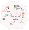
Pleural mesothelioma (PMe): The evolving molecular knowledge of a rare and aggressive cancer
- PMID: 38459714
- PMCID: PMC10994233
- DOI: 10.1002/1878-0261.13591
Pleural mesothelioma (PMe): The evolving molecular knowledge of a rare and aggressive cancer
Abstract
Mesothelioma is a type of late-onset cancer that develops in cells covering the outer surface of organs. Although it can affect the peritoneum, heart, or testicles, it mainly targets the lining of the lungs, making pleural mesothelioma (PMe) the most common and widely studied mesothelioma type. PMe is caused by exposure to fibres of asbestos, which when inhaled leads to inflammation and scarring of the pleura. Despite the ban on asbestos by most Western countries, the incidence of PMe is on the rise, also facilitated by a lack of specific symptomatology and diagnostic methods. Therapeutic options are also limited to mainly palliative care, making this disease untreatable. Here we present an overview of biological aspects underlying PMe by listing genetic and molecular mechanisms behind its onset, aggressive nature, and fast-paced progression. To this end, we report on the role of deubiquitinase BRCA1-associated protein-1 (BAP1), a tumour suppressor gene with a widely acknowledged role in the corrupted signalling and metabolism of PMe. This review aims to enhance our understanding of this devastating malignancy and propel efforts for its investigation.
Keywords: BAP1 and therapy; asbestos; mesothelioma.
© 2024 The Authors. Molecular Oncology published by John Wiley & Sons Ltd on behalf ofFederation of European Biochemical Societies.
Conflict of interest statement
The authors declare no conflicts of interest.
Figures


References
-
- Mutsaers SE, Herrick SE. Mesothelial cells. In: Geoffrey JL, Steven DS, editors. Encyclopedia of Respiratory Medicine, Four‐Volume Set. Oxford: Academic Press; 2006. p. 47–52.
-
- Valenciano AC, Rizzi TE. Abdominal, thoracic, and pericardial effusions. In: Valenciano AC, Cowell RL, editors. Cowell and Tyler's Diagnostic Cytology and Hematology of the Dog and Cat. St. Louis, MO: Elsevier Mosby; 2020. p. 229–246.
-
- Robinson BWS, Lake RA. Advances in malignant mesothelioma. N Engl J Med. 2005;353:1591–1603. - PubMed
Publication types
MeSH terms
Substances
Grants and funding
LinkOut - more resources
Full Text Sources
Medical
Research Materials
Miscellaneous

