. 2024 Jun;26(6):101117.
doi: 10.1016/j.gim.2024.101117. Epub 2024 Mar 6.

Alternative splicing expands the clinical spectrum of NDUFS6-related mitochondrial disorders

Affiliations

Alternative splicing expands the clinical spectrum of NDUFS6-related mitochondrial disorders

Camila Armirola-Ricaurte et al. Genet Med. 2024 Jun.

Abstract

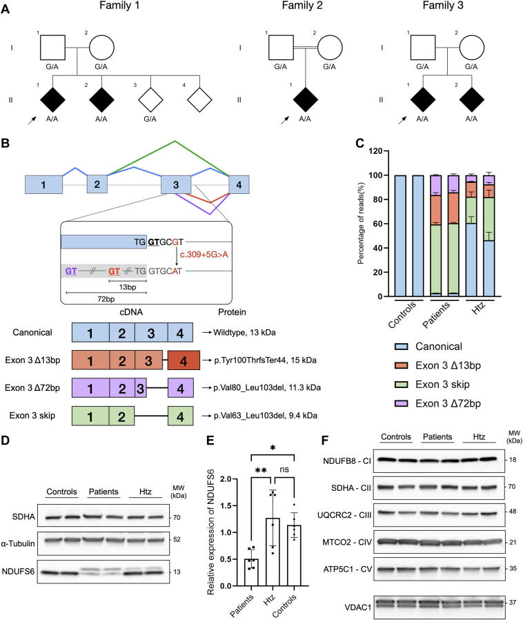
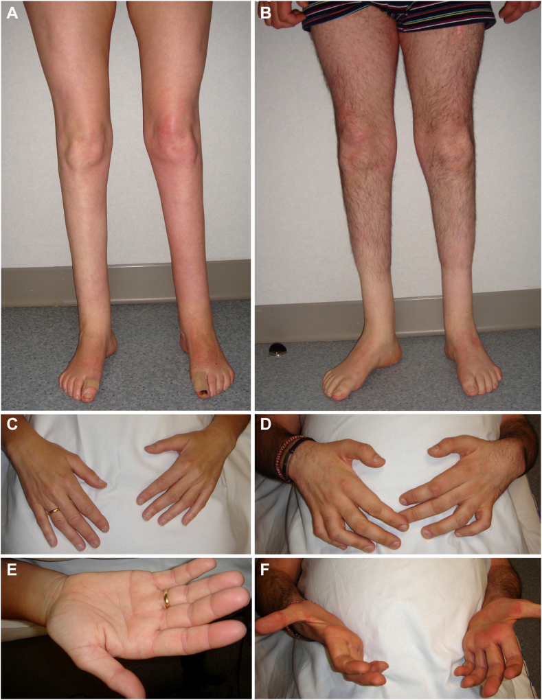
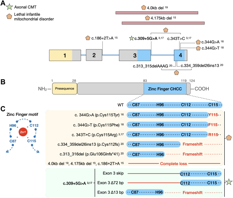
Purpose: We describe 3 families with Charcot-Marie-Tooth neuropathy (CMT), harboring a homozygous NDUFS6 NM_004553.6:c.309+5G>A variant previously linked to fatal Leigh syndrome. We aimed to characterize clinically and molecularly the newly identified patients and understand the mechanism underlying their milder phenotype.

Methods: The patients underwent extensive clinical examinations. Exome sequencing was done in 4 affected individuals. The functional effect of the c.309+5G>A variant was investigated in patient-derived EBV-transformed lymphoblasts at the complementary DNA, protein, and mitochondrial level. Alternative splicing was evaluated using complementary DNA long-read sequencing.

Results: All patients presented with early-onset, slowly progressive axonal CMT, and nystagmus; some exhibited additional central nervous system symptoms. The c.309+5G>A substitution caused the expression of aberrantly spliced transcripts and negligible levels of the canonical transcript. Immunoblotting showed reduced levels of mutant isoforms. No detectable defects in mitochondrial complex stability or bioenergetics were found.

Conclusion: We expand the clinical spectrum of NDUFS6-related mitochondrial disorders to include axonal CMT, emphasizing the clinical and pathophysiologic overlap between these 2 clinical entities. This work demonstrates the critical role that alternative splicing may play in modulating the severity of a genetic disorder, emphasizing the need for careful consideration when interpreting splice variants and their implications on disease prognosis.

Keywords: Charcot-Marie-Tooth; Mitochondrial disorders; NDUFS6; Peripheral neuropathy; Splicing.


Conflict of interest statement

Conflict of Interest The authors declare no conflicts of interest.

Figures

Figure 1
Figure 1
Genetic and functional analysis of the NDUFS6 c.309+5G>A splice variant. A. Segregation analysis of NDUFS6 c.309+5G>A in families 1-3. Black symbols indicate affected individuals. A double line indicates reported consanguinity. Index patients are indicated with a black arrowhead. B. Top panel: NDUFS6 (NM_004553.6) is schematically shown with exons 1-4 (blue rectangles) and introns (gray line). Caret-like exon connecting lines depict canonical (blue), exon 3 skip (green), exon 3 Δ72bp (purple), and exon 3 Δ13bp (red) splicing events. The 3’-end of exon 3 and intron 3 are presented in more detail to indicate the location of the NDUFS6 c.309+5G>A splice variant (red), the canonical splice-donor site (black and underlined) and the cryptic splice-donor sites (red and purple underlined) located at 13 bp and 72 bp upstream of the 3’-end of exon 3, respectively. Bottom panel: schematic representation of the NDUFS6 transcripts detected in the patients and parents from family 1. The identity of each transcript is indicated on the left, the impact of each splicing on the protein is indicated on the right. C. The relative quantification of NDUFS6 transcripts sequenced by cDNA T-LRS among the 3 genotypes of interest (heterozygotes, patients, and controls). The chart represents the percentage of reads for each transcript quantified relative to the total amount of reads per sample. The bars indicate the standard error of the mean (n = 2). D. Western blot analysis of total protein lysates isolated from lymphoblasts of heterozygotes, patients, and controls with a monoclonal rabbit anti-human NDUFS6 antibody. Monoclonal mouse anti-SDHA and anti-α-tubulin were used as controls for equal loading of mitochondrial and cytosolic fractions, respectively. E. Relative quantification of different NDUFS6 isoforms present in lymphoblasts of heterozygotes, patients, and controls. NDUFS6 band intensity is relative to α-tubulin and normalized to controls. Bar charts are represented as the standard error of the mean (n = 3 for each genotype, with 2 biological replicates for each genotype). F. Immunoblotting of mitochondrial fractions from 3 genotypes of interest (heterozygotes, patients, and controls) with monoclonal mouse anti-human NDUFB8, SDHA, UQCR2, and ATP5C1 and monoclonal rabbit anti-human MTCO2 antibodies to target 1 subunit from each OXPHOS complex. Monoclonal mouse anti-human VDAC1 was used as a control for mitochondrial proteins. All statistical analyses were performed using one-way ANOVA. Asterisks denote significance after Tukey’s multiple comparison correction. Abbreviations: Htz, heterozygotes; ∗∗P < .01, ∗P < .05; ns, not significant; SDHA, succinate dehydrogenase complex subunit A.
Figure 2
Figure 2
Clinical features of the patients from family 1. The proband, II-1, at age 46 years, exhibited marked lower leg amyotrophy (A), and incipient hand wasting involving first dorsal interossei and thenar musculature (C and E). The affected sibling II-2, at age 38 years, shows a more pronounced phenotype, with marked lower leg amyotrophy (B) and advanced wasting of hand musculature with clawing deformity (D, C, and F). Note the absence of proximal lower-limb amyotrophy in both patients (A and B).
Figure 3
Figure 3
Clinical and molecular effect of published NDUFS6 variants. A. Gene structure of NDUFS6 (NM_004553.6) with exons 1 to 4 and introns (blue line). The exons are colored according to the protein domain they encode. The location of the pathogenic variants reported in the literature is indicated on the diagram and their superscripts correspond to their number on the reference list. Symbols represent their associated clinical phenotype, as described in the legend on the left top of the figure. A dotted line in between 2 variants indicates reported compound heterozygosity. The pink elongated rectangle indicates a deletion. B. The protein domains of NDUFS6. (C) Left panel: Visual representation of the zinc-finger motif (zf-CHCC). The bound Zinc atom is represented as a red circle. Right panel: Simplified amino acid sequence of the region corresponding to the zinc-finger motif. Below the sequence of wild-type NDUFS6, the previously reported NDUFS6 variants and their consequence on the motif are shown and shaded in yellow. The effect of the aberrantly spliced transcripts found in the patients carrying the c.309+5G>A variant is shaded in green. Red residues indicate an altered amino acid sequence. Red lines indicate regions lost due to the variants. Symbols represent their associated clinical phenotype, as described in the legend on the top left panel.

Similar articles

Cited by

References

    1. Baertling F., Sánchez-Caballero L., van den Brand M.A.M., et al. NDUFA9 point mutations cause a variable mitochondrial complex I assembly defect. Clin Genet. 2018;93(1):111–118. doi: 10.1111/cge.13089. - DOI - PubMed
    1. Mimaki M., Wang X., McKenzie M., Thorburn D.R., Ryan M.T. Understanding mitochondrial complex I assembly in health and disease. Biochim Biophys Acta. 2012;1817(6):851–862. doi: 10.1016/j.bbabio.2011.08.010. - DOI - PubMed
    1. Rouzier C., Chaussenot A., Fragaki K., et al. NDUFS6 related Leigh syndrome: a case report and review of the literature. J Hum Genet. 2019;64(7):637–645. doi: 10.1038/s10038-019-0594-4. - DOI - PubMed
    1. Kmita K., Wirth C., Warnau J., et al. Accessory NUMM (NDUFS6) subunit harbors a Zn-binding site and is essential for biogenesis of mitochondrial complex I. Proc Natl Acad Sci U S A. 2015;112(18):5685–5690. doi: 10.1073/pnas.1424353112. - DOI - PMC - PubMed
    1. Fiedorczuk K., Sazanov L.A. Mammalian mitochondrial Complex I structure and disease-causing mutations. Trends Cell Biol. 2018;28(10):835–867. doi: 10.1016/j.tcb.2018.06.006. - DOI - PubMed

Publication types

MeSH terms