. 2024 Apr;300(4):107145.
doi: 10.1016/j.jbc.2024.107145. Epub 2024 Mar 7.

Purinergic signaling promotes premature senescence

Affiliations

Purinergic signaling promotes premature senescence

Daniela Volonte et al. J Biol Chem. 2024 Apr.

Abstract

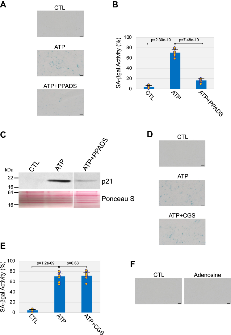
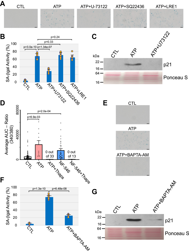
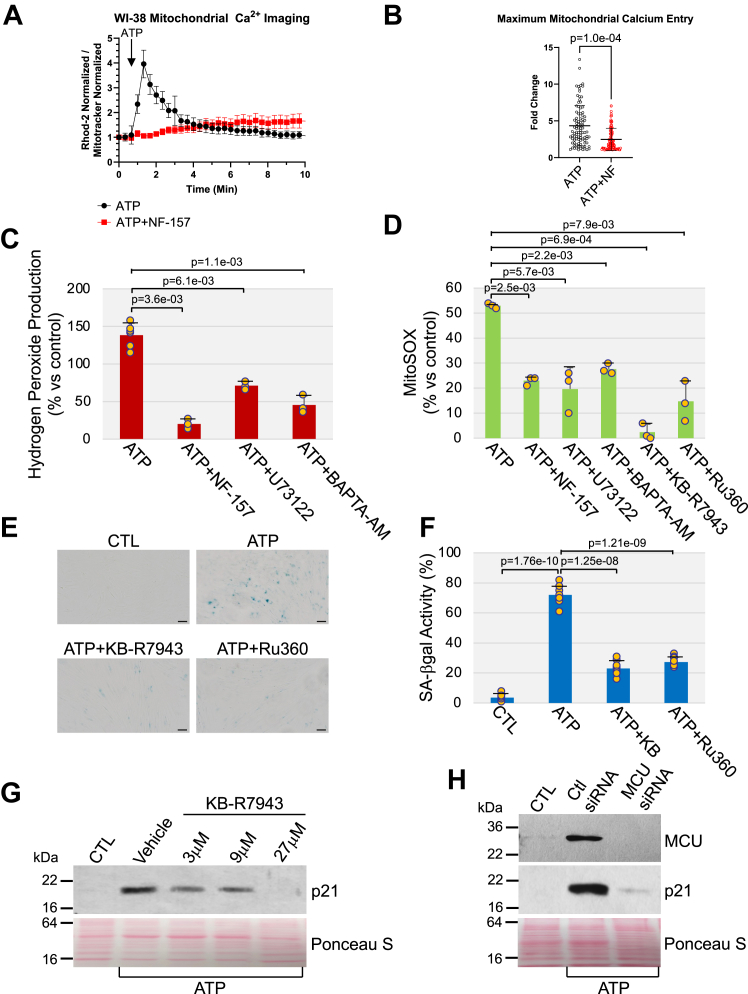
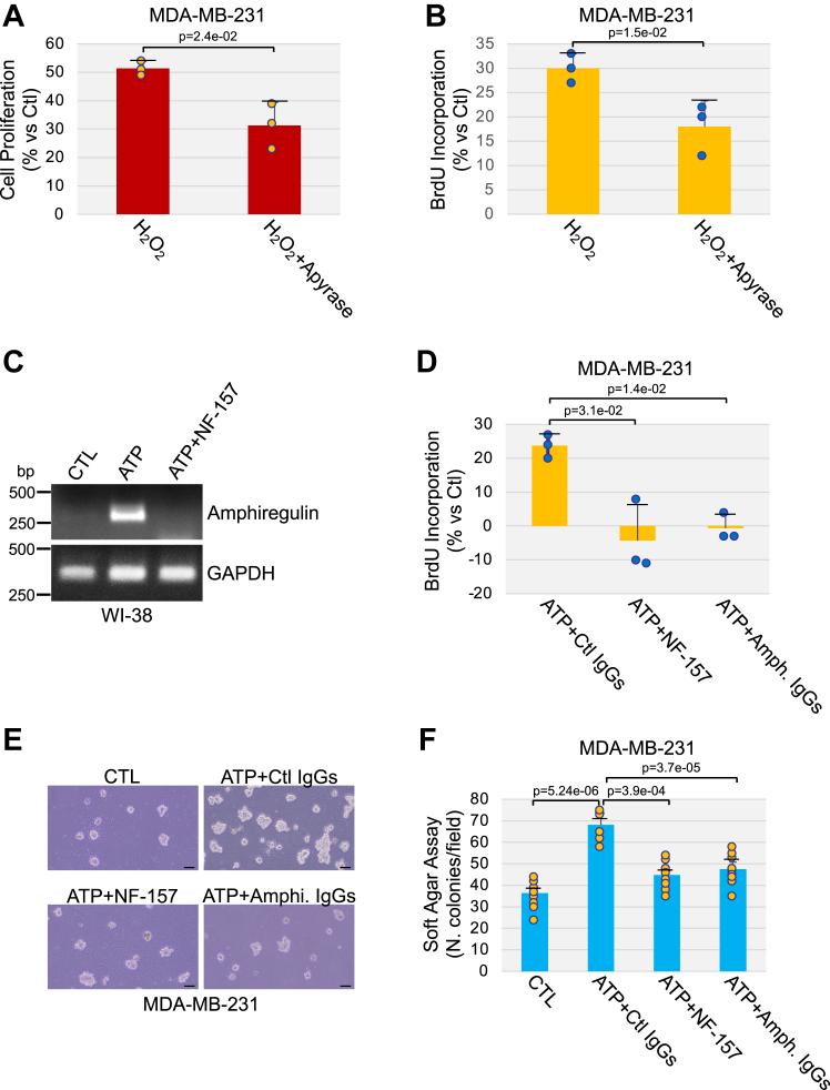
Extracellular ATP activates P2 purinergic receptors. Whether purinergic signaling is functionally coupled to cellular senescence is largely unknown. We find that oxidative stress induced release of ATP and caused senescence in human lung fibroblasts. Inhibition of P2 receptors limited oxidative stress-induced senescence, while stimulation with exogenous ATP promoted premature senescence. Pharmacological inhibition of P2Y11 receptor (P2Y11R) inhibited premature senescence induced by either oxidative stress or ATP, while stimulation with a P2Y11R agonist was sufficient to induce cellular senescence. Our data show that both extracellular ATP and a P2Y11R agonist induced calcium (Ca++) release from the endoplasmic reticulum (ER) and that either inhibition of phospholipase C or intracellular Ca++ chelation impaired ATP-induced senescence. We also find that Ca++ that was released from the ER, following ATP-mediated activation of phospholipase C, entered mitochondria in a manner dependent on P2Y11R activation. Once in mitochondria, excessive Ca++ promoted the production of reactive oxygen species in a P2Y11R-dependent fashion, which drove development of premature senescence of lung fibroblasts. Finally, we show that conditioned medium derived from senescent lung fibroblasts, which were induced to senesce through the activation of ATP/P2Y11R-mediated signaling, promoted the proliferation of triple-negative breast cancer cells and their tumorigenic potential by secreting amphiregulin. Our study identifies the existence of a novel purinergic signaling pathway that links extracellular ATP to the development of a protumorigenic premature senescent phenotype in lung fibroblasts that is dependent on P2Y11R activation and ER-to-mitochondria calcium signaling.

Keywords: ATP; purinergic signaling; reactive oxygen species; senescence; triple negative breast cancer cells.


Conflict of interest statement

Conflict of interest The authors declare that they have no conflicts of interest with the contents of this article.

Figures

Figure 1
Figure 1
Release of extracellular ATP mediates oxidative stress-induced premature senescence in human fibroblasts through P2 receptor activation. WI-38 human diploid fibroblasts were treated with sublethal hydrogen peroxide (450 μM H2O2 for 2 h). Cells were washed with PBS and recovered in complete medium for 10 days. Untreated cells were used as control. A and B, cells were stained to detect senescence-associated β-galactosidase activity. Representative images are shown in (A), quantification is shown in (B). The percentage of cells possessing enlarged and flat morphology [senescence-associated (SA) cell morphology] is shown in (C). D, cells were collected, and cell lysates were subjected to immunoblot analysis using protein-specific antibody probes. Ponceau S staining shows equal total protein loading. E, cell proliferation was quantified by BrdU incorporation assay. F, the level of extracellular ATP was quantified in the conditioned medium using an ATP bioluminescent assay kit. G and H, cells were treated with 450 μM H2O2 for 2 h and recovered in complete medium for 10 days in the presence of either 4 U/ml apyrase (G), 500 μM PPADS (H), or 5 μM CGS 15943 (H). Untreated cells served as control. Quantification of senescence-associated β-galactosidase activity is shown. Values in B, C, and (EH) represent means ± SD; statistical comparisons were made using the student’s t test. The scale bar represents 50 μm. BrdU, bromodeoxyuridine; CGS 15943, 9-chloro-2-(2-furanyl)-[1,2,4]triazolo[1,5-c]quinazolin-5-amine; H2O2, hydrogen peroxide; PPADS, pyridoxal-phosphate-6-azophenyl-2′,4′-disulfonic acid tetrasodium salt.
Figure 2
Figure 2
Stimulation with ATP is sufficient to promote premature senescence in human fibroblasts. Human diploid WI-38 fibroblasts were treated with 1.5 mM ATP for 10 days. Untreated cells were used as control. A and B, cells were subjected to senescence-associated β-galactosidase activity staining. Representative images are shown in (A), quantification is shown in (B). The percentage of cells possessing enlarged and flat morphology [senescence-associated (SA) cell morphology] is shown in (C). D, cells were collected, and cell lysates were subjected to immunoblot analysis using antibody probes specific for p21, phospho-p53, p16, and γ-H2A.X. Ponceau S staining shows equal total protein loading. EG, WI-38 cells were treated with either 400 μM ATP-γ-S or 400 μM ARL 67156 for 10 days. Untreated cells served as control. E and F, cells were stained to detect senescence-associated β-galactosidase activity. Representative images are shown in (E), quantification is shown in (F). G, the expression level of the senescence marker p21 was quantified by immunoblotting analysis using a p21-specific antibody probe. Ponceau S staining shows equal total protein loading. Values in B, C, and F represent means ± SD; statistical comparisons were made using the student’s t test. The scale bar represents 50 μm.
Figure 3
Figure 3
Activation of P2 receptors mediates premature senescence induced by ATP.AC, WI-38 cells were treated with 1.5 mM ATP for 10 days in the presence or absence of the nonspecific P2R antagonist PPADS (500 μM). Untreated cells were used as control. Cells were stained to detect senescence-associated β-galactosidase activity. Representative images are shown in (A), quantification is shown in (B). C, cells were collected and cell lysates were subjected to immunoblot analysis using an antibody probe specific for the senescence marker p21. Ponceau S staining shows equal total protein loading. D and E, WI-38 human diploid fibroblasts were treated with 1.5 mM ATP for 10 days in the presence or absence of 5 μM CGS 15943. Untreated cells served as control. Cells were subjected to senescence-associated β-galactosidase activity staining. Representative images are shown in (D), quantification is shown in (E). F, WI-38 fibroblasts were treated with 1.5 mM adenosine for 10 days. Untreated cells served as control. Senescence was quantified by senescence-associated β-galactosidase activity staining. Representative images are shown. Values in B and E represent means ± SD; statistical comparisons were made using the student’s t test. The scale bar represents 50 μm. CGS 15943, 9-chloro-2-(2-furanyl)-[1,2,4]triazolo[1,5-c]quinazolin-5-amine; PPADS, pyridoxal-phosphate-6-azophenyl-2′,4′-disulfonic acid tetrasodium salt.
Figure 4
Figure 4
P2Y11 receptor mediates ATP-induced premature senescence in human fibroblasts.A, WI-38 fibroblasts were treated with H2O2 (450 μM) for 2 h and recovered in complete medium for 10 days to induce senescence. The expression level of P2Y receptors (P2YR), P2X receptors (P2XR), pannexin channels (Panx), and connexin 43 (Cx43) was determined by RT-PCR analysis using mRNA-specific primers. B and C, WI-38 fibroblasts were treated with 1.5 mM ATP for 10 days in the presence or absence of either 40 μM NF-157 or 40 μM 5-BDBD. Untreated cells were used as control. Cells were stained to detect senescence-associated β-galactosidase activity. Representative images are shown in (B), quantification is shown in (C). D, WI-38 fibroblasts were treated with 1.5 mM ATP in the presence or absence of 40 μM NF-157. Untreated cells served as control. The expression level of the senescence marker p21 was detected by immunoblotting analysis. Ponceau S staining shows equal total protein loading. EG, WI-38 fibroblasts were treated with 80 μM NF-546 for 10 days. Untreated cells were used as control. E and F, cells were stained to detect senescence-associated β-galactosidase activity. Representative images are shown in (E), quantification is shown in (F). G, cells were collected, and cell lysates were subjected to immunoblot analysis using an antibody probe specific for the senescence marker p21. Ponceau S staining shows equal total protein loading. Values in C and F represent means ± SD; statistical comparisons were made using the student’s t test. The scale bar represents 50 μm. H2O2, hydrogen peroxide.
Figure 5
Figure 5
Inhibition of PLC-mediated calcium release from intracellular stores impairs ATP-induced premature senescence.AC, WI-38 human diploid fibroblasts were treated with 1.5 mM ATP for 10 days in the presence or absence of either U-73122 (3 μM), SQ22436 (45 μM), or LRE1 (9 μM). Untreated cells were used as control. A and B, cells were stained to detect senescence-associated β-galactosidase activity. Representative images are shown in (A), quantification is shown in (B). C, cell lysates were subjected to immunoblotting analysis with anti-p21 IgGs. Ponceau S staining shows equal total protein loading. D, intracellular calcium was quantified in WI-38 fibroblasts loaded with Fura-2 AM, before and after either ATP (100 μM) or NF-546 (80 μM) stimulation. Intracellular calcium was also quantified after intracellular stores were preemptively depleted with 1 μM thapsigargin (Thaps) treatment before agonist stimulation. EG, WI-38 fibroblasts were treated with 1.5 mM ATP for 10 days in the presence or absence of 5 μM BAPTA-AM. Untreated cells served as control. Cells were subjected to senescence-associated β-galactosidase activity staining. Representative images are shown in (E), quantification is shown in (F). G, cells were also collected and cell lysates were subjected to immunoblot analysis using an antibody probe specific for p21. Ponceau S staining shows equal total protein loading. Values in B, D, and F represent means ± SD; statistical comparisons were made using the student’s t test. The scale bar represents 50 μm. PLC, phospholipase C.
Figure 6
Figure 6
ATP stimulation promotes calcium accumulation and ROS generation in mitochondria in a P2Y11R-dependent manner. Inhibition of mitochondrial calcium accumulation impairs ATP-induced premature senescence. A, WI-38 fibroblasts were cultured overnight in the presence or absence of NF-157 (40 μM). Cells were then stimulated with ATP (100 μM) and mitochondrial Ca++ was measured by dividing the fluorescence of Rhod-2 by MitoTracker Green. B, Quantification of mitochondrial calcium uptake from (A). C, WI-38 cells were treated with 1.5 mM ATP for 10 days in the presence or absence of either 40 μM NF-157, 3 μM U-73122, or 5 μM BAPTA-AM. Untreated cells were used as control. The level of intracellular hydrogen peroxide was quantified by Amplex Red staining. D, WI-38 fibroblasts were stimulated with ATP (1.5 mM) for 10 days in the presence or absence of either NF- 157 (40 μM), U-73122 (3 μM), BAPTA-AM (5 μM), KB-R7943 (9 μM), or Ru-360 (9 μM). Untreated cells served as control. The level of mitochondrial superoxide was quantified using the MitoSOX Red superoxide indicator. E and F, human diploid WI-38 fibroblasts were treated with 1.5 mM ATP for 10 days in the presence or absence of either KB-R7943 (9 μM) or Ru-360 (9 μM). Untreated cells were used as control. Cells were subjected to senescence-associated β-galactosidase activity staining. Representative images are shown in (E), quantification is shown in (F). G, WI-38 cells were treated with 1.5 mM ATP for 10 days in the presence or absence of increasing concentrations of KB-R7943 (3, 9, and 27 μM). Untreated cells were used as control. Cells were collected and cell lysates were subjected to immunoblotting analysis using an antibody probe specific for p21. Ponceau S staining shows equal total protein loading. H, WI-38 fibroblasts were transfected with either control (Ctl) siRNA or MCU siRNA. After 24 h, cells were treated with ATP (1.5 mM) for 5 days. Untreated cells served as control. The expression level of both MCU and p21 was detected by immunoblotting analysis using specific antibody probes. Ponceau S staining shows equal total protein loading. Values in B, C, D, and F represent means ± SD; statistical comparisons were made using the student’s t test. The scale bar represents 50 μm. MCU, mitochondrial calcium uniporter; ROS, reactive oxygen species.
Figure 7
Figure 7
P2Y11R-mediated release of amphiregulin by senescent fibroblasts promotes the growth and tumorigenic potential of TNBC cells.A and B, WI-38 fibroblasts were treated with sublethal oxidative stress (450 μM H2O2) for 2 h in the presence or absence of 4 U/ml apyrase. Cells were washed with PBS and recovered in complete medium for 10 days with or without 4 U/ml apyrase. Untreated cells were used as control. Conditioned medium was used to culture MDA-MB-231 breast cancer cells for 48 h. Cell proliferation was quantified by both cell counting (A) and BrdU incorporation assay (B). C, WI-38 fibroblasts were treated with 1.5 mM ATP for 10 days in the presence or absence of 40 μM NF-157. Untreated cells were used as control. The expression level of amphiregulin was quantified by RT-PCR analysis using amphiregulin-specific primers. GAPDH expression was quantified as control. DF, WI-38 human diploid fibroblasts were treated with ATP (1.5 mM) for 10 days in the presence or absence of NF-157 (40 μM). Untreated cells served as control. Conditioned media was collected and conditioned medium from ATP-treated cells was incubated at 37 °C for 3 h with either a neutralizing amphiregulin Ab (4 μg/ml) or control IgGs (4 μg/ml). Conditioned media was then used to culture MDA-MB-231 breast cancer cells for either 2 days (D) or 10 days (E and F). In (D), MDA-MB-231 cell proliferation was quantified by BrdU incorporation assay. In (E and F), the tumorigenic potential of MDA-MB-231 cells was quantified by soft agar assay. Representative images are shown in (E), quantification is shown in (F). Values in A, B, D, and F represent means ± SD; statistical comparisons were made using the student’s t test. The scale bar represents 50 μm. BrdU, bromodeoxyuridine; H2O2, hydrogen peroxide; TNBC, triple-negative breast cancer.
Figure 8
Figure 8
Schematic diagram summarizing purinergic-dependent protumorigenic properties of senescent human fibroblasts. Exogenous stress causes the release of ATP from human diploid fibroblasts. Extracellular ATP activates the P2Y11R receptor, which promotes the release of calcium from the endoplasmic reticulum in a Gq/11/PLC/IP3-dependent manner. Calcium that is released from the ER accumulates in mitochondria through MCU. Mitochondrial calcium overload causes mitochondrial ROS generation. Increased ROS levels promote premature senescence through the activation of the p53/p21 pathway. Senescent fibroblasts release amphiregulin in an ATP/P2Y11R-dependent manner, which promotes the proliferation and tumorigenic potential of triple-negative breast cancer cells. ER, endoplasmic reticulum; IP3, inositol-1,4,5-trisphosphate; MCU, mitochondrial calcium uniporter; PLC, phospholipase C; ROS, reactive oxygen species.

References

    1. Dimri G.P., Lee X., Basile G., Acosta M., Scott G., Roskelley C., et al. A biomarker that identifies senescent human cells in culture and in aging skin in vivo. Proc. Natl. Acad. Sci. U. S. A. 1995;92:9363–9367. - PMC - PubMed
    1. Lundberg A.S., Hahn W.C., Gupta P., Weinberg R.A. Genes involved in senescence and immortalization. Curr. Opin. Cell Biol. 2000;12:705–709. - PubMed
    1. Sherr C.J., DePinho R.A. Cellular senescence: mitotic clock or culture shock? Cell. 2000;102:407–410. - PubMed
    1. Wynford-Thomas D. Cellular senescence and cancer. J. Pathol. 1999;187:100–111. - PubMed
    1. Chen Q., Fischer A., Reagan J.D., Yan L.J., Ames B.N. Oxidative DNA damage and senescence of human diploid fibroblast cells. Proc. Natl. Acad. Sci. U. S. A. 1995;92:4337–4341. - PMC - PubMed

Publication types

Substances

LinkOut - more resources