. 2024 Mar 9;14(1):5797.
doi: 10.1038/s41598-024-56520-5.

A high-throughput gut-on-chip platform to study the epithelial responses to enterotoxins

Affiliations

A high-throughput gut-on-chip platform to study the epithelial responses to enterotoxins

Moran Morelli et al. Sci Rep. .

Abstract

Enterotoxins are a type of toxins that primarily affect the intestines. Understanding their harmful effects is essential for food safety and medical research. Current methods lack high-throughput, robust, and translatable models capable of characterizing toxin-specific epithelial damage. Pressing concerns regarding enterotoxin contamination of foods and emerging interest in clinical applications of enterotoxins emphasize the need for new platforms. Here, we demonstrate how Caco-2 tubules can be used to study the effect of enterotoxins on the human intestinal epithelium, reflecting toxins' distinct pathogenic mechanisms. After exposure of the model to toxins nigericin, ochratoxin A, patulin and melittin, we observed dose-dependent reductions in barrier permeability as measured by TEER, which were detected with higher sensitivity than previous studies using conventional models. Combination of LDH release assays and DRAQ7 staining allowed comprehensive evaluation of toxin cytotoxicity, which was only observed after exposure to melittin and ochratoxin A. Furthermore, the study of actin cytoskeleton allowed to assess toxin-induced changes in cell morphology, which were only caused by nigericin. Altogether, our study highlights the potential of our Caco-2 tubular model in becoming a multi-parametric and high-throughput tool to bridge the gap between current enterotoxin research and translatable in vivo models of the human intestinal epithelium.


Conflict of interest statement

M.M., M.C.R., and K.Q. are employees of Mimetas B.V., which markets OrganoPlate, OrganoTEER, and OrganoFlow, and holds the registered trademarks OrganoPlate, OrganoTEER, and OrganoFlow.

Figures

Figure 1
Figure 1
The OrganoPlate and OrganoTEER allow perfused culturing of Caco-2 cell tubules. (A) Photograph of the bottom and top of the OrganoPlate, showing 64 microfluidic channel chips embedded in a standard 384-well microtiter plate and a zoomed-in view of a single chip. (B) Schematic picture depicting the structure of a single microfluidic chip, which consists of three channels: the left channel (1), middle channel (2) and right channel (3), which are accessible via inlets (1a, 2a, 3a) and outlets (1b, 3b). Squares represent the access wells of the 384-well plate. Imaging and direct observation of the culture is possible via the observation window (2b). A top view phase-contrast image of the observation window and a transversal view schematic diagram of the observation window is provided. These pictures show the right channel where the Caco-2 cells form the epithelial tubule, the middle channel seeded with extracellular matrix (ECM) and the left channel filled with medium. The right and left channels are separated from the middle channel thanks to the PhaseGuides, which allow barrier-free channel separation. (C) Diagram picturing the tridimensional structure of a perfused chip, indicating how the Caco-2 tubule channel and the medium channel undergo bidirectional medium flow. (D) Photographs of the OrganoFlow rocker device, on top of which OrganoPlates are placed to induce medium flow. (E) Transversal view of a channel that shows how bidirectional medium flow is generated by continuous angular tilting of the OrganoPlate by the OrganoFlow rocker.
Figure 2
Figure 2
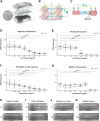
Exposure to enterotoxins leads to dose-dependent loss of barrier integrity. (A) Photograph of the OrganoTEER setup: the plate holder (1) holds the OrganoPlate (2) in which the electrode board (3) is positioned and connected to the measuring device (4). An expanded view of the electrodes is shown. (B) Schematic diagram showing the electrode positioning in a single OrganoPlate chip. Two electrode pairs are inserted into the chip wells. Current-carrying electrodes (blue) impose an AC voltage across the chip and voltage-sensing electrodes (green) measure the resulting current. (C) Schematic diagram showing a transversal view of the OrganoPlate chip and the electrode pair positioning across the medium channel and the Caco-2 tube channel containing the medium dilution with/without enterotoxin. (DG) Dose-dependent decrease in TEER after a 5 h exposure to nigericin (D) and patulin (E), and a 24 h exposure to ochratoxin A (F) and melittin (G). Caco-2 tubules were incubated with various concentrations of enterotoxin diluted in medium. TEER values were normalized to baseline TEER values taken before exposure (t = 0). EC50 values obtained after fitting the TEER data with a dose–response regression model are indicated. Three independent experiments (N = 3) were performed and 2–4 technical replicates (n = 2–4) were included per enterotoxin concentration tested. Significance was determined using Wallis test followed by Dunn’s post hoc test, where **** p < 0.0001; *** p < 0.001; ** p < 0.01; * p < 0.05. H–K Phase contrast images of the tubules before (t = 0) after a 5 h exposure to the highest concentrations of nigericin (H) and patulin (I), and a 24 h exposure to ochratoxin A (J) and melittin (K). Pictures are representative of three independent experiments (N = 3) with 2–4 technical replicates (n = 2–4) per enterotoxin concentration tested. Scale bar in white = 300 µm.
Figure 3
Figure 3
Only Melittin showed dose-dependent increase in LDH. LDH release was quantified in the Caco-2 channel medium after a 5 h exposure to nigericin (A) and patulin (B), and a 24 h exposure to ochratoxin A (C) and melittin (D). LDH concentrations in enterotoxin-exposed tubules were normalized with those of the vehicle control. Three independent experiments (N = 3) were performed and 2–4 technical replicates (n = 2–4) were included per enterotoxin concentration tested. Significance was determined using Kruskal–Wallis or ANOVA test followed by Dunn’s or Dunnett’s post hoc tests respectively, where *** p < 0.001; ** p < 0.01; * p < 0.05.
Figure 4
Figure 4
DRAQ7 staining allows assessment of the effect on cytotoxicity and cell permeability induced by ochratoxin A and melittin. Caco-2 tubules exposed to ochratoxin A and melittin during 24 h were stained with DRAQ7 nuclear dye, which only penetrates permeabilized or dead cells. Nuclei were stained with NucBlue Fixed Cell ReadyProbes Reagent. (A) A CellProfiler pipeline was designed to identify and quantify DRAQ-positive (DRAQ +) objects and nuclei from confocal images. DRAQ7 + objects were filtered to only consider those overlapping nuclear objects, which were identified as DRAQ7 + cells. The number of DRAQ7 + cells was normalized against the cell number, obtaining the output measurement %DRAQ7 + , which was considered a measure of enterotoxin cytotoxicity and cell permeability. Pictures are representative of three independent experiments (N = 3) with 2–4 technical replicates (n = 2–4) per enterotoxin concentration tested. Scale bar in white = 300 µm. (B,C) Dose-response increase in %DRAQ7 + cells after a 24 h exposure to ochratoxin A (B) and melittin (C). Three independent experiments (N = 3) were performed and 2-4 technical replicates (n = 2–4) were included per enterotoxin concentration tested. Significance was determined using Kruskal-Wallis test followed by Dunn’s post hoc test, where **** p < 0.0001; *** p < 0.001; ** p < 0.01; * p < 0.05.
Figure 5
Figure 5
Quantification of nigericin-induced morphological changes in the actin cytoskeleton. Caco-2 tubules exposed to nigericin during 24 h were stained with ActinGreen 488 ReadyProbes Reagent. Nuclei were stained with NucBlue Fixed Cell ReadyProbes Reagent. (A) A CellProfiler pipeline was designed to identify actin clumps from confocal images and quantify actin clump number and area. Nuclei were also identified, which allowed quantification of cell number. Actin clump number and area were normalized against the cell number, obtaining the output measurements actin clump number/cell number and actin clump area/cell number, which were used as a measure of enterotoxin-induced actin disorganization and effect on cell morphology. Pictures are representative of three independent experiments (N = 3) with 2–4 technical replicates (n = 2–4) per enterotoxin concentration tested. Scale bar in white = 300 µm. (B,C) Dose–response increase in actin clump number (B) and actin clump area (C) after a 5 h exposure to nigericin. Three independent experiments (N = 3) were performed and 2–4 technical replicates (n = 2–4) were included per enterotoxin concentration tested. Significance was determined using Kruskal–Wallis test followed by Dunn’s post hoc test, where **** p < 0.0001; *** p < 0.001; ** p < 0.01; * p < 0.05.

Similar articles

Cited by

References

    1. Vancamelbeke M, Vermeire S. The intestinal barrier: A fundamental role in health and disease. Expert Rev. Gastroenterol. Hepatol. 2017;11:821–834. doi: 10.1080/17474124.2017.1343143. - DOI - PMC - PubMed
    1. Zihni C, Mills C, Matter K, Balda MS. Tight junctions: From simple barriers to multifunctional molecular gates. Nat. Rev. Mol. Cell Biol. 2016;17:564–580. doi: 10.1038/nrm.2016.80. - DOI - PubMed
    1. Dal Peraro M, van der Goot FG. Pore-forming toxins: Ancient, but never really out of fashion. Nat. Rev. Microbiol. 2016;14:77–92. doi: 10.1038/nrmicro.2015.3. - DOI - PubMed
    1. Guha S, et al. Applications and evolution of melittin, the quintessential membrane active peptide. Biochem. Pharmacol. 2021;193:114769. doi: 10.1016/j.bcp.2021.114769. - DOI - PMC - PubMed
    1. Eichner M, Protze J, Piontek A, Krause G, Piontek J. Targeting and alteration of tight junctions by bacteria and their virulence factors such as Clostridium perfringens enterotoxin. Pflugers Arch. 2017;469:77–90. doi: 10.1007/s00424-016-1902-x. - DOI - PubMed