. 2024 Mar 9;20(1):98.
doi: 10.1186/s12917-024-03933-z.

Histological, immunohistochemical and serological investigations of the ovary during follicular phase of estrous cycle in Saidi sheep

Affiliations

Histological, immunohistochemical and serological investigations of the ovary during follicular phase of estrous cycle in Saidi sheep

Mahmoud Abd-Elkareem et al. BMC Vet Res. .

Abstract

Background: Saidi sheep are the most abundant ruminant livestock species in Upper Egypt, especially in the Assiut governorate. Sheep are one of the most abundant animals raised for food in Egypt. They can convert low-quality roughages into meat and milk in addition to producing fiber and hides therefore; great opportunity exists to enhance their reproduction. Saidi breed is poorly known in terms of reproduction. So this work was done to give more information on some hormonal, oxidative, and blood metabolites parameters in addition to histological, histochemical and immunohistochemical investigations of the ovary during follicular phase of estrous cycle. The present study was conducted on 25 healthy Saidi ewes for serum analysis and 10 healthy ewes for histological assessment aged 2 to 5 years and weighted (38.5 ± 2.03 kg).

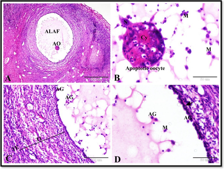
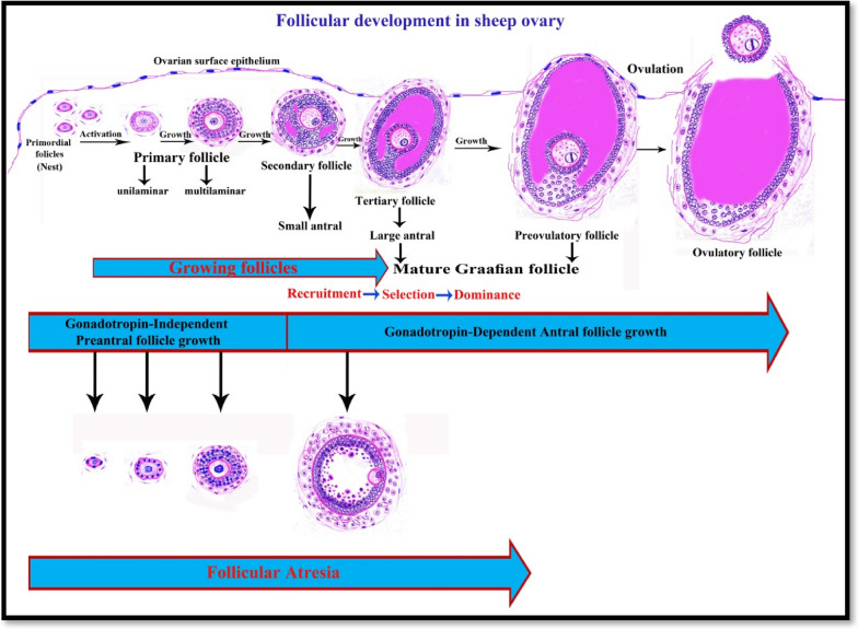
Results: The follicular phase of estrous cycle in Saidi sheep was characterized by the presence of ovarian follicles in different stages of development and atresia in addition to regressed corpus luteum. Interestingly, apoptosis and tissue oxidative markers play a crucial role in follicular and corpus luteum regression. The most prominent features of the follicular phase were the presence of mature antral (Graafian) and preovulatory follicles as well as increased level of some blood metabolites and oxidative markers. Here we give a new schematic sequence of ovarian follicles in Saidi sheep and describing the features of different types. We also clarified that these histological pictures of the ovary was influenced by hormonal, oxidative and blood metabolites factors that characterizes the follicular phase of estrous cycle in Saidi sheep.

Conclusion: This work helps to understanding the reproduction in Saidi sheep which assist in improving the reproductive outcome of this breed of sheep. These findings are increasingly important for implementation of a genetic improvement program and utilizing the advanced reproductive techniques as estrous synchronization, artificial insemination and embryo transfer.

Keywords: Antioxidants; Estrous cycle; Ovarian follicles; Ovary; PRA; SOD2; Saidi sheep.


Conflict of interest statement

The authors declare no competing interests.

Figures

Fig. 1
Fig. 1
Photomicrograph of the sheep ovary during the follicular phase. A Ovarian cortex showing preovulatory follicles (POF) and large antral follicles (LAF). B Ovarian cortex showing growing follicles (GF), small antral follicles (SAF) and preovulatory follicles (POF). C Ovarian cortex showing ovarian surface epithelium (S) which formed of single layers of flattened epithelial cells, dense irregular connective tissue of tunica albugenia (TA) and primordial follicles (PF). D Ovarian cortex showing growing follicles (GF), atretic follicles (AF) and interstitial glands (ISG). E The wall of preovulatory follicles showing dissociated granulosa cells (G), thin basement membrane (arrow), theca interna (TI) and theca externa (TE). F Showing atretic antral follicle (AAF). G Showing corpus albicans (CA). J Showing the structure of the corpus albicans which composed of dense irregular connective tissue (CT) and some blood vessels (BV). K Showing the ovarian medulla formed of loose connective tissue (LCT) and numerous blood vessels (BV). Original magnification; A & B: X12.5, scale bar = 1mm, C & J: X200, scale bar = 100 µm, D, F & K: X100, scale bar = 200 µm, E: X 400, scale bar = 50 µm, G: X 40, scale bar = 500 µm, haematoxylin and eosin stain
Fig. 2
Fig. 2
Photomicrograph of the sheep ovary during the follicular phase showing follicular atresia. A Showing atretic large antral follicle (ALAF) with apoptotic oocyte (AO). B Higher magnification of apoptotic oocyte in A; the oocyte have deeply stained acidophilic cytoplasm (Cy) and fragmented pyknotic nucleus (N) and surrounded by several macrophages (M). C & D Showing atretic large antral follicle with thickening and proliferation of theca interna (TI), apoptotic granulosa cells (AG), recruitment of several macrophages (M), thin interrupted follicular basement membrane (arrow) and normal theca externa (TE). Note the zona granulosa formed of single layer. Original magnification; A: X40, scale bar = 500 µm and B-D: X400, scale bar = 50 µm, haematoxylin and eosin stain
Fig. 3
Fig. 3
Photomicrograph illustrating the collagen fibers distribution in the sheep ovary during the follicular phase. A Showing ovarian surface epithelium (arrowhead) and tunica albuginea formed of dense irregular collagenous connective tissue. B Showing atretic follicle with a collagenous connective tissue core (AF), growing follicle (GF) surrounded by theca folliculi with few collegen fibers, and loose connective tissue of ovaian cortex with collgen fibers (C). C Showing corpus albicans (CA) which composed of dense irregular collegen fibers. D & E Showing mature graafian follicle which consisted of oocyte (O), zona pellucida (arrowhead), corona radiate (CR), cumulus oopherous (CO), large follicular cavity (FC) filled with follicular fluid, zona granulosa (ZG), Basement membrane (arrow), theca folliculi (TF) which formed of theca interna (TI) and theca externa (TE) with many collagen fibers. F Showing atretic anteral follicle with thinning of zona granulosa (become single layer) and thickening and proliferation of theca interna with proliferation of collagen fibers (arrow). Original magnification; A & D: X100, scale bar = 200 µm, B: X40, scale bar = 500 µm, C & E: X200, scale bar = 100 µm, F: X 400, scale bar = 50 µm, Crossmon's trichrome technique
Fig. 4
Fig. 4
Photomicrograph illustrating the collagen fibers type I (A-C) and elastic fibers (D-F) distribution in the sheep ovary during the follicular phase. A-C: Showing collagen fibers type I in tunica albuginea (TA), theca externa (TE) of large antral follicle (LAF), and of preovulatory follicle (POF), surrounded the regressed corpus luteum (RCL), in corpus albicans (CA), surrounded the blood vessles (BV) and in ovarian stroma (OS). D-F: Showing elastic fibers in tunica albuginea (TA), ovarian stroma ( formula image 
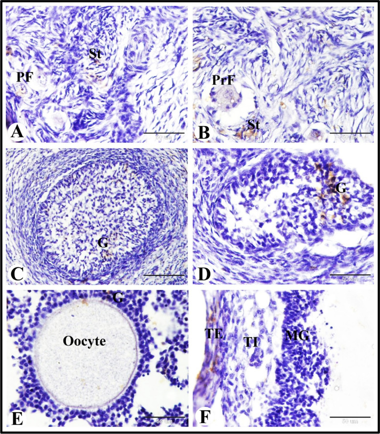
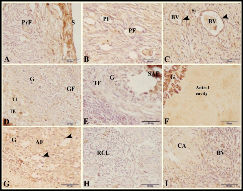
) theca externa (arrow) of growing (GF) and antral follicles (AF). Note the orcein positive granulosa cells (G) of antral follicles and the orcein negative zona pellucida (Z) and the cytoplasm of the oocyte. Original magnification; A, B & D: X40, scale bar = 500 µm, C & E: X100, scale bar = 200 µm, F: X 400, scale bar = 50 µm, A-C: Picro-Sirius red technique and D-F: Orcien stain
Fig. 5
Fig. 5
Photomicrograph of the sheep ovary during the follicular phase. A Ovarian cortex showing preovulatory follicles (POF) and regressed corpus luteum (RCL). B Ovarian cortex showing two large antral follicles (LAF1 & LAF2) surrounded by the same theca folliculi (TF) and connected by zona garnulosa with the same basement membrane (arrows). C Ovarian cortex showing atretic antral follicles (AAF) with thickening in the PAS positive follicular basement membrane (arrow) and zona pellucida (arrowhead). D The wall of preovulatory follicles showing dissociated granulosa cells (G), thin interrupted PAS positive follicular basement membrane (arrow), theca interna (TI) and theca externa (TE) with proliferation of fibroblasts (arrowhead). E Showing atretic antral follicle with hyalinization of the follicular basement membrane (BM), proliferation of theca interna cells (TI) and angiogenesis (BV). Note the central region contained few apoptotic granulosa cells (AG) and eliminated oocyte. F Showing regressed corpus luteum with autophagic lutein cells (ALC); some had PAS positive material (star) and degeneration of blood vessels with thick PAS positive endothelial basement membrane (BV). Original magnification; A: X12.5, scale bar = 1mm, B & C: X40, scale bar = 500 µm, D-F: X200, scale bar = 100 µm, PAS & Hx
Fig. 6
Fig. 6
Photomicrograph of GR immunostaining in the sheep ovary during the follicular phase. A Showing slight GR immunostaining in the ovarian surface epithelium (S), negative GR immunostaining in the primordial follicles (PF) and growing follicle (GF). B Showing slight GR immunostaining in the stroma cells (St) and oocyte of the primary follicle (arrow). C Showing moderate GR immunostaining in granulosa cells (G) and negative GR immunostaining in oocyte (O) of the antral follicles. D Showing negative GR immunostaining in the granulosa cells (G), theca interna cells (TI) and theca externa cells of mature graafian follicles. E Showing moderate GR immunostaining in the apoptotic granulosa cells (arrows) and negative GR immunostaining in the theca interna cells (TI) and theca externa cells of atretic antral follicles. F Showing strong GR immunostaining in the lutein cells (arrows) of the regressed corpus luteum. Original magnification; A-F: X400, scale bar = 50 µm
Fig. 7
Fig. 7
Photomicrograph of SOD2 immunostaining in the sheep ovary during the follicular phase. A & B Showing slight SOD2 immunostaining in the stroma cells (St) and negative SOD2 immunostaining in the primordial (PF) and primary (PrF) follicles. C-E Showing moderate SOD2 immunostaining in the granulosa cells (G) of the growing (C & D) and antral (E) follicles and negative SOD2 immunostaining in the oocyte. F Showing moderate SOD2 immunostaining in theca externa cells (TE) and negative SOD2 immunostaining in the granulosa cells (G) and theca interna cells (TI) of preovulatory follicles. Original magnification; A-B & D-F: X400, scale bar = 50 µm, C: X200, scale bar = 100 µm
Fig. 8
Fig. 8
Photomicrograph of PRA immunoexpression in the sheep ovary during the follicular phase. A Showing strong PRA immunoexpression in the surface epithelial cells (S) and slight PRA immunoexpression in the primary (PrF) follicles. B slight PRA immunoexpression in the primordial (PF). C Showing strong PRA immunoexpression in the endothelial cells (arrowheads) of blood vessels (BV) and moderate PRA immunoexpression in the stroma cells (St). D Showing slight PRA immunoexpression in the granulosa cells (G), theca interna cells (TI) and theca externa cells (TE) of the growing follicles (GF). E Showing slight PRA immunoexpression in the granulosa cells (G) and theca folliculi cells (TF) of the small antral follicles (SAF). F Showing strong PRA immunoexpression in the granulosa cells (G) of the preovulatory follicles. Note the large antral cavity. G Showing slight PRA immunoexpression in the granulosa cells (G) and negative PRA immunoexpression in the apoptotic granulosa cells (arrowheads) of the atretic follicles (AF). H Showing negative PRA immunoexpression in the fibrous tissue (fibrocytes and collagen fibers) of the regressed corpus luteum (RCL). I Showing slight PRA immunoexpression in the fibrous tissue (fibrocytes and collagen fibers) and blood vessels (BV) of the corpus albicans (CA). Original magnification; A-I: X400, scale bar = 50 µm
Fig. 9
Fig. 9
Photomicrograph of immunofluorescence of TUNEL assay in the sheep ovary during the follicular phase. A Showing apoptotic granulosa cells (AGC) in the atretic antral follicle (AAF). B Showing apoptotic interstitial gland cells (AISG). C Showing apoptotic basal granulosa cells (arrow) of membrana granulosa (MG) and apoptotic theca interna cells (ATI) of atretic grafiaan follicles. D Showing apoptotic lutein cells (ALC) of the regressed corpus luteum. E Showing apoptotic apical (antral) granulosa cells (AGC) of membrana granulosa (MG) of atretic grafiaan follicles. F Showing apoptotic granulosa cells (AGC), apoptotic theca interna cells (ATI) and apoptotic theca externa cells (ATE) of atretic grafiaan follicles. Original magnification; A-F X400, scale bar = 50 µm
Fig. 10
Fig. 10
Drawing diagram showing the follicular development and follicular stages in sheep ovary. Preantral phase: Formation of primordial follicles which present in nests; Primordial follicle has a single layer of flattened follicular cells. Follicular activation of the primordial follicles to form unilaminar primary follicles; unilaminar primary follicle has a single layer of cuboidal follicular cells. Then growth of unilaminar primary follicles to form multilaminar primary follicles; multilaminar primary follicles have several layers of granulosa cells and few theca cells. All the preantral follicles have a primary oocyte surrounded by follicular cells and separated from the follicular cells by increased thickness zona pellucida. Antral phase: Follicle growth continues through the phases of recruitment, selection, dominance, and preovulatory stage of follicular waves. Secondary follicle has several layers of granulosa cells with small accumulations (pools) of follicular fluid in the intracellular spaces and a small number of theca cells. Tertiary follicle has several granulosa cell layers, corona radiata, cumulous oopherous, theca folliculi, and primary oocyte and is characterized by a single large antral cavity which contains follicular fluid. Preovulatory (Mature Graafian) follicle is the last stage of follicle development; these follicles are larger, have more antral fluid and may contain a secondary oocyte and protrude from ovarian surface epithelium and separated from them by very thin layer of granulosa cells and tunica albuginea. Ovulatory follicle is preovulatory from which the ovum and its surrounding cells released to the peritoneal catvity by rupture of the thin follicular wall

Similar articles

Cited by

References

    1. Wang H, Feng X, Muhatai G, Wang L. Expression profile analysis of sheep ovary after superovulation and estrus synchronisation treatment. Vet Med Sci. 2022;8:1276–1287. doi: 10.1002/vms3.783. - DOI - PMC - PubMed
    1. Germot A, Khodary MG, Othman OE-M, Petit D. Shedding Light on the Origin of Egyptian Sheep Breeds by Evolutionary Comparison of Mitochondrial D-Loop. Animals. 2022;12:2738. doi: 10.3390/ani12202738. - DOI - PMC - PubMed
    1. El-Homosi FF, Abd El-Hafiz GA. Reproductive perpormance of Ossimi and Saidi sheep under two pre pubertal planes of nutrition. Assiut Vet Med J. 1982;10:59–66.
    1. Teleb D Teleb D, Ahmed N, El –Din TH, Abou El Soud S, Hassan OM. STUDY ON LEVELS OF SOME BLOOD HORMONAL AND BIOCHEMICAL CONSTITUENTS DURING DIFFERENT REPRODUCTIVE STATUS IN SAIDI EWES. Egypt J Sheep Goats Sci. 2019;9:1–10.
    1. Teleb D, El-Sayed E, Sallam A.E.A. Characterizing the estrous and ovarian activity of Saidi sheep throughout the year. Egypt J Basic Appl Physiol. 2008;7(2):325–39.