. 2024 Mar 9;15(1):2165.
doi: 10.1038/s41467-024-46509-z.

TERRA-LSD1 phase separation promotes R-loop formation for telomere maintenance in ALT cancer cells

Affiliations

TERRA-LSD1 phase separation promotes R-loop formation for telomere maintenance in ALT cancer cells

Meng Xu et al. Nat Commun. .

Abstract

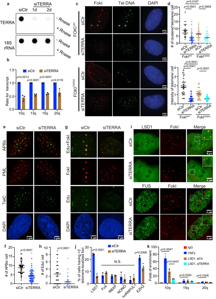
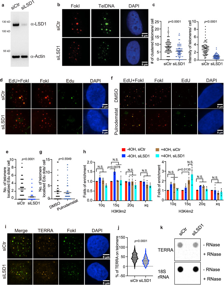
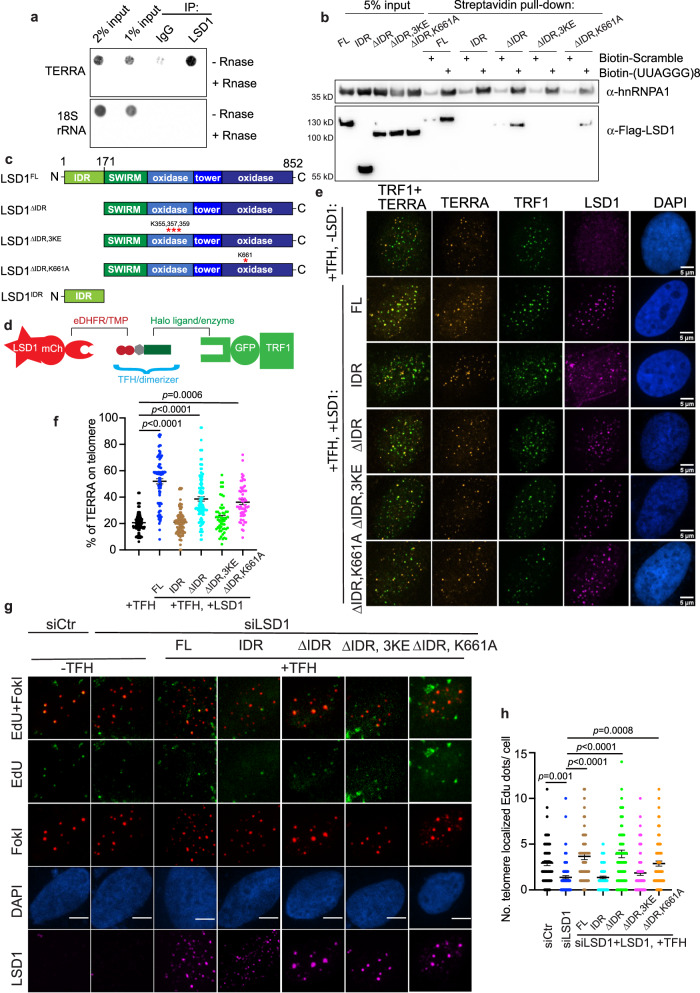
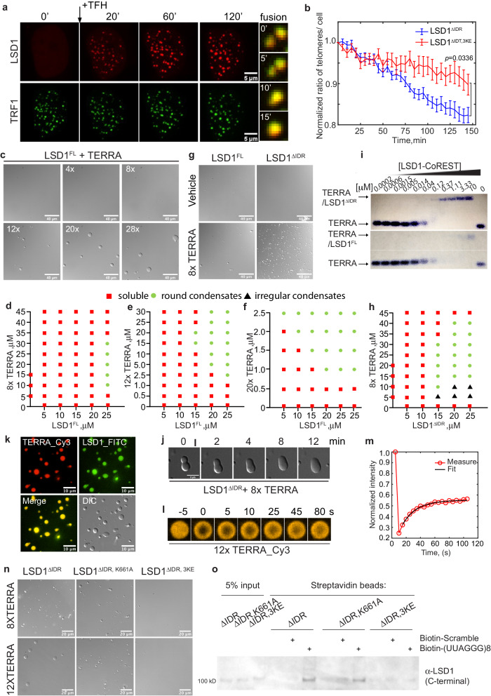
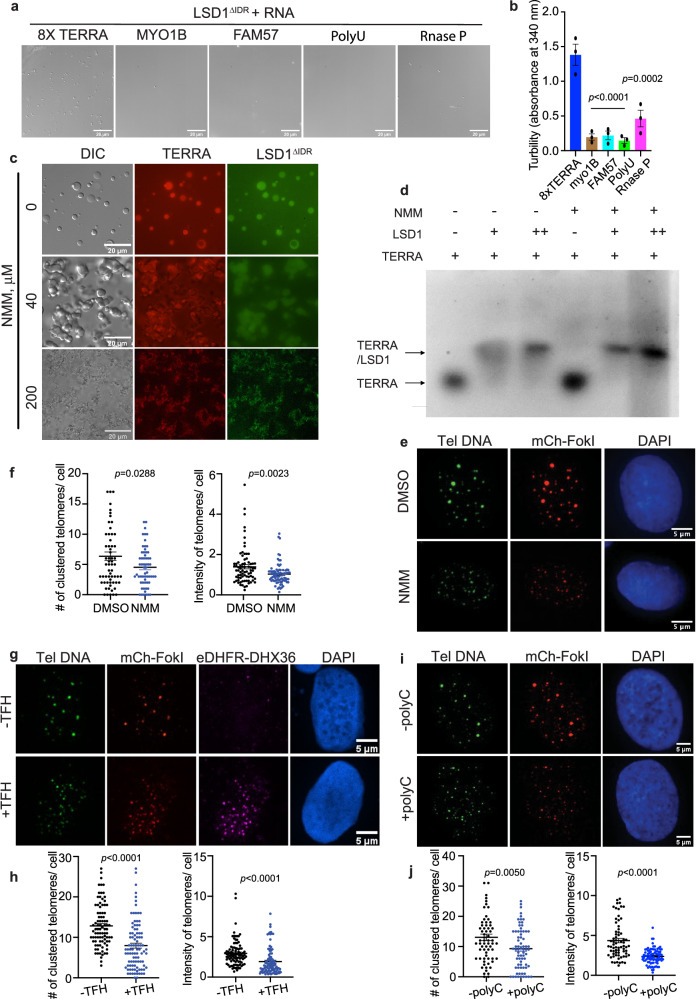
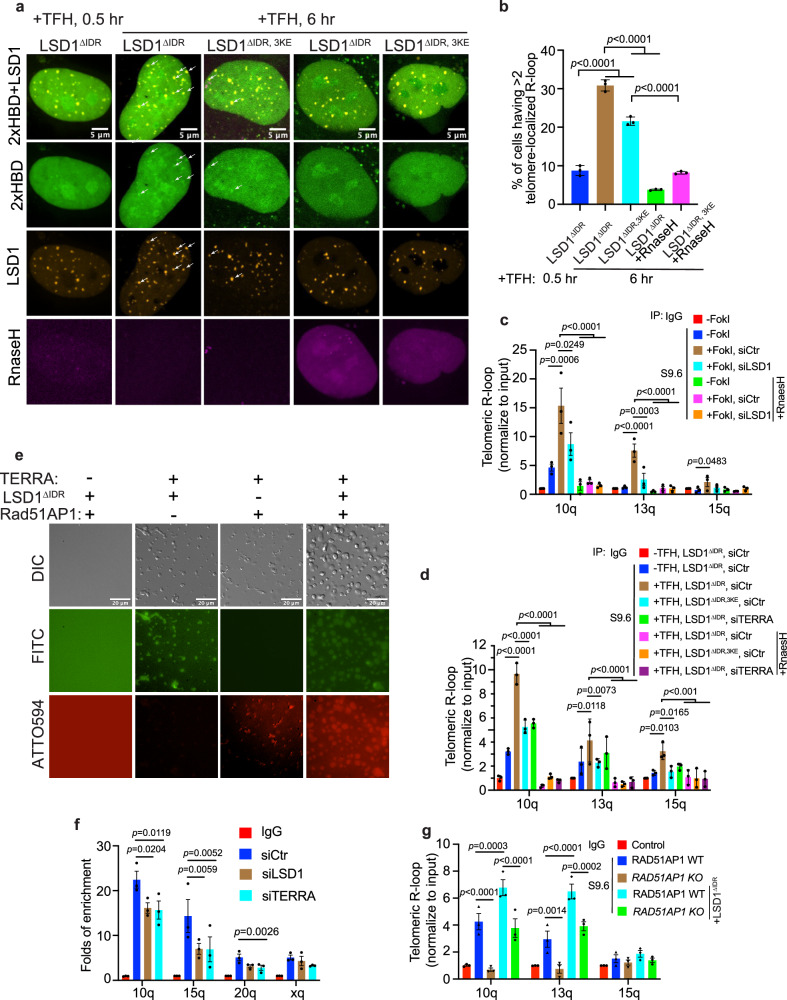
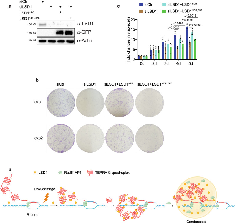
The telomere repeat-containing RNA (TERRA) forms R-loops to promote homology-directed DNA synthesis in the alternative lengthening of telomere (ALT) pathway. Here we report that TERRA contributes to ALT via interacting with the lysine-specific demethylase 1A (LSD1 or KDM1A). We show that LSD1 localizes to ALT telomeres in a TERRA dependent manner and LSD1 function in ALT is largely independent of its demethylase activity. Instead, LSD1 promotes TERRA recruitment to ALT telomeres via RNA binding. In addition, LSD1 and TERRA undergo phase separation, driven by interactions between the RNA binding properties of LSD1 and the G-quadruplex structure of TERRA. Importantly, the formation of TERRA-LSD1 condensates enriches the R-loop stimulating protein Rad51AP1 and increases TERRA-containing R-loops at telomeres. Our findings suggest that LSD1-TERRA phase separation enhances the function of R-loop regulatory molecules for ALT telomere maintenance, providing a mechanism for how the biophysical properties of histone modification enzyme-RNA interactions impact chromatin function.


Conflict of interest statement

The authors declare no competing interests.

Figures

Fig. 1
Fig. 1. LSD1 localization on ALT telomeres requires TERRA.
a Total TERRA levels were evaluated by northern-dot-blot assay, following transient knockdown of TERRA in U2OS for 24 and 48 h (n = two independent experiments). b TERRA levels from chromosome 10, 13, 15, 20 were quantitated by qRT-PCR, following transient knockdown of TERRA in U2OS for 48 h (n = three independent experiments, two-tailed unpaired t test). c Representative images and d quantification of telomere clustering from DNA FISH after siTERRA in U2OS cells stably expressing TRF1-FokI with treatment of 4-Hydroxyestradiol (4-OH, 6 h) (n = 50 cells for FokIWT siCtr, n = 69 cells for FokIWT siTERRA, n = 58 cells for FokID450A siCtr, n = 76 cells for FokID450A siTERRA over two independent experiments, two-tailed unpaired t test). e Representative images and f quantification of APB bodies (defined by PML and telomere FISH colocalization) after siTERRA in FokI cells (n = 110 cells for siCtr, n = 122 cells for siTERRA over three independent experiments, two-tailed unpaired t test). g Representative images and h quantification of Edu foci on telomeres after siTERRA in FokI cells (n = 51 cells for siCtr, n = 70 cells for siTERRA over two independent experiments, two-tailed unpaired t test). i Representative images for GFP-LSD1 and GFP-FUS and j quantification of telomeric localization for GFP-tagged LSD1, FUS, NONO, RBXP, hnRNPUL1, and immunostained EZH2 with siControl and siTERRA in U2OS cells (more than 100 cells per group, n = three independent experiments, two-way ANOVA). k ChIP-qRCR assay was performed to detect telomeric LSD1 with or without siTERRA from indicated chromatin U2OS cells synchronized in G2 (n = three independent experiments, one-way ANOVA). Error bars are mean ± SEM. NS not significant. Source data are provided as a Source Data file.
Fig. 2
Fig. 2. Knocking down LSD1 leads to loss of ALT phenotypes.
a Western blot of siLSD1 in U2OS cells (n = three independent experiments). b Representative images and c quantification of telomere clustering (DNA FISH) after siLSD1 in FokI stable cell line with treatment of 4-hydroxyestradiol (4-OH, 6 h) (n = 72 cells examined for siCtr, n = 90 cells for siLSD1 over two independent experiments, two-tailed unpaired t test). d Representative images and e quantification of Edu foci on telomeres after siLSD1 in FokI cells (n = 70 cells for siCtr, n = 102 cells examined for siLSD1 over three independent experiments, two-tailed unpaired t test). f Representative images and g quantification of Edu foci on telomeres after treatment of Pulrodemstat (200 nM, 16 h) in FokI cells (n = 46 cells for DMSO, n = 70 cells for Pulrodemstat over two independent experiments, two-tailed unpaired t test). h ChIP-qRCR assay was performed to detect telomeric H3K4m2 and H3K9m2 from indicated chromatin in siCtr- or siLSD1-transfected FokI cells treated with and without 4-hydroxyestradiol (4-OH, 6 h) (n = three independent experiments, one-way ANOVA). i Representative images and j quantification of TERRA (RNA FISH) localization on telomeres after siLSD1 in FokI expressing U2OS cells (n = 136 cells for siCtr, n = 127 cells for siLSD1 over three independent experiments, two-tailed unpaired t test). k Northern-dot-blot was used to detect total TERRA levels in U2OS cells treated with siCtr or siLSD1 for 48 h (n = two independent experiments).18S rRNA serves as loading control. Error bars are mean ± SEM. NS not significant. Source data are provided as a Source Data file.
Fig. 3
Fig. 3. Chemical dimerization of LSD1 to telomeres is sufficient to enrich TERRA and rescue ALT phenotypes.
a IP-recovered RNA was detected via dot-blot with probes annealing with TERRA or 18S rRNA (refer to Supplementary Fig. 4a for IP efficiency). b RNA pull-down assay was used to evaluate the binding of Streptavidin bead-bound biotinylated (UUAGGG)8 oligonucleotide to flag-tagged LSD1FL, LSD1IDR, LSD1ΔIDR, LSD1ΔIDR, 3KE, and LSD1ΔIDR, K661A from U2OS cell lysates (n = three independent experiments). c Schematic of LSD1: full-length (LSD1FL), C-terminus (LSD1ΔIDR,), N-terminus (LSD1IDR), mutant for nucleic acid binding (LSD1ΔIDR, 3KE), mutant for demethylation activity (LSD1ΔIDR, K661A). d Dimerization schematic: LSD1 is fused to mCherry and eDHFR, and TRF1 is fused to Halo enzyme and GFP. The dimerizer is TFH, TMP(trimethoprim)- Fluorobenzamide-Halo ligand. e Representative images and f quantification of TERRA (RNA FISH) localization on telomeres after dimerizing overexpressed LSD1FL, LSD1IDR, LSD1ΔIDR, and LSD1ΔIDR, 3KE, and LSD1ΔIDR, K661A to Halo-TRF1 in U2OS cells with dimerizer TFH (n = 108 cells for +TFH alone, n = 87 cells for LSD1FL, n = 103 cells for LSD1IDR, n = 113 cells for LSD1ΔIDR, n = 55 cells for LSD1ΔIDR, 3KE, n = 53 cells for LSD1ΔIDR, K661A over three independent experiments, one-way ANOVA). g Representative images and h quantification of Edu assay showing the rescue effect of newly synthesized telomeric DNA after dimerizing LSD1FL, LSD1IDR, LSD1ΔIDR, LSD1ΔIDR, 3KE, and LSD1ΔIDR, K661A to telomeres in siLSD1 cells (n = 69 cells for siCtr, n = 89 cells for siLSD1, n = 67 cells for LSD1FL, n = 77 cells for LSD1IDR, n = 90 cells for LSD1ΔIDR, n = 75 cells for LSD1ΔIDR, 3KE, n = 81 cells for LSD1ΔIDR, K661A over two independent experiments, one-way ANOVA). Error bars are mean ± SEM. NS not significant. Source data are provided as a Source Data file.
Fig. 4
Fig. 4. TERRA drives LSD1 phase separation.
a Representative images of U2OS cells expressing mCh-eDHFR-LSD1 and Halo-GFP-TRF1 after adding TFH to induce dimerization at indicated time points. Montage on the right shows the fusion event of LSD1 foci over time, every 5 min between images. b Mean telomere number per cell after dimerizing mCh-eDHFR-LSD1 (WT and 3KE mutant) to Halo-GFP-TRF1 stably expressed in U2OS cells (n = 20 cells for LSD1ΔIDR, n = 18 cells for LSD1ΔIDR, 3KE over three independent experiments, two-tailed unpaired t test). c Representative DIC images of condensates formed by a mixture of purified LSD1 at 25 μM and different UUAGGG repeats as mimics of TERRA at 2.5 μM (n = three independent experiments). d-f Phase diagram of full-length LSD1 with the addition of 8× (d), 12× (e), 20× (f), TERRA. g Representative DIC images and h phase diagram of LSD1ΔIDR with 8× TERRA. i Representative EMSA showing (UUAGGG)8U RNA binding of truncated LSD1 CoREST (top) and full-length LSD1-CoREST (bottom) (n= two independent experiments). j Fusion of condensates containing 25 μM LSD1 and 25 μM 8× TERRA. k Fluorescent images of condensates containing Cy3-labeled 10 μM 12× TERRA and FITC-labeled 50 μM LSD1 (n = three independent experiments). l FRAP of condensates containing 10 μM 12× Cy3-TERRA and 50 μM LSD1 condensates. m A representative FRAP curve for TERRA is shown. The fit to an exponential curve generates mobile fraction and recovery time. Mean values of 19 condensates from three independent experiments are shown in Supplementary Table 1. n Representative DIC images of LSD1ΔIDR, LSD1ΔIDR, 3KE, and LSD1ΔIDR, K661A with the addition of TERRA (n = three independent experiments). o In vitro pull-down assay was used to evaluate the binding of biotinylated (UUAGGG)8 oligonucleotide to recombinant LSD1ΔIDR, LSD1ΔIDR, 3KE, and LSD1ΔIDR, K661A from E. coli. (n = three independent experiments). Source data are provided as a Source Data file.
Fig. 5
Fig. 5. TERRA G-quadruplex structure is required for LSD1 phase separation and ALT functions.
Representative DIC images (a) and turbidity assay (b) of LSD1ΔIDR (20 μM) with the addition of different RNAs (20 μM). Turbidity measurements are shown as mean ± SEM (n = three independent experiments, one-way ANOVA). c Representative images of fluorescent LSD1ΔIDR (50 μM) with 12× TERRA (25 μM) pretreated with G-quadruplex binding ligand, N-methyl mesoporphyrin IX, (NMM) (n= three independent experiments). d In vitro EMSA of 8X TERRA GG(UUAGGG)8U (+ = 2 μM) alone and in the presence of LSD1 (+ = 10 μM and ++ = 20 μM) and NMM (+ = 200 μM). The 0.75% agarose gel contained Syber Safe dye (10,000:1, Invitrogen) and was performed under the following conditions (60 V, 1 h, 4 °C). The fluorescence data were visualized at 520 nm using an Amersham Imager 600 (AI600) instrument (n = two independent experiments). e Representative images and f quantification of telomere clustering (DNA FISH) after treatment of NMM (200 nM, 24 h) in FokI stable cell line (n = 77 cells for DMSO, n = 80 cells examined for NMM over two independent experiments, two-tailed unpaired t test). g Representative images and h quantification of telomere clustering (DNA FISH) after co-transfection of eDHFR-DHX36, Halo-TRF1 and mCh-FokI with or without TFH treatment in U2OS cells (n = 96 cells for -TFH, n = 99 cells examined for +TFH over three independent experiments, two-tailed unpaired t test). DHX36 are indicated by antibody-based immunostaining. i Representative images and j quantification of telomere clustering (DNA FISH) after co-transfection of polyC with mCh-FokI in U2OS cells (n = 69 cells for -polyC, n = 84 cells for +polyC over three independent experiments, two-tailed unpaired t test). Error bars are mean ± SEM. N.S., not significant. Source data are provided as a Source Data file.
Fig. 6
Fig. 6. LSD1 promotes R-loop formation at ALT telomeres.
a Representative images and b quantification of R-loop (indicated by 2× HBD-GFP) on telomeres after dimerization of LSD1ΔIDR, LSD1ΔIDR, 3KE to Halo-TRF1 in U2OS cells (n = three independent experiments, one-way ANOVA). Because the telomeres are not labeled, LSD1 foci after adding TFH for 0.5 h (shortly after dimerization completion) were used to infer telomere locations. c DRIP assay shows R-loop level on telomeres from chromatin 10q, 13q, 15q after siLSD1 or siTERRA (100 nM, 48 h) in FokI stable cell line (n = three experiments, one-way ANOVA). d DRIP assay shows R-loop level on telomeres from chromatin 10q, 13q, 15q after dimerization of LSD1ΔIDR and LSD1ΔIDR, 3KE to Halo-TRF1 in siCtr or siTERRA U2OS cells (n = three independent experiments, two-way ANOVA). e Fluorescent images of condensates after combination of ATTO594-labeled Rad51AP1 (30 μM), FITC-labeled LSD1ΔIDR (50 μM) and 8× TERRA (50 μM). f ChIP-qRCR assay detecting Rad51AP1 binding to telomere DNA from indicated chromatin in siCtr-, siLSD1-, and siTERRA- transfected U2OS cells (n= three independent experiments, one-way ANOVA). g DRIP assay shows R-loop level on telomeres from chromatin 10q, 13q, 15q after dimerization of LSD1ΔIDR to Halo-TRF1 in U2OS RAD51AP1 WT and KO cells (n = three independent experiments, two-way ANOVA). Error bars are mean ± SEM. NS not significant. Source data are provided as a Source Data file.
Fig. 7
Fig. 7. LSD1-TERRA interaction affects ALT cell viability.
a Western blot of GFP-tagged LSD1ΔIDR and LSD1ΔIDR,3KE expression in siLSD1 cells (n = three independent experiments). b Images of colony formation assays with U2OS cell after indicated treatment. c Quantification of viable U2OS cells after indicated treatment using CellTiter-Blue assay (n = three independent experiments, one-way ANOV. Error bars are mean ± SEM). d A model for TERRA-LSD1 phase separation at ALT telomeres. In response to DNA damage signaling in the ALT pathway, TERRA recruits LSD1 to telomeres. TERRA-LSD1 interaction at telomeres, likely enhanced by DNA damage, may further enrich TERRA on telomeres, leading to local condensate formation. Hereafter, other proteins, such as Rad51AP1, may partition into TERRA-LSD1 condensates to help stimulate R-loop formation for telomere maintenance in ALT cancer cells. Created with BioRender.com. Source data are provided as a Source Data file.

Similar articles

Cited by

References

    1. Mattick, J. S. et al. Long non-coding RNAs: definitions, functions, challenges and recommendations. Nat. Rev. Mol. Cell Biol. 24, 430–447 (2023). - PMC - PubMed
    1. Hegazy YA, Fernando CM, Tran EJ. The balancing act of R-loop biology: The good, the bad, and the ugly. J. Biol. Chem. 2020;295:905–913. doi: 10.1016/S0021-9258(17)49903-0. - DOI - PMC - PubMed
    1. Thakur J, Henikoff S. Architectural RNA in chromatin organization. Biochem. Soc. Trans. 2020;48:1967–1978. doi: 10.1042/BST20191226. - DOI - PMC - PubMed
    1. Davidovich C, Cech TR. The recruitment of chromatin modifiers by long noncoding RNAs: lessons from PRC2. RNA. 2015;21:2007–2022. doi: 10.1261/rna.053918.115. - DOI - PMC - PubMed
    1. Tsai M-C, et al. Long noncoding RNA as modular scaffold of histone modification complexes. Science. 2010;329:689–693. doi: 10.1126/science.1192002. - DOI - PMC - PubMed