. 2024 Mar 9;22(1):259.
doi: 10.1186/s12967-024-05028-7.

Amino acid profile alteration in age-related atrial fibrillation

Affiliations

Amino acid profile alteration in age-related atrial fibrillation

Yunying Huang et al. J Transl Med. .

Abstract

Background: Amino acids (AAs) are one of the primary metabolic substrates for cardiac work. The correlation between AAs and both atrial fibrillation (AF) and aging has been documented. However, the relationship between AAs and age-related AF remains unclear.

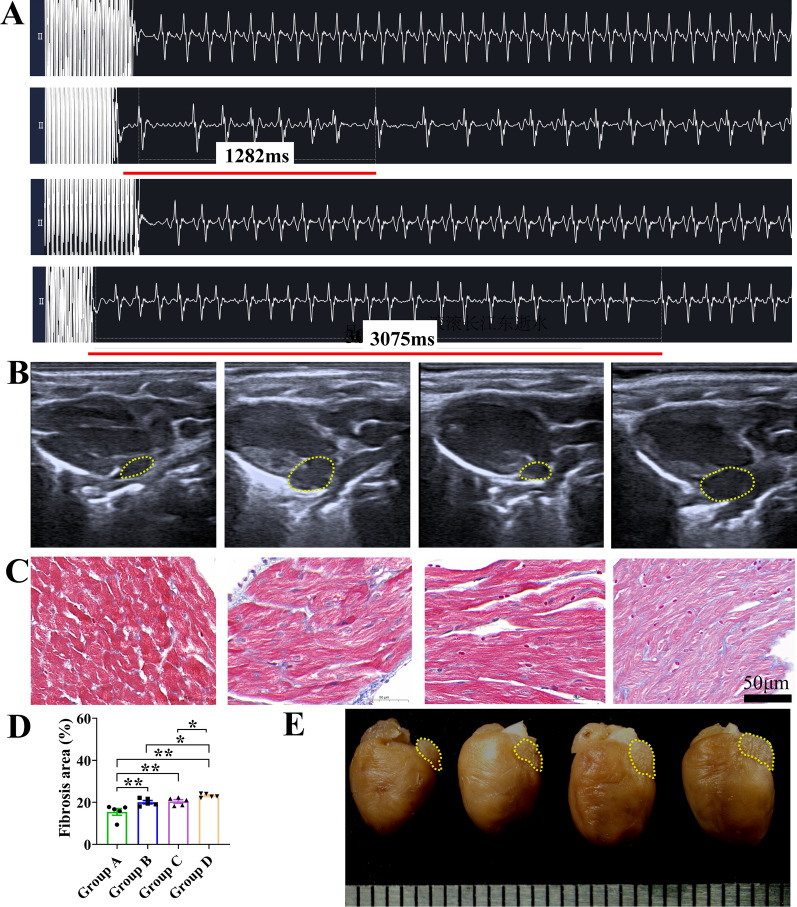
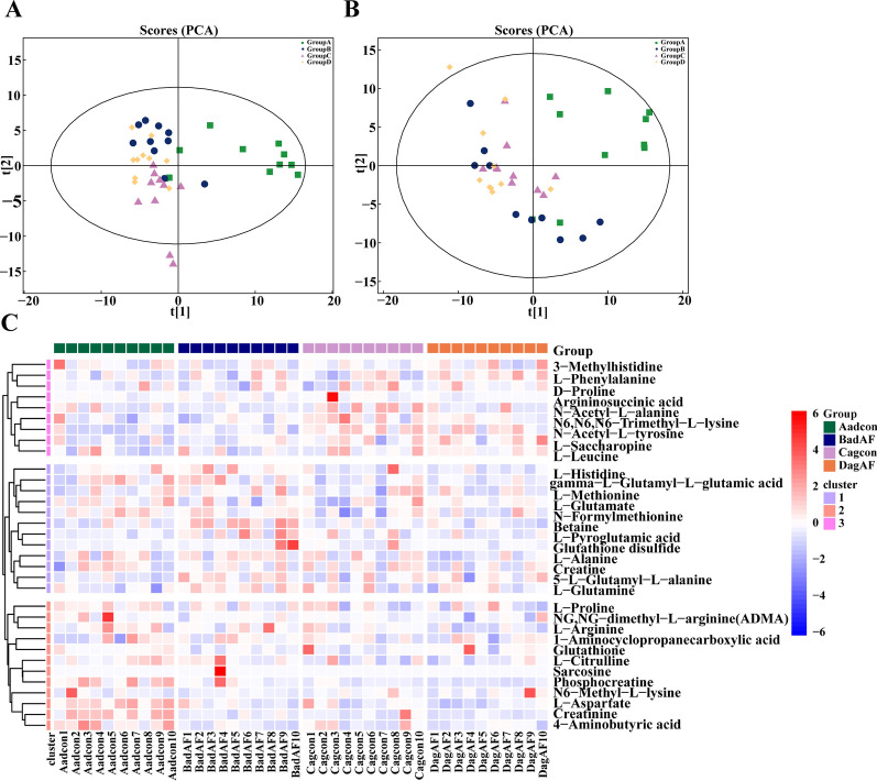
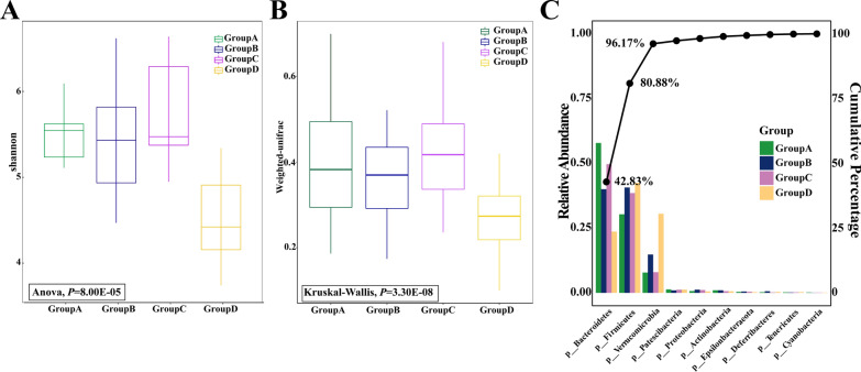
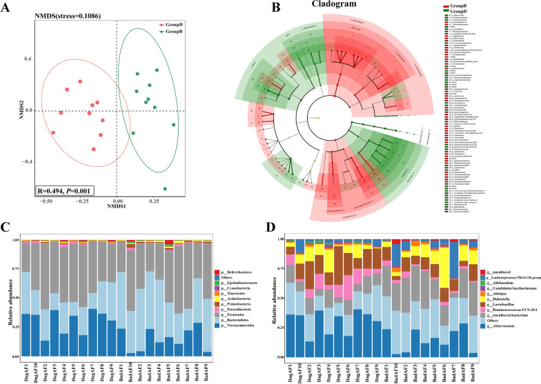
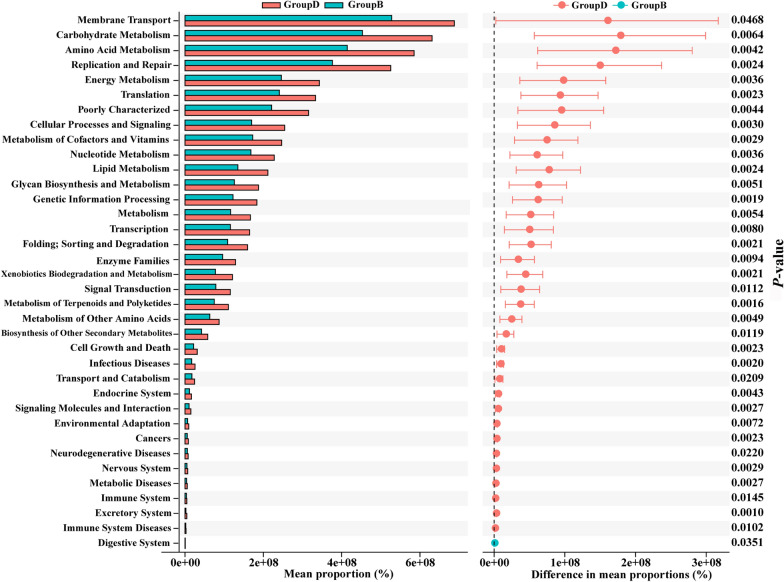
Methods: Initially, the plasma AA levels of persistent AF patients and control subjects were assessed, and the correlations between AA levels, age, and other clinical indicators were explored. Subsequently, the age-related AF mouse model was constructed and the untargeted myocardial metabolomics was conducted to detect the level of AAs and related metabolites. Additionally, the gut microbiota composition associated with age-related AF was detected by a 16S rDNA amplicon sequencing analysis on mouse fecal samples.

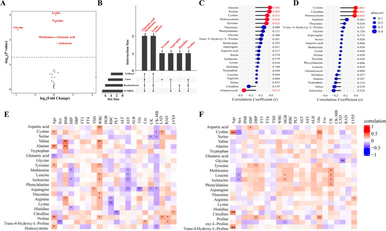
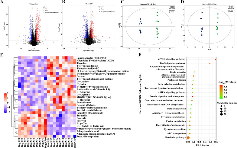
Results: Higher circulation levels of lysine (Student's t-test, P = 0.001), tyrosine (P = 0.002), glutamic acid (P = 0.008), methionine (P = 0.008), and isoleucine (P = 0.014), while a lower level of glycine (P = 0.003) were observed in persistent AF patients. The feature AAs identified by machine learning algorithms were glutamic acid and methionine. The association between AAs and age differs between AF and control subjects. Distinct patterns of AA metabolic profiles were observed in the myocardial metabolites of aged AF mice. Aged AF mice had lower levels of Betaine, L-histidine, L-alanine, L-arginine, L-Pyroglutamic acid, and L-Citrulline compared with adult AF mice. Aged AF mice also presented a different gut microbiota pattern, and its functional prediction analysis showed AA metabolism alteration.

Conclusion: This study provided a comprehensive network of AA disturbances in age-related AF from multiple dimensions, including plasma, myocardium, and gut microbiota. Disturbances of AAs may serve as AF biomarkers, and restoring their homeostasis may have potential benefits for the management of age-related AF.

Keywords: Age-related atrial fibrillation; Aging; Amino acids; Atrial fibrillation; Gut microbiota; Metabolomics.


Conflict of interest statement

The authors declared no competing interests.

Figures

Fig. 1
Fig. 1
Association of plasma amino acids level and clinical indicators of patients. A Volcano plot of the different amino acids between persistent atrial fibrillation patients and control subjects (Student’s t-test, P < 0.05). B The distribution of feature amino acids screened by each machine learning method. The number of amino acids identified in each subset is represented in the histogram. C–D The association of plasma amino acids level and age of C control subjects and D persistent atrial fibrillation patients. EF The association of plasma amino acids level and clinical indicators of E control subjects and F persistent atrial fibrillation patients. *P < 0.05 and **P < 0.01. BMI, body mass index. SBP, systolic blood pressure. DBP, diastolic blood pressure. FT3, free triiodothyronine. FT4, free thyroxine. TSH, thyroid-stimulating hormone. WBC, white blood cell. HGB, hemoglobin. RBC, red blood cell. PLT, platelets. ALT, alanine aminotransferase. AST, aspartate aminotransferase. ALB, albumin. Glu, blood glucose. BUN, blood urea nitrogen. Cre, creatinine. CK, creatine kinase. CK-MB, creatine kinase isoenzyme. LAD, left atrial diameter. RAD, right atrial diameter. LVEF, Left ventricular ejection fraction
Fig. 2
Fig. 2
Atrial fibrillation mouse model establishment. A Surface electrocardiogram of each group of mice, and the sequence from top to bottom was adult control (Group A), adult AF (Group B), aged control (Group C), and aged AF (Group D). The ECG of Group B and Group D mice showed the atrial fibrillation waveform. B Left atrial area detected by M-mode echocardiogram, and the sequence from left to right was Group A-D. Yellow dashed lines encircle the left atrium. C, D Representative images of Masson's trichrome staining of the atrial area and measurements of the fibrosis area, and the sequence from left to right was Group AD (n = 5 in each group), *P < 0.05; **P < 0.01. E Heart gross specimens and the sequence from left to right was Group A–D. Yellow dashed lines encircle the left atrium
Fig. 3
Fig. 3
Myocardial untargeted metabolomics and amino acids related metabolites profiles among four mice groups. A, B Principal component analysis (PCA) score plots of adult control (Group A, Aadcon), adult AF (Group B, BadAF), aged control (Group C, Cagcon), and aged AF (Group D, DagAF) in A positive ion mode and B negative ion mode. C Hierarchical clustering heatmap of amino acids and related metabolites profile of four groups in positive ion mode. n = 10 in each group
Fig. 4
Fig. 4
Myocardial untargeted metabolomics between adult AF (Group B, BadAF) and aged AF (Group D, DagAF) mice. AB Volcano plot of the different metabolites in univariate analysis (FC > 1.5 or < 0.67, and t-test P-value < 0.05) in A positive ion mode and B negative ion mode. C–D Orthogonal partial least-squares discriminant analysis (OPLS-DA) score plot in C positive ion mode and D negative ion mode. E Hierarchical clustering heatmap of metabolites with significant differences (OPLS-DA VIP value > 1 and t-test P-value < 0.05, with qualitative names) in positive ion mode. F Top 20 enriched KEGG pathways of the significantly different metabolites in positive ion mode. n = 10 in each group
Fig. 5
Fig. 5
Gut microbiota diversity and relative abundance among four mice groups. A Gut microbiota alpha diversity was different among groups based on the Shannon index (P = 8.00E-05). B Gut microbiota beta diversity was different among groups based on Weighted Unifrac distance (P = 3.30E-08). C Pareto chart illustrating the relative abundance of the top 10 gut microbiota at the phylum level among four groups. Bars are arranged in ascending order of frequency. The Pareto line indicates cumulative percentage, highlighting that the top 3 gut microbiota contribute to 96.17% of the total abundance. Group A: adult control, Group B: adult AF, Group C: aged control, and Group aged AF, n = 10 in each group
Fig. 6
Fig. 6
Gut microbiota composition of adult AF (Group B, BadAF) and aged AF (Group D, DagAF) mice. A Non-metric multi-dimensional scaling (NMDS) and ANOSIM test (the box within NMDA plot). B The taxonomic cladogram plotted from LEfSe analysis. The red and green nodes represent species with significantly more abundance (LEfSe LDA score > 2 and P-value < 0.05) in Group B and Group D, respectively. C-D The community composition of the top 10 representative species and their relative abundance at the phylum level (C) and the genus level (D). n = 10 in each group
Fig. 7
Fig. 7
KEGG functional predictive analysis of gut microbiota between adult AF (Group B) and aged AF (Group D) mice conducted by STAMP differential analysis

Similar articles

Cited by

References

    1. Hindricks G, Potpara T, Dagres N, et al. 2020 ESC Guidelines for the diagnosis and management of atrial fibrillation developed in collaboration with the European Association for Cardio-Thoracic Surgery (EACTS): the task force for the diagnosis and management of atrial fibrillation of the European Society of Cardiology (ESC) Developed with the special contribution of the European Heart Rhythm Association (EHRA) of the ESC. Eur Heart J. 2021;42(5):373–498. doi: 10.1093/eurheartj/ehaa612. - DOI - PubMed
    1. Staerk L, Sherer JA, Ko D, Benjamin EJ, Helm RH. Atrial fibrillation: epidemiology, pathophysiology, and clinical outcomes. Circ Res. 2017;120(9):1501–1517. doi: 10.1161/CIRCRESAHA.117.309732. - DOI - PMC - PubMed
    1. Kornej J, Borschel CS, Benjamin EJ, Schnabel RB. Epidemiology of atrial fibrillation in the 21st Century: novel methods and new insights. Circ Res. 2020;127(1):4–20. doi: 10.1161/CIRCRESAHA.120.316340. - DOI - PMC - PubMed
    1. Jahangir A, Lee V, Friedman PA, et al. Long-term progression and outcomes with aging in patients with lone atrial fibrillation: a 30-year follow-up study. Circulation. 2007;115(24):3050–3056. doi: 10.1161/CIRCULATIONAHA.106.644484. - DOI - PubMed
    1. Potpara TS, Stankovic GR, Beleslin BD, et al. A 12-year follow-up study of patients with newly diagnosed lone atrial fibrillation: implications of arrhythmia progression on prognosis: the Belgrade Atrial Fibrillation study. Chest. 2012;141(2):339–347. doi: 10.1378/chest.11-0340. - DOI - PubMed

Publication types