. 2024 Mar 11;11(1):16.
doi: 10.1186/s40779-024-00512-z.

Targeting vulnerable microcircuits in the ventral hippocampus of male transgenic mice to rescue Alzheimer-like social memory loss

Affiliations

Targeting vulnerable microcircuits in the ventral hippocampus of male transgenic mice to rescue Alzheimer-like social memory loss

Hui-Yang Lei et al. Mil Med Res. .

Abstract

Background: Episodic memory loss is a prominent clinical manifestation of Alzheimer's disease (AD), which is closely related to tau pathology and hippocampal impairment. Due to the heterogeneity of brain neurons, the specific roles of different brain neurons in terms of their sensitivity to tau accumulation and their contribution to AD-like social memory loss remain unclear. Therefore, further investigation is necessary.

Methods: We investigated the effects of AD-like tau pathology by Tandem mass tag proteomic and phosphoproteomic analysis, social behavioural tests, hippocampal electrophysiology, immunofluorescence staining and in vivo optical fibre recording of GCaMP6f and iGABASnFR. Additionally, we utilized optogenetics and administered ursolic acid (UA) via oral gavage to examine the effects of these agents on social memory in mice.

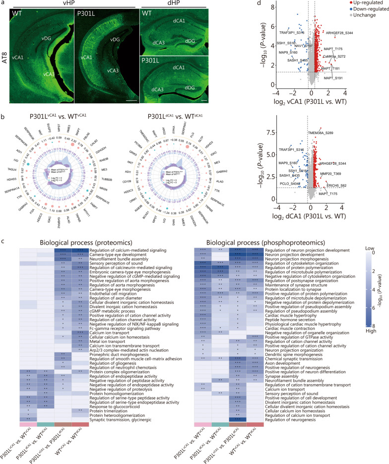
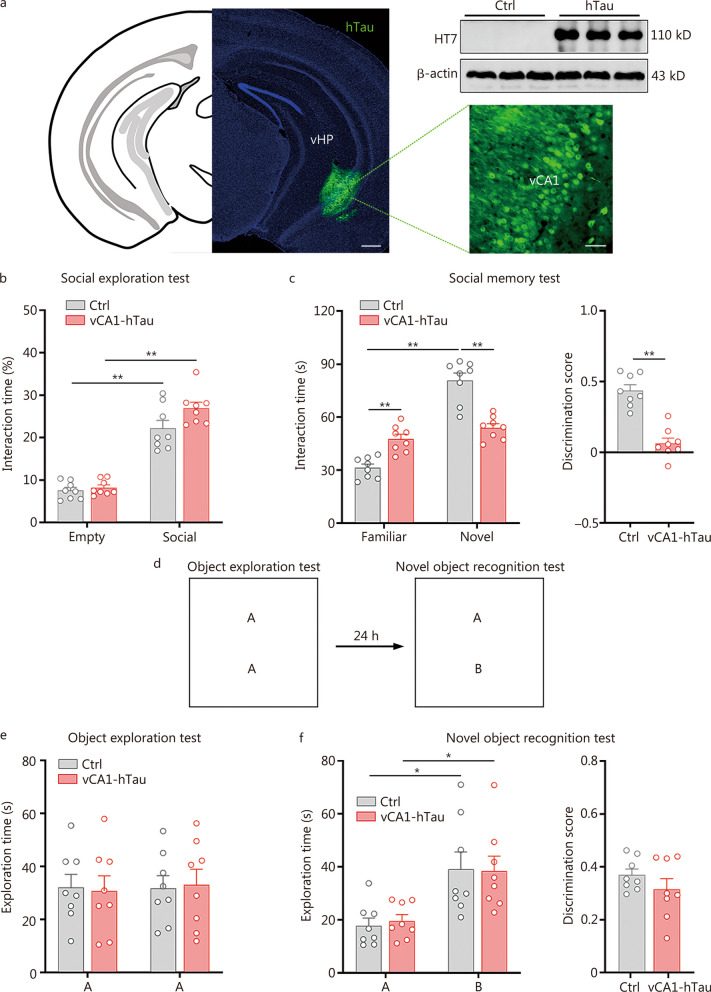
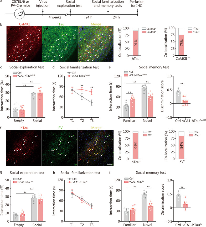
Results: The results of proteomic and phosphoproteomic analyses revealed the characteristics of ventral hippocampal CA1 (vCA1) under both physiological conditions and AD-like tau pathology. As tau progressively accumulated, vCA1, especially its excitatory and parvalbumin (PV) neurons, were fully filled with mislocated and phosphorylated tau (p-Tau). This finding was not observed for dorsal hippocampal CA1 (dCA1). The overexpression of human tau (hTau) in excitatory and PV neurons mimicked AD-like tau accumulation, significantly inhibited neuronal excitability and suppressed distinct discrimination-associated firings of these neurons within vCA1. Photoactivating excitatory and PV neurons in vCA1 at specific rhythms and time windows efficiently ameliorated tau-impaired social memory. Notably, 1 month of UA administration efficiently decreased tau accumulation via autophagy in a transcription factor EB (TFEB)-dependent manner and restored the vCA1 microcircuit to ameliorate tau-impaired social memory.

Conclusion: This study elucidated distinct protein and phosphoprotein networks between dCA1 and vCA1 and highlighted the susceptibility of the vCA1 microcircuit to AD-like tau accumulation. Notably, our novel findings regarding the efficacy of UA in reducing tau load and targeting the vCA1 microcircuit may provide a promising strategy for treating AD in the future.

Keywords: Alzheimer’s disease; Social memory; Tau protein; Transcription factor EB (TFEB); Ursolic acid; Ventral hippocampus.


Conflict of interest statement

The authors declare that they have no competing interests.

Figures

Fig. 1
Fig. 1
Proteomics and phosphoproteomics of dorsal hippocampal CA1 (dCA1) and ventral hippocampal CA1 (vCA1) in P301L mice. a Representative images showing abundant hyper-phosphorylated tau (p-Tau, AT8) in the vCA1 but not in the dCA1 of 3-month-old male P301L mice. Scale bar = 500 μm (vHP) or 200 μm (dHP). b Radar graph depicting differentially expressed proteins in the vCA1 (left) or dCA1 (right) between P301L mice and WT mice. From the outside to the inside, the first circle represents the name of protein. The orange arrow in the second circle represents the sequence of the differentially expressed proteins according to their P-values, arranged from smallest to largest. The third circle represents the ratio of differential expression changes in the comparison groups, namely, P301LvCA1 vs. WTvCA1 and P301LdCA1 vs. WTdCA1, with log2 transformation. In this representation, the colour pink indicates upregulation, while blue denotes down-regulation. The size of the data point corresponds to the magnitude of the difference. The fourth circle represents the average quantitative value of the two groups. c Heatmap displaying the functional enrichment clusters of differentially expressed proteins and phosphorylated proteins according to biological function. d Volcano plot of phosphoproteomic data revealing changes in the levels of phosphorylated proteins in P301LvCA1 vs. WTvCA1 and P301LdCA1 vs. WTdCA1. The red and blue dots represent phosphorylated proteins whose abundance significantly increased and decreased, respectively. n = 3 per group. *P < 0.05; **P < 0.01; ***P < 0.001. WT wild type, vDG ventral dentate gyrus, dDG dorsal dentate gyrus, SERPINA3K serine protease inhibitor A3K, SERPINA3N serine protease inhibitor A3N, ADI1 1,2-dihydroxy-3-keto-5-methylthiopentene dioxygenase, LVD isovaleryl-CoA dehydrogenase, TAGLN transgelin, HDHD3 haloacid dehalogenase-like hydrolase domain-containing protein 3, SERPINA1A serine protease inhibitor A1A, TTR transthyretin, GABRA3 γ-aminobutyric acid receptor subunit alpha-3, FGA fibrinogen alpha chain, HDDC3 guanosine-3’,5’-bis (diphosphate) 3’-pyrophosphohydrolase MESH1, IGKC immunoglobulin kappa constant, GABRA2 γ-aminobutyric acid receptor subunit alpha-2, ALAD delta-aminolevulinic acid dehydratase, TRPM5 transient receptor potential cation channel subfamily M member 5, MYH11 myosin-11, FABP7 fatty acid-binding protein type 7, SERPINA1E serine protease inhibitor A1E, NRGN neurogranin, TUBB2B tubulin beta-2B, ME3 NADP-dependent malic enzyme, RAB3B ras-related protein Rab-3B, D2HGDH D-2-hydroxyglutarate dehydrogenase, CALB2 calretinin, FBLN5 fibulin-5, MAPT microtubule-associated protein tau, NME3 nucleoside diphosphate kinase 3, MPST mercaptopyruvate sulfurtransferase, SUGP2 SURP and G-patch domain-containing protein 2, NNT nicotinamide nucleotide transhydrogenase, TMEM254C transmembrane protein 254c, CD109 CD109 antigen, P01864 Ig gamma-2A chain C region, BCAS1 breast carcinoma-amplified sequence 1 homolog, D2HGDH D-2-hydroxyglutarate dehydrogenase, ITPKA inositol-trisphosphate 3-kinase A, MAP9 microtubule-associated protein 9, SASH1 SAM and SH3 domain-containing protein 1, ARHGEF2 Rho guanine nucleotide exchange factor 2, CAMKIIa calcium/calmodulin-dependent protein kinase type II subunit alpha, TRAF3IP3 TNF receptor-associated factor interacting protein 3, PCLO protein piccolo, TMEM38A trimeric intracellular cation channel type A, ARHGEF28 rho guanine nucleotide exchange factor 28, MMP20 matrix metalloproteinase-20, ERICH5 glutamate-rich protein 5, S serine, T threonine
Fig. 2
Fig. 2
Tau accumulation in vCA1 impairs social memory. a Expression of exogenous hTau in vCA1 by the AAV-hSyn-hTau-eGFP virus. Scale bar = 500 μm (vHP) or 50 μm (vCA1). b Overexpression of hTau in vCA1 had no effect on sociability. c Overexpression of hTau in vCA1 significantly disrupted social preference for novel conspecifics and decreased the discrimination score in social memory test. d Behavioural schematic of novel objective recognition tests. e There was no difference in the exploration time of object A between the two groups. f Both vCA1-hTau and control mice spent more time investigating object B in the test. No difference was detected in the discrimination score between control and vCA1-hTau mice. n = 8 per group. *P < 0.05; **P < 0.01, as determined by two-way ANOVA or two-tailed unpaired t test. The data are presented as the mean ± SEM. vHP ventral hippocampal, vCA1 ventral hippocampal CA1, hTau human tau
Fig. 3
Fig. 3
Overexpression of hTau in excitatory and PV neurons impairs social memory. a Timeline of the experimental procedures. b Representative images showing the expression of AAV-CaMKII-Cre-mCherry and AAV-DIO-hTau-eGFP in the vCA1 of C57BL/6 mice. The arrows indicate colabelled cells. Scale bar = 100 μm. Quantitative analysis revealed that 91% of the hTau+ neurons were colocalized with CaMKII and 75% of the CaMKII+ neurons were colocalized with hTau. c vCA1-hTauCaMKII and control mice spent more time investigating conspecifics than empty cups. d vCA1-hTauCaMKII mice failed to familiarize themselves with the same social partner in trials 1 to 3 (T1–T3). e In the social memory test, vCA1-hTauCaMKII mice spent similar amounts of time investigating familiar and novel conspecifics. The social discrimination score was lower in the vCA1-hTauCaMKII group. f Representative images showing AAV-DIO-hTau-mCherry virus infection (red) and anti-PV immunofluorescence staining (green) in the vCA1 of PV-Cre mice. The arrows indicate colabelled cells. Scale bar = 100 μm. Quantitative analysis revealed that 94% of the neurons with hTau were PV+ and 84% of the PV+ neurons were colocalized with hTau. g Both vCA1-hTauPV group and control group exhibited a preference for conspecifics over empty cups. h vCA1-hTauPV mice succeeded in familiarizing themselves with the same social partner in trials 1 to 3 (T1–T3). i In the social memory test, vCA1-hTauPV mice spent similar amounts of time investigating familiar and novel conspecifics. The social discrimination score was lower in the vCA1-hTauPV group than in the control group. n = 8 per group. **P < 0.01, as determined by two-way ANOVA, two-way repeated ANOVA or two-tailed unpaired t test. The data are presented as the mean ± SEM. IHC immunohistochemistry, CaMKII calcium/calmodulin-dependent protein kinase II, PV parvalbumin, vCA1 ventral hippocampal CA1, hTau human tau
Fig. 4
Fig. 4
Tau accumulation disrupts the discharge of excitatory and PV neurons. a Timeline of the experimental procedures. b Schematic of AAV-CaMKII-hTau-mCherry injection into the vCA1 of C57BL/6 mice. c Representative traces of action potentials of CaMKII+ neurons from the control and vCA1-hTauCaMKII groups following the injection of currents. Ctrl: n = 19 neurons from 4 mice; vCA1-hTauCaMKII: n = 22 neurons from 5 mice. d Schematic of AAV-DIO-hTau-mCherry injection into the vCA1 of PV-Cre mice. e Representative traces of action potentials of PV neurons from the control and vCA1-hTauPV groups following the injection of currents. Ctrl: 21 neurons from 5 mice; vCA1-hTauPV: 24 neurons from 5 mice. Representative image of GCaMP6f expression in CaMKII-hTau+ (f) and PV-hTau+ (g) neurons in the vCA1. The arrows indicate colabelled cells. Scale bar = 100 μm. h Representative traces of GCaMP6f signals in the control and vCA1-hTauCaMKII groups. i Per-bout stacked plots of CaMKII-GCaMP6f signals were aligned to the start of the interaction event. The presence of “#” indicates a significant increase in the average calcium signals during the social exploration period (the first 2 s) compared to the baseline period (2 s before social exploration). n = 8 per group. j Quantitative analysis of peak dF/F of Ca2+ signals in CaMKII+ neurons during the social memory test. k Representative traces of GCaMP6f signals in the control and vCA1-hTauPV groups. l Per-bout stacked plots of PV-GCaMP6f signals aligned to the start of the interaction event. The presence of “#” indicates a significant increase in the average calcium signals during the social exploration period (the first 2 s) compared to those during the baseline period (2 s before social exploration). n = 6 per group. m Quantitative analysis of peak dF/F of Ca2+ signals in PV+ neurons during the social memory test. n = 6 mice per group. *P < 0.05; **P < 0.01, as determined by two-way ANOVA, two-way repeated ANOVA, two-tailed unpaired t test or two-tailed t test. The data are presented as the mean ± SEM. IHC immunohistochemistry, CaMKII calcium/calmodulin-dependent protein kinase II, PV parvalbumin, vCA1 ventral hippocampal CA1, GCaMP6f genetically encoded calcium indicators, hTau human tau
Fig. 5
Fig. 5
vCA1-hTauPV accumulation disinhibits excitatory neurons during novel conspecific identification. a Schematic of GABA signal recording in the social memory test. b Representative image of iGABASnFR and hTau expression in the vCA1 of PV-Cre mice. Scale bar = 100 μm. c Representative trace of GABA signals. d Per-bout stacked plots of GABA signals on CaMKII+ neurons aligned to the start of the interaction event. The presence of “#” indicates a significant increase in the average GABA signals during the social exploration period (the first 2 s) compared to those during the baseline period (2 s before social exploration). e Quantitative analysis of peak dF/F of GABA signals into CaMKII+ neurons during social memory test. f Schematic of Ca2+ signal recording in the social memory test. g Representative image of GCaMP6f and hTau expression in the vCA1 of PV-Cre mice. Scale bar = 100 μm. h Representative trace of GCaMP6f signals. i Per-bout stacked plots of GCaMP6f signals of CaMKII+ neurons aligned to the start of the interaction event. The presence of “#” indicates a significant increase in the average calcium signals during the social exploration period (the first 2 s) compared to those during the baseline period (2 s before social exploration). j Quantitative analysis of peak dF/F of Ca2+ signals in CaMKII+ neurons during the social memory test. n = 6 per group. *P < 0.05; **P < 0.01 as determined by two-way ANOVA or two-tailed paired t test. The data are presented as the mean ± SEM. CaMKII calcium/calmodulin-dependent protein kinase II, PV parvalbumin, vCA1 ventral hippocampal CA1, iGABASnFR intensity-based GABA-sensing fluorescence reporters, GCaMP6f genetically encoded calcium indicators
Fig. 6
Fig. 6
Photoactivation of excitatory and PV neurons rescues tau accumulation-impaired discrimination in social memory. Schematic of stimulation while vCA1-hTau mice were investigating familiar (a) and novel (c) conspecifics in the social memory test. b When vCA1-hTauCaMKII mice were exploring a familiar mouse, photoactivation of excitatory neurons in the vCA1 significantly increased the preference for the novel mouse and concurrently raised the discrimination score. n = 9 per group. d Photostimulation of PV neurons in the vCA1 at 40 Hz, as in which vCA1-hTauPV mice were exposed a novel mouse, efficiently increased the social interaction time with the novel mouse and increased the discrimination score. n = 8 per group. **P < 0.01, as determined by two-way ANOVA or two-tailed unpaired t test. The data are expressed as the mean ± SEM. CaMKII calcium/calmodulin-dependent protein kinase II, PV parvalbumin, vCA1 ventral hippocampal CA1, eYFP enhanced yellow fluorescent protein, ChR2 channelrhodopsins-2
Fig. 7
Fig. 7
Ursolic acid (UA) reduces the pathological tau load to improve social memory via TFEB. a Treatment with 30 μmol/L UA markedly reduced the total tau (tau5, HT7) and phosphorylated tau (p-Tau, AT8) levels in primary neurons instantly transferred with hTau, determined by Western blotting. b Treatment with 30 μmol/L UA for 12 h increased the translocation of TFEB from the cytoplasm to the nucleus, as detected by immunostaining. Green: TFEB; blue: DAPI. Scale bar = 25 μm. c UA treatment significantly increased LC3B-II and decreased p62 levels in HEK293-hTau cells, as determined by Western blotting. d Increased colocalization of mRFP and GFP was observed in HEK293-hTau cells transfected with the tandem fluorescent mRFP-GFP-LC3 (tfLC3) construct, indicating that an increased number of autophagosomes did not fuse with the lysosome. The number of red puncta (mature autolysosomes) increased in the UA-treated group, suggesting that UA promoted autophagy. Scale bar = 10 μm. e Timeline of the experimental procedures. f Knocking down TFEB effectively decreased TFEB levels in vCA1-hTau mice. g, h Knocking down TFEB attenuated UA-induced increases in LC3B-II, decreases in p62, total tau (tau5, HT7) and phosphorylated tau (p-Tau, AT8) in vCA1-hTau mice. i Knocking down TFEB had no significant effect on sociability. j Knocking down TFEB effectively attenuated the UA-induced increase in interaction time with novel mice and social discrimination score in vCA1-hTau mice. hTau + shNT + UA (L): n = 9; hTau + shTFEB + UA (L): n = 8. *P < 0.05; **P < 0.01, as determined by one-way ANOVA, two-way ANOVA or two-tailed unpaired t test. The data are expressed as the mean ± SEM. L low-dose, TFEB transcription factor EB, mRFP mCherry red fluorescence protein, GFP green fluorescence protein, LC3B microtubule-associated proteins 1A/1B light chain 3B, p62 sequestosome-1, i.g. intragastric administration
Fig. 8
Fig. 8
Proposed working model of how to target vulnerable microcircuits in the ventral hippocampus to ameliorate Alzheimer-like social memory loss. Proteomic and phosphoproteomic analyses revealed the characteristics of ventral hippocampal CA1 (vCA1) under both physiological conditions and in the presence of AD-like tau pathology. Excitatory and PV neurons in the vCA1 are vulnerable to tau accumulation. Accumulated tau suppresses excitability and disrupts the firing patterns of excitatory and PV neurons, impairing social memory. Intragastric administration of UA efficiently promoted autophagy to reduce the accumulation of tau and improve social memory in a TFEB-dependent manner. vCA1 ventral hippocampal CA1, PV parvalbumin, TFEB transcription factor EB, UA ursolic acid, AD alzheimer’s disease

References

    1. Li MZ, Liu EJ, Zhou QZ, Li SH, Liu SJ, Yu HT, Pan QH, et al. Intracellular accumulation of tau inhibits autophagosome formation by activating TIA1-amino acid-mTORC1 signaling. Mil Med Res. 2022;9(1):38. - PMC - PubMed
    1. Ranganath C, Ritchey M. Two cortical systems for memory-guided behaviour. Nat Rev Neurosci. 2012;13(10):713–726. doi: 10.1038/nrn3338. - DOI - PubMed
    1. Ritchey M, Libby LA, Ranganath C. Cortico-hippocampal systems involved in memory and cognition: the PMAT framework. Prog Brain Res. 2015;219:45–64. doi: 10.1016/bs.pbr.2015.04.001. - DOI - PubMed
    1. Kim S, Nilakantan AS, Hermiller MS, Palumbo RT, Vanhaerents S, Voss JL. Selective and coherent activity increases due to stimulation indicate functional distinctions between episodic memory networks. Sci Adv. 2018;4(8):eaar2768. doi: 10.1126/sciadv.aar2768. - DOI - PMC - PubMed
    1. Berron D, Neumann K, Maass A, Schütze H, Fliessbach K, Kiven V, et al. Age-related functional changes in domain-specific medial temporal lobe pathways. Neurobiol Aging. 2018;65:86–97. doi: 10.1016/j.neurobiolaging.2017.12.030. - DOI - PubMed