

Effects of Weight Bearing on Marrow Adipose Tissue and Trabecular Bone after Anterior Cruciate Ligament Reconstruction in the Rat Proximal Tibial Epiphysis
- PMID: 38463204
- PMCID: PMC10918432
- DOI: 10.1267/ahc.23-00060
Effects of Weight Bearing on Marrow Adipose Tissue and Trabecular Bone after Anterior Cruciate Ligament Reconstruction in the Rat Proximal Tibial Epiphysis
Abstract
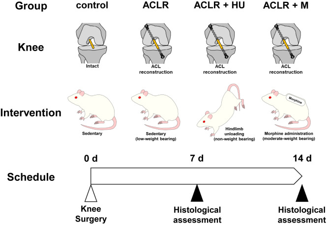
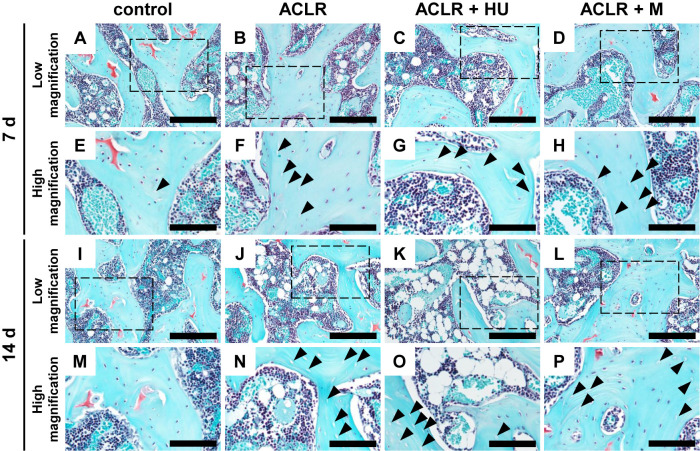
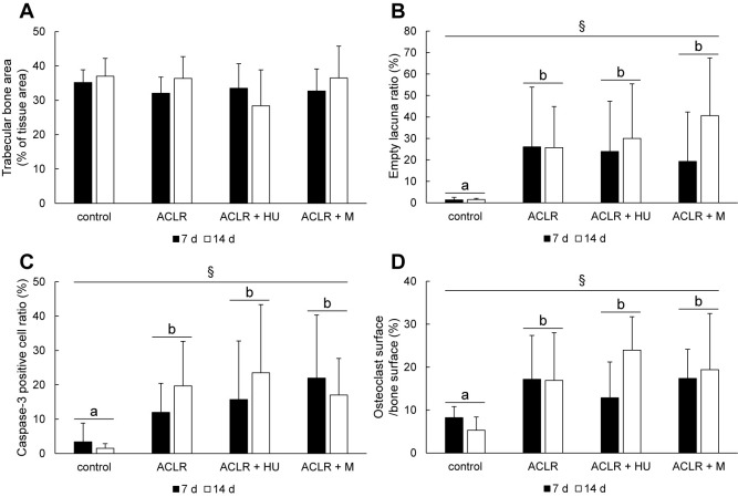
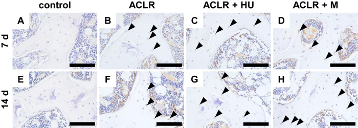
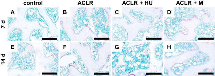
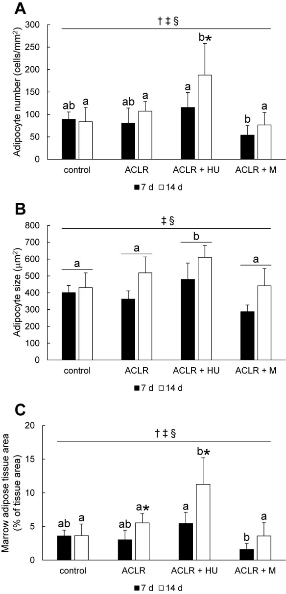
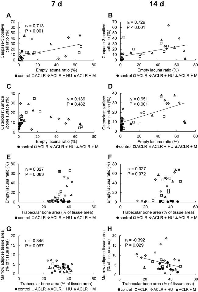
The effects of mechanical unloading after anterior cruciate ligament (ACL) reconstruction on bone and marrow adipose tissue (MAT) are unclear. We investigated weight bearing effects on bone and MAT after ACL reconstruction. Rats underwent unilateral knee ACL transection and reconstruction, followed by hindlimb unloading (non-weight bearing), no intervention (low-weight bearing, the hindlimb standing time ratio (STR; operated/contralateral) during treadmill locomotion ranging from 0.55 to 0.91), or sustained morphine administration (moderate-weight bearing, STR ranging from 0.80 to 0.95). Untreated rats were used as controls. At 7 or 14 days after surgery, changes in trabecular bone and MAT in the proximal tibial were assessed histologically. Histological assessments at 7 or 14 days after surgery showed that ACL reconstruction without post-operative intervention did not significantly change trabecular bone and MAT areas. Hindlimb unloading after ACL reconstruction induced MAT accumulation with adipocyte hyperplasia and hypertrophy within 14 days, but did not significantly affect trabecular bone area. Increased weight bearing through morphine administration did not affect trabecular bone and MAT parameters. Our results suggest that early weight bearing after ACL reconstruction is important in reducing MAT accumulation, and that reduction in weight bearing alone is not sufficient to induce bone loss early after ACL reconstruction.
Keywords: ACL reconstruction; apoptosis; marrow adipose tissue; trabecular bone; weight bearing.
2024 The Japan Society of Histochemistry and Cytochemistry.
Conflict of interest statement
VThe authors declare that there are no conflicts of interest.
Figures
Similar articles
-
Effects of joint immobilization and treadmill exercise on marrow adipose tissue and trabecular bone after anterior cruciate ligament reconstruction in the rat proximal tibial epiphysis.Acta Histochem. 2023 Apr;125(3):152012. doi: 10.1016/j.acthis.2023.152012. Epub 2023 Feb 9. Acta Histochem. 2023. PMID: 36773546
-
Marrow adipose tissue accumulation and dysgenesis of the trabecular bone after anterior cruciate ligament transection and reconstruction in the rat proximal tibial epiphysis.Acta Histochem. 2022 May;124(4):151891. doi: 10.1016/j.acthis.2022.151891. Epub 2022 Mar 31. Acta Histochem. 2022. PMID: 35367815
-
Long-term observation of marrow adipose tissue and trabecular bone in the rat proximal tibial epiphysis after anterior cruciate ligament reconstruction: effects of immobilization and non-weightbearing.Biotech Histochem. 2025 Feb;100(2):72-82. doi: 10.1080/10520295.2025.2470622. Epub 2025 Feb 26. Biotech Histochem. 2025. PMID: 40008463
-
[Combined posterior and anterior cruciate ligament reconstruction : Arthroscopic treatment with the GraftLink® system].Oper Orthop Traumatol. 2019 Feb;31(1):20-35. doi: 10.1007/s00064-018-0580-6. Epub 2018 Dec 18. Oper Orthop Traumatol. 2019. PMID: 30564843 Review. German.
-
Anterior cruciate ligament strain and tensile forces for weight-bearing and non-weight-bearing exercises: a guide to exercise selection.J Orthop Sports Phys Ther. 2012 Mar;42(3):208-20. doi: 10.2519/jospt.2012.3768. Epub 2012 Feb 29. J Orthop Sports Phys Ther. 2012. PMID: 22387600 Review.
Cited by
-
Roles of osteoclasts in pathological conditions.Pathol Int. 2025 Feb;75(2):55-68. doi: 10.1111/pin.13500. Epub 2024 Dec 20. Pathol Int. 2025. PMID: 39704061 Free PMC article. Review.
References
-
- Ahdjoudj, S., Lasmoles, F., Holy, X., Zerath, E. and Marie, P. J. (2002) Transforming growth factor beta2 inhibits adipocyte differentiation induced by skeletal unloading in rat bone marrow stroma. J. Bone. Miner. Res. 17; 668–677. - PubMed
-
- Anderson, M. J., Diko, S., Baehr, L. M., Baar, K., Bodine, S. C. and Christiansen, B. A. (2016) Contribution of mechanical unloading to trabecular bone loss following non-invasive knee injury in mice. Journal of Orthopaedic Research: official publication of the Orthopaedic Research Society. 34; 1680–1687. - PMC - PubMed