

Intercellular crosstalk between cancer cells and cancer-associated fibroblasts via exosomes in gastrointestinal tumors
- PMID: 38463229
- PMCID: PMC10920350
- DOI: 10.3389/fonc.2024.1374742
Intercellular crosstalk between cancer cells and cancer-associated fibroblasts via exosomes in gastrointestinal tumors
Abstract
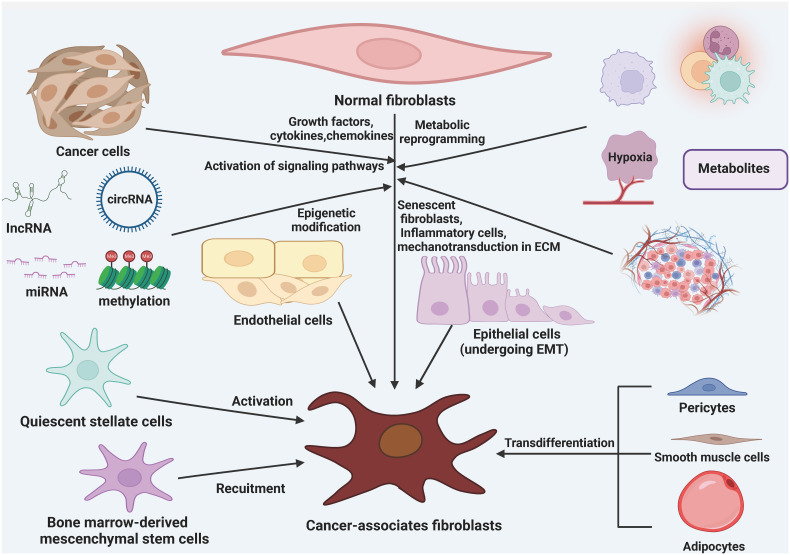
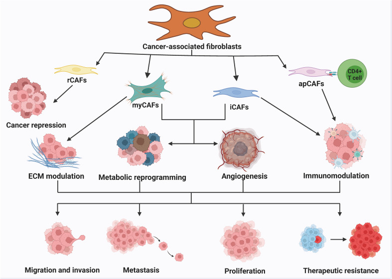
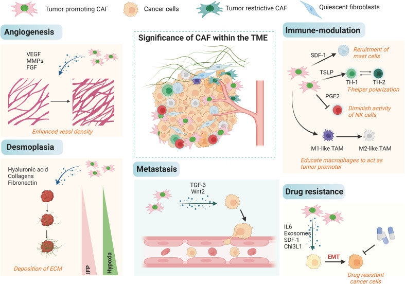
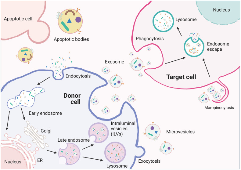
Gastrointestinal (GI) tumors are a significant global health threat, with high rates of morbidity and mortality. Exosomes contain various biologically active molecules like nucleic acids, proteins, and lipids and can serve as messengers for intercellular communication. They play critical roles in the exchange of information between tumor cells and the tumor microenvironment (TME). The TME consists of mesenchymal cells and components of the extracellular matrix (ECM), with fibroblasts being the most abundant cell type in the tumor mesenchyme. Cancer-associated fibroblasts (CAFs) are derived from normal fibroblasts and mesenchymal stem cells that are activated in the TME. CAFs can secrete exosomes to modulate cell proliferation, invasion, migration, drug resistance, and other biological processes in tumors. Additionally, tumor cells can manipulate the function and behavior of fibroblasts through direct cell-cell interactions. This review provides a summary of the intercellular crosstalk between GI tumor cells and CAFs through exosomes, along with potential underlying mechanisms.
Keywords: cancer-associated fibroblasts (CAFs); exosomes; gastrointestinal (GI) tumors; review; tumor microenvironment (TME).
Copyright © 2024 Cao and Ouyang.
Conflict of interest statement
The authors declare that the research was conducted in the absence of any commercial or financial relationships that could be construed as a potential conflict of interest.
Figures
Similar articles
-
Cancer associated-fibroblast-derived exosomes in cancer progression.Mol Cancer. 2021 Dec 1;20(1):154. doi: 10.1186/s12943-021-01463-y. Mol Cancer. 2021. PMID: 34852849 Free PMC article. Review.
-
Crosstalk between cancer-associated fibroblasts and immune cells in the tumor microenvironment: new findings and future perspectives.Mol Cancer. 2021 Oct 11;20(1):131. doi: 10.1186/s12943-021-01428-1. Mol Cancer. 2021. PMID: 34635121 Free PMC article. Review.
-
Exosomal microRNAs mediating crosstalk between cancer cells and cancer-associated fibroblasts in the tumor microenvironment.Pathol Res Pract. 2022 Nov;239:154159. doi: 10.1016/j.prp.2022.154159. Epub 2022 Oct 10. Pathol Res Pract. 2022. PMID: 36244248 Review.
-
Cancer-associated fibroblasts and its derived exosomes: a new perspective for reshaping the tumor microenvironment.Mol Med. 2023 May 22;29(1):66. doi: 10.1186/s10020-023-00665-y. Mol Med. 2023. PMID: 37217855 Free PMC article. Review.
-
Exploring the multifaceted role of direct interaction between cancer cells and fibroblasts in cancer progression.Front Mol Biosci. 2024 May 28;11:1379971. doi: 10.3389/fmolb.2024.1379971. eCollection 2024. Front Mol Biosci. 2024. PMID: 38863965 Free PMC article. Review.
Cited by
-
Decellularized tissues as platforms for digestive system cancer models.Heliyon. 2024 May 21;10(11):e31589. doi: 10.1016/j.heliyon.2024.e31589. eCollection 2024 Jun 15. Heliyon. 2024. PMID: 38845895 Free PMC article. Review.
-
Heterocellular Adhesion in Cancer Invasion and Metastasis: Interactions between Cancer Cells and Cancer-Associated Fibroblasts.Cancers (Basel). 2024 Apr 24;16(9):1636. doi: 10.3390/cancers16091636. Cancers (Basel). 2024. PMID: 38730588 Free PMC article. Review.
-
GPD1L may inhibit the development of esophageal squamous cell carcinoma through the PI3K/AKT signaling pathway: bioinformatics analysis and experimental exploration.Mol Biol Rep. 2024 Nov 13;51(1):1149. doi: 10.1007/s11033-024-10070-1. Mol Biol Rep. 2024. PMID: 39535578
-
Beneficial and challenges of exosome application in ischemic heart disease.Stem Cell Res Ther. 2025 May 19;16(1):247. doi: 10.1186/s13287-025-04363-w. Stem Cell Res Ther. 2025. PMID: 40390086 Free PMC article. Review.
-
Bioinformatics analysis identifies NT5M modulates immune evasion through PD-L1 via CXCL8 in pancreatic adenocarcinoma.Sci Rep. 2025 Jul 19;15(1):26207. doi: 10.1038/s41598-025-10098-8. Sci Rep. 2025. PMID: 40683920 Free PMC article.
References
-
- Morgan E, Soerjomataram I, Rumgay H, Coleman HG, Thrift AP, Vignat J, et al. . The global landscape of esophageal squamous cell carcinoma and esophageal adenocarcinoma incidence and mortality in 2020 and projections to 2040: New estimates from GLOBOCAN 2020. Gastroenterology. (2022) 163:649–658.e2. doi: 10.1053/j.gastro.2022.05.054 - DOI - PubMed
-
- Nesteruk K, Spaander MCW, Leeuwenburgh I, Peppelenbosch MP, Fuhler GM. Achalasia and associated esophageal cancer risk: What lessons can we learn from the molecular analysis of Barrett's-associated adenocarcinoma? Biochim Biophys Acta Rev Cancer. (2019) 1872:188291. doi: 10.1016/j.bbcan.2019.04.007 - DOI - PubMed
Publication types
LinkOut - more resources
Full Text Sources