

Postoperative encapsulated hemoperitoneum in a patient with gastric stromal tumor treated by exposed endoscopic full-thickness resection: A case report
- PMID: 38463350
- PMCID: PMC10921194
- DOI: 10.4240/wjgs.v16.i2.601
Postoperative encapsulated hemoperitoneum in a patient with gastric stromal tumor treated by exposed endoscopic full-thickness resection: A case report
Abstract
Background: Gastric stromal tumors, originating from mesenchymal tissues, are one of the most common tumors of the digestive tract. For stromal tumors originating from the muscularis propria, compared with conventional endoscopic submucosal dissection (ESD), endoscopic full-thickness resection (EFTR) can remove deep lesions and digestive tract wall tumors completely. However, this technique has major limitations such as perforation, postoperative bleeding, and post-polypectomy syndrome. Herein, we report a case of postoperative serous surface bleeding which formed an encapsulated hemoperitoneum in a patient with gastric stromal tumor that was treated with exposed EFTR. Feasible treatment options to address this complication are described.
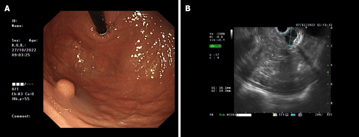
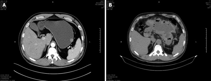
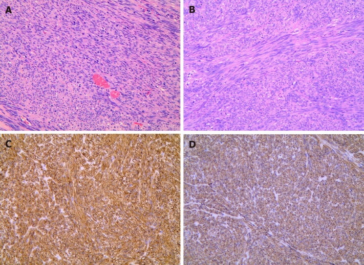
Case summary: A 47-year-old male patient had a hemispherical protrusion found during gastric endoscopic ultrasonography, located at the upper gastric curvature adjacent to the stomach fundus, with a smooth surface mucosa and poor mobility. The lesion was 19.3 mm × 16.1 mm in size and originated from the fourth ultrasound layer. Computed tomography (CT) revealed no significant evidence of lymph node enlargement or distant metastasis. Using conventional ESD technology for mucosal pre-resection, exposed EFTR was performed to resect the intact tumor in order to achieve a definitive histopathological diagnosis. Based on its morphology and immunohistochemical expression of CD117 and DOG-1, the lesion was proven to be consistent with a gastric stromal tumor. Six days after exposed EFTR, CT showed a large amount of encapsulated fluid and gas accumulation around the stomach. In addition, gastroscopy suggested intracavitary bleeding and abdominal puncture drainage indicated serosal bleeding. Based on these findings, the patient was diagnosed with serosal bleeding resulting in encapsulated abdominal hemorrhage after exposed EFTR for a gastric stromal tumor. The patient received combined treatments, such as hemostasis under gastroscopy, gastrointestinal decompression, and abdominal drainage. All examinations were normal within six months of follow-up.
Conclusion: This patient developed serous surface bleeding in the gastric cavity following exposed EFTR. Serosal bleeding resulting in an encapsulated hemoperitoneum is rare in clinical practice. The combined treatment may replace certain surgical techniques.
Keywords: Abdominal infection; Case report; Complication; Exposed endoscopic full-thickness resection; Gastric stromal tumors; Hemoperitoneum; Postoperative bleeding.
©The Author(s) 2024. Published by Baishideng Publishing Group Inc. All rights reserved.
Conflict of interest statement
Conflict-of-interest statement: The authors declare that they have no conflict of interest to disclose.
Figures
Similar articles
-
The application of endoscopic loop ligation in defect repair following endoscopic full-thickness resection of gastric submucosal tumors originating from the muscularis propria layer.Scand J Gastroenterol. 2022 Jan;57(1):119-123. doi: 10.1080/00365521.2021.1981994. Epub 2021 Dec 2. Scand J Gastroenterol. 2022. PMID: 34854324
-
[A novel and simplified closure method for defect closure after endoscopic full-thickness resection of gastric submucosal tumors: short-term outcomes of "Shao-Mai" closure method].Zhonghua Wei Chang Wai Ke Za Zhi. 2020 Feb 25;23(2):183-187. doi: 10.3760/cma.j.issn.1671-0274.2020.02.015. Zhonghua Wei Chang Wai Ke Za Zhi. 2020. PMID: 32074800 Chinese.
-
[Risk factors for complications of endoscopic full-thickness resection of upper gastrointestinal submucosal tumors].Zhonghua Wei Chang Wai Ke Za Zhi. 2023 Apr 25;26(4):365-371. doi: 10.3760/cma.j.cn441530-20220715-00312. Zhonghua Wei Chang Wai Ke Za Zhi. 2023. PMID: 37072314 Chinese.
-
Gastric duplication cysts: literature review and a case report of rare multiple gastric duplication cysts treated by endoscopic submucosal dissection.Postgrad Med. 2023 Nov;135(8):775-783. doi: 10.1080/00325481.2023.2274308. Epub 2024 Jan 10. Postgrad Med. 2023. PMID: 37877305 Review.
-
Efficacy and safety of gastric exposed endoscopic full-thickness resection without laparoscopic assistance: a systematic review.Endosc Int Open. 2020 Sep;8(9):E1173-E1182. doi: 10.1055/a-1198-4357. Epub 2020 Aug 31. Endosc Int Open. 2020. PMID: 32904958 Free PMC article. Review.
References
Publication types
LinkOut - more resources
Full Text Sources
Miscellaneous
