

Treatment of hemolymphangioma by robotic surgery: A case report
- PMID: 38463376
- PMCID: PMC10921197
- DOI: 10.4240/wjgs.v16.i2.596
Treatment of hemolymphangioma by robotic surgery: A case report
Abstract
Background: Hemolymphangioma of the jejunum is rare and lacks clinical specificity, and can manifest as gastrointestinal bleeding, abdominal pain, and intestinal obstruction. Computed tomography, magnetic resonance imaging, and other examinations show certain characteristics of the disease, but lack accuracy. Although capsule endoscopy and enteroscopy make up for this deficiency, the diagnosis also still requires pathology.
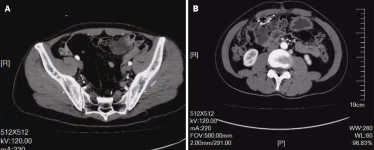
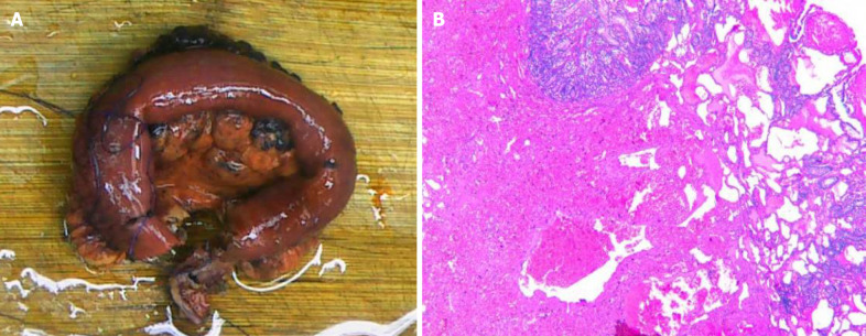
Case summary: A male patient was admitted to the hospital due to abdominal distension and abdominal pain, but a specific diagnosis by computed tomography examination was not obtained. Partial resection of the small intestine was performed by robotic surgery, and postoperative pathological biopsy confirmed the diagnosis of hemolymphangioma. No recurrence in the follow-up examination was observed.
Conclusion: Robotic surgery is an effective way to treat hemolymphangioma through minimally invasive techniques under the concept of rapid rehabilitation.
Keywords: Case report; Enteroscopy; Hemolymphangioma; Rehabilitation; Robotic surgery.
©The Author(s) 2024. Published by Baishideng Publishing Group Inc. All rights reserved.
Conflict of interest statement
Conflict-of-interest statement: The authors have no conflicts of interest to declare.
Figures
Similar articles
-
Small intestinal hemolymphangioma treated with enteroscopic injection sclerotherapy: A case report and review of literature.World J Gastroenterol. 2020 Apr 7;26(13):1540-1545. doi: 10.3748/wjg.v26.i13.1540. World J Gastroenterol. 2020. PMID: 32308353 Free PMC article. Review.
-
Jejunal hemolymphangioma: A case report.Medicine (Baltimore). 2020 Jan;99(4):e18863. doi: 10.1097/MD.0000000000018863. Medicine (Baltimore). 2020. PMID: 31977886 Free PMC article.
-
Small intestinal hemolymphangioma with bleeding: a case report.World J Gastroenterol. 2012 May 7;18(17):2145-6. doi: 10.3748/wjg.v18.i17.2145. World J Gastroenterol. 2012. PMID: 22563205 Free PMC article.
-
A case of invasive hemolymphangioma of the pancreas.World J Gastroenterol. 2008 May 14;14(18):2932-4. doi: 10.3748/wjg.14.2932. World J Gastroenterol. 2008. PMID: 18473426 Free PMC article. Review.
-
A very rare case of duodenal hemolymphangioma presenting with iron deficiency anemia.Int J Surg Case Rep. 2014;5(3):118-21. doi: 10.1016/j.ijscr.2013.12.026. Epub 2014 Jan 14. Int J Surg Case Rep. 2014. PMID: 24503337 Free PMC article.
Cited by
-
Case Report: Giant mesenteric hemolymphangioma enveloping the ileum.Front Oncol. 2025 Jun 18;15:1557916. doi: 10.3389/fonc.2025.1557916. eCollection 2025. Front Oncol. 2025. PMID: 40606978 Free PMC article.
References
-
- Fan Z, Li Y, Yan K, Wu W, Yin S, Yang W, Xing B, Li X, Zhang X. Application of contrast-enhanced ultrasound in the diagnosis of solid pancreatic lesions--a comparison of conventional ultrasound and contrast-enhanced CT. Eur J Radiol. 2013;82:1385–1390. - PubMed
Publication types
LinkOut - more resources
Full Text Sources
