. 2024 Mar 9;5(3):e508.
doi: 10.1002/mco2.508. eCollection 2024 Mar.

Current clinical findings of acute neurological syndromes after SARS-CoV-2 infection

Affiliations

Current clinical findings of acute neurological syndromes after SARS-CoV-2 infection

Minjin Wang et al. MedComm (2020). .

Abstract

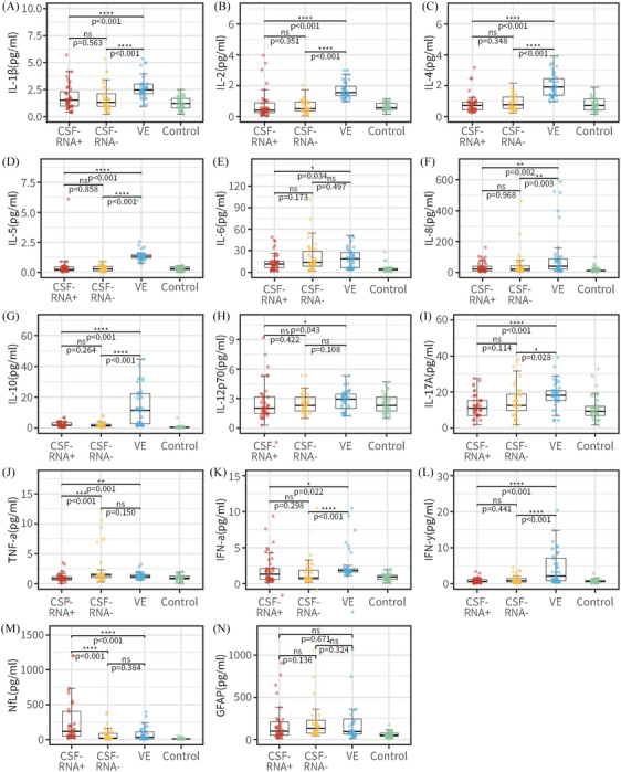
Neuro-COVID, a condition marked by persistent symptoms post-COVID-19 infection, notably affects various organs, with a particular focus on the central nervous system (CNS). Despite scant evidence of SARS-CoV-2 invasion in the CNS, the increasing incidence of Neuro-COVID cases indicates the onset of acute neurological symptoms early in infection. The Omicron variant, distinguished by heightened neurotropism, penetrates the CNS via the olfactory bulb. This direct invasion induces inflammation and neuronal damage, emphasizing the need for vigilance regarding potential neurological complications. Our multicenter study represents a groundbreaking revelation, documenting the definite presence of SARS-CoV-2 in the cerebrospinal fluid (CSF) of a significant proportion of Neuro-COVID patients. Furthermore, notable differences emerged between RNA-CSF-positive and negative patients, encompassing aspects such as blood-brain barrier integrity, extent of neuronal damage, and the activation status of inflammation. Despite inherent limitations, this research provides pivotal insights into the intricate interplay between SARS-CoV-2 and the CNS, underscoring the necessity for ongoing research to fully comprehend the virus's enduring effects on the CNS. The findings underscore the urgency of continuous investigation Neuro-COVID to unravel the complexities of this relationship, and pivotal in addressing the long-term consequences of COVID-19 on neurological health.

Keywords: Neuro‐COVID; SARS‐CoV‐2; central nerve injury; neurological syndromes; neurotropic invasion.


Conflict of interest statement

The authors declare no conflict of interest.

Figures

FIGURE 1
FIGURE 1
A flowchart of the study subjects. aIn order to ensure the integrity of WGS analysis outcomes, we exclusively opt for samples that exhibit positive results through RT‐PCR detection with a Ct < 25 for WGS analysis. bIncluding CSF cytokines levels, total protein levels, oligoclonal bands, serum levels of neurofilament light protein and glial fibrillary acidic protein.
FIGURE 2
FIGURE 2
Images of brain MRI scans of Neuro‐COVID patients. (A and B) A 20‐year‐old woman presented with gradually worsening delirium after an upper respiratory tract infection. MR imaging of the brain demonstrated prominent T2‐FLAIR abnormalities in the splenium of the corpus callosum (Arrowheads). (C and D) A 55‐year‐old man with recurrent acute myelitis presented with progressively worsening numbness and weakness in the extremities. Contrast‐enhanced MRI of the cervical and thoracic spine exhibited abnormal enhancement of the cervical spinal cord and enhancement of the cervicothoracic meninges (Arrowheads). (E and F) A 19‐year‐old man presented with psychosis and behavior disorder. MRI of the brain prominent T2‐FLAIR abnormalities in the bilateral hippocampus (Arrowheads).
FIGURE 3
FIGURE 3
Genome statistics of SARS‐CoV‐2 isolates. Schematic representation of the genome organization and genetic mutations in the genome of SARS‐CoV‐2. Yellow dots indicate mutations found in both CSF and NPS samples. Mapping and semi‐log depth of coverage of sequencing reads in CSF and NPS samples against the first Wuhan SARS‐CoV‐2 genome.
FIGURE 4
FIGURE 4
Comparison of the SARS‐CoV‐2 related parameters and laboratory parameters in Neuro‐COVID patients, patients with viral encephalitis and controls. CSF Il‐1β (A), IL‐2 (B), IL‐4 (C), IL‐5 (D), IL‐6 (E), IL‐8 (F), IL‐10 (G), IL‐12p70 (H), IL‐17A (I), TNF‐α (J), IFN‐α (K), and IFN‐γ (L) determined by immunofluorescence technology. Plasma NfL (M) and GFAP (N) from SiMoA Platform analysis. Comparisons between RNACSF‐positive group (red), RNACSF‐negative group (yellow), viral encephalitis group (blue) and control (green) done using the Mann–Whitney U test. *p < 0.05; **p < 0.01, ***p < 0.001, ****p < 0.0001. CSF, cerebrospinal fluid; IL, interleukin; TNF, tumor necrosis factor; IFN, interferon; NfL, neurofilament light chain; GFAP, glial fibrillary acidic protein.

Similar articles

References

    1. Monje M, Iwasaki A. The neurobiology of long COVID. Neuron. 2022;110(21):3484‐3496. - PMC - PubMed
    1. Shimohata T. Neuro‐COVID‐19. Clin Exp Neuroimmunol. 2022;13(1):17‐23. - PMC - PubMed
    1. Mao L, Jin H, Wang M, et al. Neurologic manifestations of hospitalized patients with coronavirus disease 2019 in Wuhan, China. JAMA Neurol. 2020;77(6):683‐690. - PMC - PubMed
    1. Douaud G, Lee S, Alfaro‐Almagro F, et al. SARS‐CoV‐2 is associated with changes in brain structure in UK Biobank. Nature. 2022;604(7907):697‐707. - PMC - PubMed
    1. Koralnik IJ, Tyler KL. COVID‐19: a global threat to the nervous system. Ann Neurol. 2020;88(1):1‐11. - PMC - PubMed