. 2024 Mar 5:36:185-202.
doi: 10.1016/j.bioactmat.2024.02.029. eCollection 2024 Jun.

Cell-homing and immunomodulatory composite hydrogels for effective wound healing with neovascularization

Affiliations

Cell-homing and immunomodulatory composite hydrogels for effective wound healing with neovascularization

Hayeon Byun et al. Bioact Mater. .

Abstract

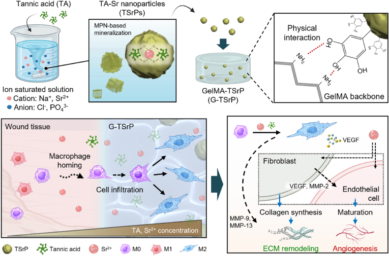
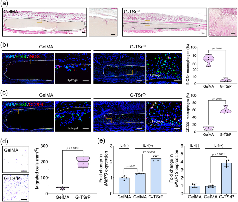
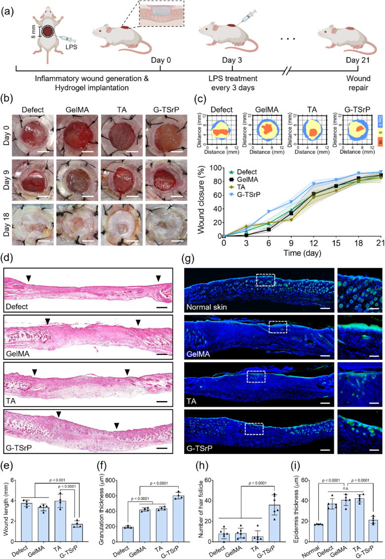
Wound healing in cases of excessive inflammation poses a significant challenge due to compromised neovascularization. Here, we propose a multi-functional composite hydrogel engineered to overcome such conditions through recruitment and activation of macrophages with adapted degradation of the hydrogel. The composite hydrogel (G-TSrP) is created by combining gelatin methacryloyl (GelMA) and nanoparticles (TSrP) composed of tannic acid (TA) and Sr2+. These nanoparticles are prepared using a one-step mineralization process assisted by metal-phenolic network formation. G-TSrP exhibits the ability to eliminate reactive oxygen species and direct polarization of macrophages toward M2 phenotype. It has been observed that the liberation of TA and Sr2+ from G-TSrP actively facilitate the recruitment and up-regulation of the expression of extracellular matrix remodeling genes of macrophages, and thereby, coordinate in vivo adapted degradation of the G-TSrP. Most significantly, G-TSrP accelerates angiogenesis despite the TA's inhibitory properties, which are counteracted by the released Sr2+. Moreover, G-TSrP enhances wound closure under inflammation and promotes normal tissue formation with strong vessel growth. Genetic analysis confirms macrophage-mediated wound healing by the composite hydrogel. Collectively, these findings pave the way for the development of biomaterials that promote wound healing by creating regenerative environment.

Keywords: Composite hydrogels; Immunomodulation; Multi-functional nanoparticles; Neovascularization; Wound healing.


Conflict of interest statement

None.

Figures

Image 1
Graphical abstract
Fig. 1
Fig. 1
Schematic illustration of the fabrication of gelatin methacryloyl (GelMA) hydrogel with tannic acid-strontium nanoparticle (G-TSrP) and its wound-healing mechanism. TSrPs were fabricated by mixing tannic acid (TA) with ion-saturated solution in one-step process. The nanoparticles were then incorporated in the GelMA hydrogel via physical interactions. In an inflammatory wound, the G-TSrP releases TA and Sr2+ to recruit and polarize macrophages into the M2 state to accelerate regenerative pathway. In addition, the polarized M2 macrophages, along with Sr2+, stimulates fibroblasts and endothelial cells, leading to increased collagen synthesis and matrix metalloproteinase (MMP) secretion, and the maturation of endothelial cells, essential for extracellular matrix (ECM) remodeling and angiogenesis, respectively.
Fig. 2
Fig. 2
Fabrication and characterization of TSrP. (a) Scanning electron microscopy (SEM) (top, scale bar: 500 nm) and transmission electron microscopy (bottom, scale bar: 250 nm) images of TSrP fabricated with 1 mg/mL TA. (b) Size distribution of nanoparticles made of different concentrations of TA. (c) Fourier transform infrared spectroscopy spectra of the nanoparticles. (d) Energy dispersive X-ray (EDX) elemental mapping and analysis of the TSrP fabricated with 1 mg/mL TA. Scale bar: 250 nm. (e) Results of a Folin-Ciocalteu assay of total phenol content of TSrPs prepared with different TA concentrations. (f) ABTS radical-scavenging effect of different concentrations of TSrP fabricated with 1 mg/mL TA.
Fig. 3
Fig. 3
Characterization and biocompatibility of nanocomposite hydrogels. (a) Optical images of hydrogels with different concentrations of TSrP. Scale bar: 4 mm. (b) SEM images of cross-sectioned hydrogels after lyophilization. Scale bar: 50 μm. (c) Swelling ratio of hydrogels with different concentrations of TSrP and their (d) storage modulus. (e) Young's modulus obtained from tensile test and (f) o-phthalaldehyde assay result of GelMA and G-TSrP hydrogels (8 mg/mL TSrP) with the same storage modulus. (g) ABTS radical-scavenging effect of hydrogels with different TSrP concentrations. (h) Representative live/dead staining images of human dermal fibroblasts (HDFBs) (scale bar: 250 μm) and (i) viability of various cell types cultured with hydrogel extracts for 24 h.
Fig. 4
Fig. 4
Intracellular reactive oxygen species (ROS)-scavenging effect of G-TSrP and release profile of TA and Sr2+. 2′,7′-dichlorofluorescein diacetate (DCFDA) assay images and their quantification using (a) HDFBs (scale bar: 100 μm) and (b) RAW264.7 cells (scale bar: 50 μm) treated with different hydrogel extracts and H2O2. (c) DCFDA assay images and their quantification using RAW264.7 cells after treatment of hydrogel extracts and lipopolysaccharide (LPS). Scale bar: 50 μm. (d) Release profile of TA and Sr2+ from G-TSrP for 14 days.
Fig. 5
Fig. 5
Modulation of macrophage polarization by G-TSrP. (a) Percentage of iNOS-positive cells from immunofluorescence (IF) images of RAW264.7 cells after treatment with LPS and hydrogel extracts. (b) F-actin staining images of RAW264.7 cells after treatment with LPS and hydrogel extracts, along with measurement of their cell area. Scale bar: 25 μm. (c) Percentage of CD206-positive cells from IF images of RAW264.7 cells after treatment with interleukin (IL)-4 and hydrogel extracts. (d) F-actin staining images of RAW264.7 cells after treatment with IL-4 and hydrogel extracts, along with measurement of their aspect ratio. Scale bar: 25 μm. Relative gene expression of (e) M1 polarization markers and (f) M2 polarization markers of RAW264.7 cells cultured using hydrogel extracts with their induction factors (M1: LPS, M2: IL-4).
Fig. 6
Fig. 6
Effect of G-TSrP on angiogenesis. (a) Relative expression of vascular endothelial growth factor (VEGF) and MMP-2 genes of HDFBs cultured with different hydrogel extracts for 7 days. (b) Representative images and the total branching length of human umbilical vein endothelial cells (HUVECs) after treatment of hydrogel extracts. Scale bar: 250 μm. (c) Scratch assay and wound closure after 24 h of incubation with hydrogel extracts. Scale bar: 250 μm. (d) Representative images of transwell migration assay using HUVECs cultured with different hydrogel extracts for 24 h. Scale bar: 125 μm.
Fig. 7
Fig. 7
In vivo and in vitro cell homing and biodegradation of composite hydrogels. (a) Hematoxylin and eosin (H&E) staining images (scale bar: 250 μm) and their magnified images (scale bar: 50 μm) of mouse subcutaneous tissues 14 days after hydrogel implantation. (b) Representative dual IF staining (F4/80 and iNOS) images (scale bar: 250 μm) and their magnified images (scale bar: 50 μm) of mouse subcutaneous tissues 14 days after implantation, along with their quantification. (c) Representative dual IF staining (F4/80 and CD206) images (scale bar: 250 μm) and their magnified images (scale bar: 50 μm) of mouse subcutaneous tissues 14 days after implantation, along with their quantification. (d) Images of migrated RAW264.7 cells cultured with GelMA and G-TSrP hydrogel for 24 h and their quantification. Scale bar: 250 μm. (e) Relative gene expression of MMP9 and MMP13 of RAW264.7 cells cultured with IL-4 and each hydrogel extract.
Fig. 8
Fig. 8
In vivo inflammatory wound healing. (a) Schematic illustration of the establishment of the inflammatory wound-healing model and the timeline. (b) Representative optical images of wound area after implantation of different hydrogels. Scale bar: 4 mm. (c) Schematic diagram and the quantification of closed wounds. (d) Representative H&E staining images and analysis of the (e) wound length and (f) granulation thickness of wound tissue at day 21. Scale bar: 250 μm. (g) Representative IF staining images (scale bar: 250 μm) and their magnified images (scale bar: 125 μm) of cytokeratin in wounds 21 days after implantation. Analysis of (h) number of hair follicles and (i) epidermis thickness using the IF staining images of cytokeratin.
Fig. 9
Fig. 9
Tissue remodeling and angiogenesis of inflammatory wound. (a) Masson's trichrome staining and immunohistochemistry (IHC) staining of collagen type Ⅰ (COL Ⅰ) images of the wound tissue on day 21. Scale bar: 125 μm. (b) Representative IF images of CD31 and α-SMA (scale bar: 125 μm) of wound tissues on day 21 and their magnified images (scale bar: 30 μm). (c) CD31-positive and (d) α-SMA-positive area analyzed by IF images. (e) Analysis of vessels in wound tissue based on α-SMA-positive vessels in IF images.
Fig. 10
Fig. 10
Genetic analysis of macrophage-mediated inflammatory wound healing. (a) Total heatmap data of RAW264.7 cells after treatment of different hydrogel extracts in the presence of LPS. (b) Volcano plots representing gene expression in the G-TSP group compared with the GelMA group. (c) Principal component analysis (PCA) plot displaying the variances of the genes in macrophages of GelMA, TA and G-TSrP group. (d) Up-regulated and down-regulated gene ontology (GO) analysis. (e) Schematic illustration of the mechanism of macrophage-mediated wound healing process promoted by G-TSrP. GO enrichment analysis of (f) positive regulation of immune response, (g) cell migration, and (h) angiogenesis.

References

    1. Gurtner G.C., Werner S., Barrandon Y., Longaker M.T. Wound repair and regeneration. Nature. 2008;453:314–321. doi: 10.1038/nature07039. - DOI - PubMed
    1. Fu Y.J., Shi Y.F., Wang L.Y., Zhao Y.F., Wang R.K., Li K., Zhang S.T., Zha X.J., Wang W., Zhao X., Yang W. All-natural immunomodulatory bioadhesive hydrogel promotes angiogenesis and diabetic wound healing by regulating macrophage heterogeneity. Adv. Sci. 2023;10:1–15. doi: 10.1002/advs.202206771. - DOI - PMC - PubMed
    1. Pasparakis M., Haase I., Nestle F.O. Mechanisms regulating skin immunity and inflammation. Nat. Rev. Immunol. 2014;14:289–301. doi: 10.1038/nri3646. - DOI - PubMed
    1. Kim S.Y., Nair M.G. Macrophages in wound healing: activation and plasticity. Immunol. Cell Biol. 2019;97:258–267. doi: 10.1111/IMCB.12236. - DOI - PMC - PubMed
    1. Mao J., Chen L., Cai Z., Qian S., Liu Z., Zhao B., Zhang Y., Sun X., Cui W. Advanced biomaterials for regulating polarization of macrophages in wound healing. Adv. Funct. Mater. 2022;32:1–25. doi: 10.1002/adfm.202111003. - DOI

LinkOut - more resources